Fluoroscopic tracking and visualization system

Inactive Publication Date: 2005-02-15
STRYKER EURO HLDG I LLC +1
View PDF29 Cites 261 Cited by
  • Summary
  • Abstract
  • Description
  • Claims
  • Application Information

AI Technical Summary

Benefits of technology

A single tracking element may be affixed to each of the fluoroscope, the patient, and the surgical tool. One presently preferred embodiment of a tracking element employs a magnetic field element, such as one configured with three mutually orthogonal coils, that otherwise operates as a substantially point-origin field generator or field sensor. The element may have a rigid or oriented housing, so that when attached to a rigid object, the tracked coordinates of the element yield all coordinates, with only a defined constant offset, of the object itself. The element may be energized as a field generator, or sampled as a field sensor, to produce or detect a field modulated in phase, frequency or time so that some or all of the x-, y-, z-, roll-, pitch-, and yaw coordinates of each tracking element, and thus its associated object, are quickly and accurately determined. A table of position correction factors or characteristics may be compiled for one or more of the tracki

Problems solved by technology

Intraoperative x-ray images taken by C-arm fluoroscopes alone have both a high degree of distortion and a low degree of repeatability, due largely to deformations of the basic source and camera assembly, and to intrinsic variability of positioning and image distortion properties of the camera.
In an intraoperative sterile field, such devices must be draped, which may impair optical or acoustic signal paths of the signal elements they employ to track the patient, tool or camera.
The procedure of correlating the lesser quality and non-planar fluoroscopic images with planes in the 3-D image data sets may be time-consuming, and in those techniques that rely on fiducials or added markers, the processing necessary to identify and correlate markers between various sets of images may require the surgeon to follow a lengthy initialization protocol, or may be a slow and computationally intensive procedure.
Correlation of patient anatomy or intraoperative fluoroscopic images with precompiled 3-D diagnostic image data sets may also be complicated by intervening movement of the imaged structures, particularly soft tissue structures, between the times of original imaging and the intraoperative procedure.
In cases where a growing tumor or evolving condition actually changes the tissue dimension or position between imaging sessions, further confounding factors may appear.
While various jigs and proprietary subassemblies have been devised to make each individual coordinate sensing or ima

Method used


Image

  • Fluoroscopic tracking and visualization system
  • Fluoroscopic tracking and visualization system
  • Fluoroscopic tracking and visualization system

Examples


Embodiment Construction

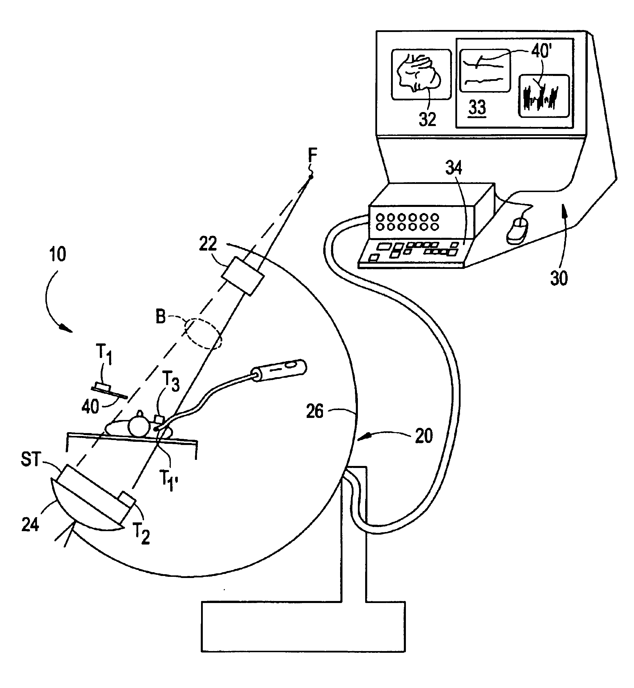
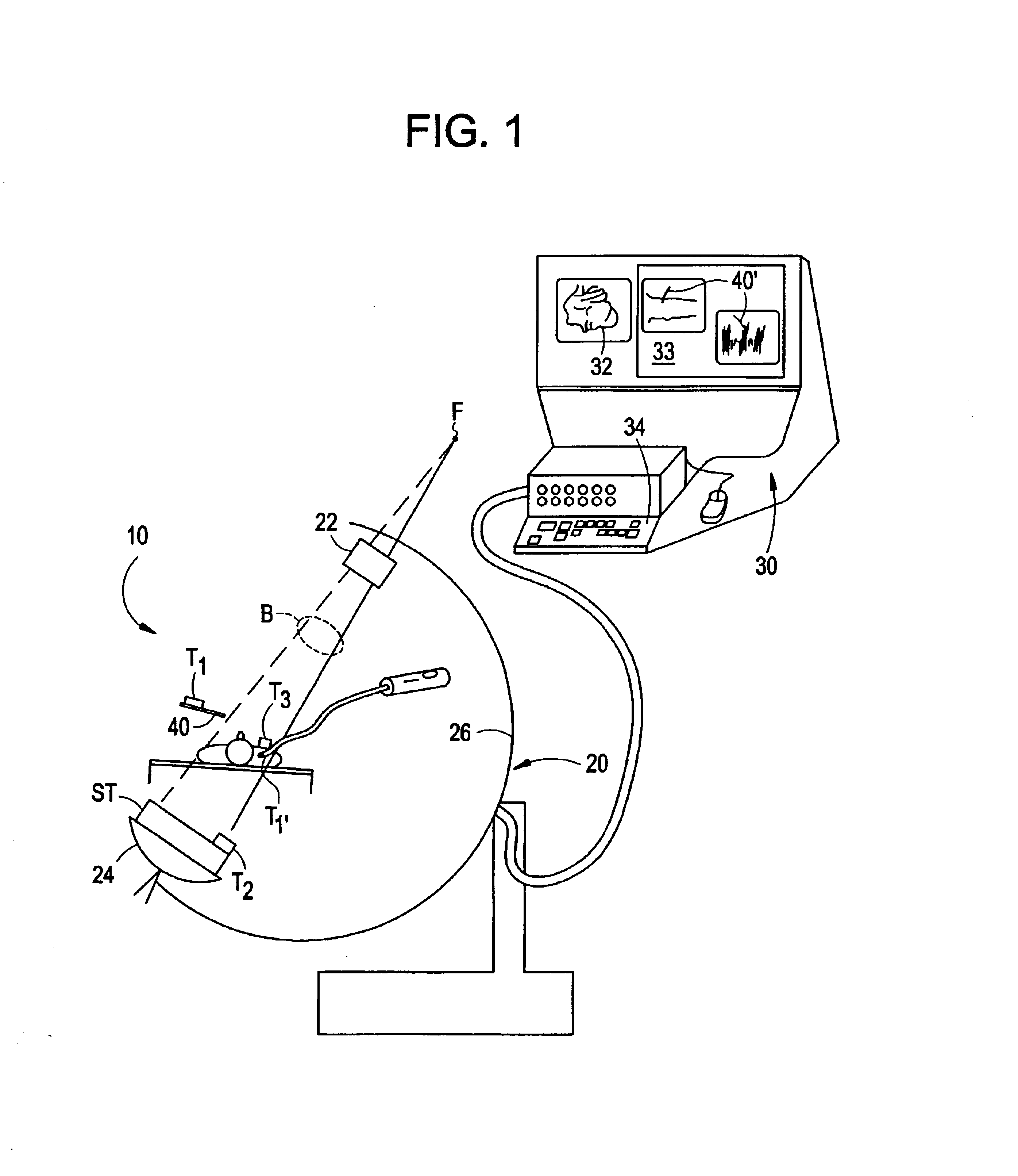
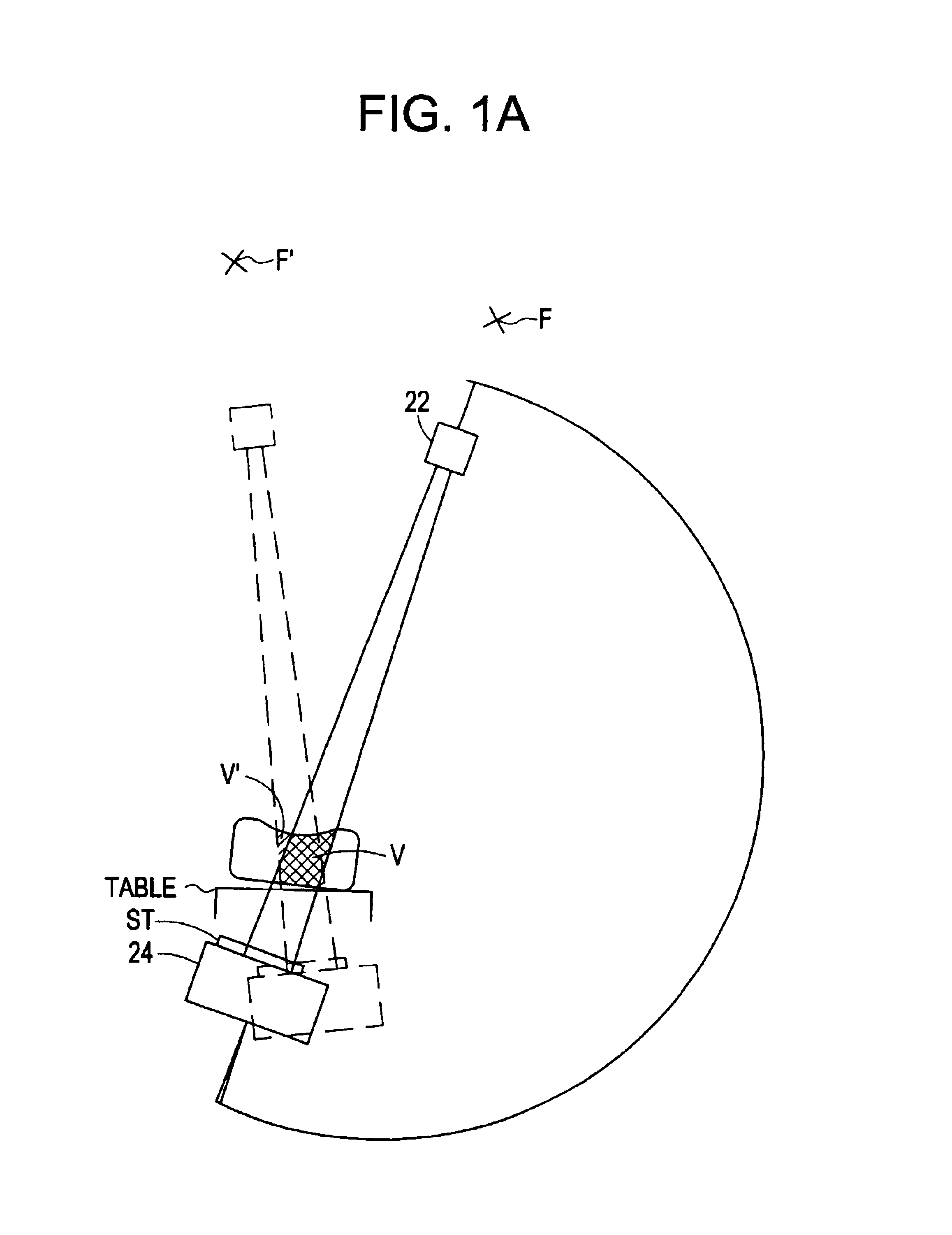
FIG. 1 illustrates elements of a basic embodiment of a system 10 in accordance with the present invention for use in an operating room environment. As shown, the system 10 includes a fluoroscope 20, a work station 30 having one or more displays 32 and a keyboard / mouse or other user interface 34, and a plurality of tracking elements T1, T2, T3. The fluoroscope 20 is illustrated as a C-arm fluoroscope in which an x-ray source 22 is mounted on a structural member or C-arm 26 opposite to an x-ray receiving and detecting unit, referred to herein as an imaging assembly 24. The C-arm moves about a patient for producing two dimensional projection images of the patient from different angles The patient remains positioned between the source and the camera, and may, for example, be situated on a table or other support, although the patient may move. The tracking elements, described further below, are mounted such that one element T1 is affixed to, incorporated in or otherwise secured against m...


PUM


Abstract

A method for surgical imaging and display including (i.) positioning a defined set of markers disposed in a pattern so as to be imaged in each pose or view of an imaging assembly, the set of markers being fixed in pre-determined positions in a rigid carrier, (ii.) securing a first tracking element against motion with respect to the rigid carrier so that determining a position of the first tracking element in a single measurement determines positions of all the markers of the set, and (iii.) identifying images of at least a subset of the markers in a first view.

Description

BACKGROUNDThe present invention relates to medical and surgical imaging, and in particular to intraoperative or perioperative imaging in which images are formed of a region of the patient's body and a surgical tool or instrument is applied thereto, and the images aid in an ongoing procedure. It is of a special utility in surgical procedures such as brain surgery and arthroscopic procedures on the knee, wrist, shoulder or spine, as well as certain types of angiography, cardiac procedures, interventional radiology and biopsies in which x-ray images may be taken to display, correct the position of, or otherwise navigate a tool or instrument involved in the procedure.Several areas of surgery have required very precise planning and control for the placement of an elongated probe or other article in tissue or bone that is internal or difficult to view directly. In particular, for brain surgery, stereotactic frames to define the entry point, probe angle and probe depth are used to access a...

Claims


Application Information

Patent Timeline
IPC IPC(8): A61B19/00A61B5/06A61B6/12A61B6/00
CPCA61B5/06A61B6/12A61B6/5235A61B6/547A61B19/52A61B19/5244A61B6/583A61B6/5247A61B2019/5272A61B2019/5238A61B2019/5251A61B2019/5255A61B5/062A61B6/4441A61B6/465A61B6/487A61B6/584A61B34/20A61B90/36A61B2034/2051A61B2034/2055A61B2034/2072A61B2090/376
Inventor SEELEY, TERESALIN, FAITHKAPUR, TINAGREGERSON, GENE
Owner STRYKER EURO HLDG I LLC
Why Eureka
Try Eureka