Artery- and vein-specific proteins and uses therefor

a technology applied in the field of artery and vein specific proteins, can solve the problems of morbidity and mortality in modern society, disassembly of the circulatory system, etc., and achieve the effect of enhancing or inhibiting the

Inactive Publication Date: 2005-05-03
CALIFORNIA INST OF TECH
View PDF26 Cites 38 Cited by
  • Summary
  • Abstract
  • Description
  • Claims
  • Application Information

AI Technical Summary

Benefits of technology

[0004]The present invention relates to methods of distinguishing and separating arterial cells from vein cells, and more specifically, distinguishing and separating arterial endothelial cells from venous endothelial cells based on their respective molecular markers; methods of selectively targeting or delivering drugs or agents to arteries or veins; methods of altering (enhancing or inhibiting, where “inhibiting” includes partially or complet...

Problems solved by technology

Disorders of the circulatory system, such as coronary artery di...

Method used


Image

  • Artery-  and vein-specific proteins and uses therefor
  • Artery-  and vein-specific proteins and uses therefor

Examples


example 1

Targeted Mutagenesis of EphrinB2 in Mice

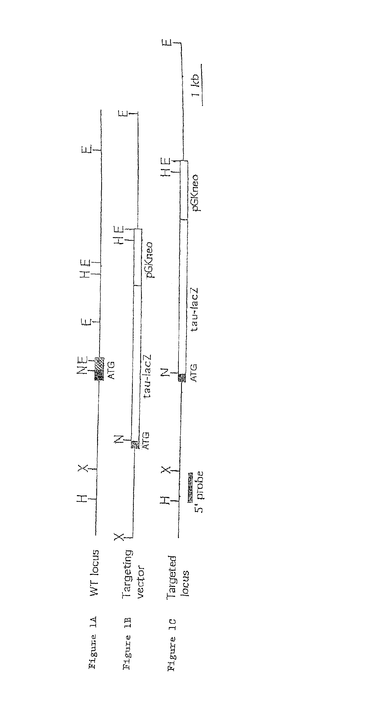
[0082]Targeted disruption of the EphrinB2 gene was achieved by homologous recombination in embryonic stem cells. The targeting strategy involved deleting the signal sequence and fusing a tau-lacZ indicator gene in frame with the initiation codon. The expression pattern of β-galactosidase in heterozygous (EphrinB2tlacZ / +) embryos was indistinguishable from that previously reported for the endogenous gene. (Bennett, B. D. et al. Proc. Natl. Acad. Sci. USA 92: 1866-1870 (1995); Bergemann, A. D. et al. Mol. Cell. Bio. 1995:4921-4929 (1995); Wang, H. U. and Anderson, D. J. Neuron 18:383-396 (1997)). While prominent expression was detected in the hindbrain and somites, lower levels were observed in the aorta and heart as early as E8.25. Expression in the yolk sac was first detected at E8.5. Heterozygous animals appeared phenotypically normal. In homozygous embryos, growth retardation was evident at E10 and lethality occurred with 100% penetrance aro...

example 2

Reciprocal Expression Pattern of EphrinB2 and EphB4 in Arteries and Veins

[0083]The enlarged heart observed in dying mutant embryos prompted examination of the expression of EphrinB2tlacZ in the vascular system in detail. Expression was consistently observed in arteries but not veins. In the yolk sac, for example, the posterior vessels connected to the vitelline artery, but not the vitelline vein, expressed the gene, as detected by lacZ staining. In the trunk, labeling was detected in the dorsal aorta, vitelline artery, umbilical artery and its allantoic vascular plexus, but not the umbilical, anterior and common cardinal veins (the umbilical vein is labeled with anti-PECAM-1 antibody). In the head, labeling was seen in branches of the internal carotid artery, but not in those of the anterior cardinal vein. In situ hybridization with EphrinB2 cDNA probes confirmed that the selective expression of tau-lacZ in arteries correctly reflected the pattern of expression of the endogenous gen...

example 3

Vasculogenesis Occurs Normally in EphrinB2 Mutant Embryos

[0084]The formation of the major vessels in the trunk was unaffected by the lack of EphrinB2, as examined by lacZ and PECAM-1 double staining of 9 somite embryos. Expression of EphrinB2-lacZ was seen in the dorsal aorta and vitelline artery, but not the umbilical and posterior cardinal veins. The dorsal aorta, vitelline artery, posterior cardinal and umbilical veins, for example, formed, although some dilation and wrinkling of the vessel wall was observed. Similarly the intersomitic vessels originating from the dorsal aorta formed at this stage. Between E8.5 and E9.0, the primitive endocardium appeared only mildly perturbed in mutants, while a pronounced disorganization was apparent at E10. Red blood cells developed and circulated normally up to E9.5 in both the mutant yolk sac and embryo proper.


PUM

PropertyMeasurementUnit
Solubility (mass)aaaaaaaaaa
Fluorescenceaaaaaaaaaa

Abstract

Arterial and venous endothelial cells are molecularly distinct from the earliest stages of angiogenesis. This distinction is revealed by expression on arterial cells of a transmembrane ligand, called EphrinB2 whose receptor EphB4 is expressed on venous cells. Targeted disruption of the EphrinB2 gene prevents the remodeling of veins from a capillary plexus into properly branched structures. Moreover, it also disrupts the remodeling of arteries, suggesting that reciprocal interactions between pre-specified arterial and venous endothelial cells are necessary for angiogenesis. This distinction can be used to advantage in methods to alter angiogenesis, methods to assess the effect of drugs on artery cells and vein cells, and methods to identify and isolate artery cells and vein cells, for example.

Description

RELATED APPLICATIONS[0001]This application is a continuation-in-part of PCT Application Number PCT / US99 / 08098 filed Apr. 13, 1999, which is a continuation-in-part of U.S. application Ser. No. 09 / 085,820 filed on May 28, 1998, which is a continuation-in-part of U.S. application Ser. No. 09 / 083,546 filed on May 22, 1998 (abandoned). This application also claims the benefit of U.S. Provisional Application No. 60 / 081,757 filed on Apr. 13, 1998. The teachings of each of these applications are incorporated herein by reference in their entirety.BACKGROUND OF THE INVENTION[0002]The process of blood vessel formation is fundamental in both development and disease. The circulatory system is the first organ system to emerge during embryogenesis, and is necessary to nourish the developing fetus. Disorders of the circulatory system, such as coronary artery disease, are a major cause of morbidity and mortality in modern society. Thus, repairing, replacing and promoting the growth of blood vessels ...

Claims


Application Information

Patent Timeline
IPC IPC(8): A01K67/033A01K67/027A01K67/00A61K35/12A61K38/00A61K38/17A61K39/00A61K45/00A61K38/19A61K39/395A61K47/48A61K47/46A61P35/00A61P9/10A61P9/00C07H21/00C07H21/04C07K14/00C07K16/00C07K16/28C07K16/18C12Q1/68C12Q1/04C12N5/00C12N5/02C12N15/09C12N5/10C12N5/06C12N5/08G01N33/53G01N33/50G01N33/48G01N33/567G01N33/574C07K14/705C12N5/071C12N15/85C12P21/02
CPCA01K67/0276A61K47/48561A61K49/0002C07K14/705C07K16/2866C07K16/30C12N5/0691C12N15/8509A01K67/0275A61K39/3955A61K47/48538A61K49/00G01N33/50A01K2227/105A01K2267/03A01K2267/0375C07K2316/96C12N2503/02G01N2333/52A01K2217/075C07K2317/76A61K49/0008G01N33/573G01N33/6863A61P35/00A61P9/00A61P9/10
Inventor WANG, HAI U.CHEN, ZHOUFENGANDERSON, DAVID J.
Owner CALIFORNIA INST OF TECH
Why Eureka
Try Eureka