Methods for disease detection

a disease detection and method technology, applied in the field of disease detection, can solve the problems proteins, other cellular components in healthy individuals, and relatively large cellular components, and achieve the effect of reducing the integrity (intactness) of nucleic acids

Inactive Publication Date: 2005-07-19
ESOTERIX GENETIC LAB
View PDF218 Cites 72 Cited by
  • Summary
  • Abstract
  • Description
  • Claims
  • Application Information

AI Technical Summary

Benefits of technology

[0006]The present invention provides that the integrity of nucleic acids, proteins, and / or other cellular components in a biological sample comprising shed cellular material indicates the disease status of the patient from whom the sample was obtained. According to the invention, tissue or body fluid samples, especially those described below, contain shed cellular debris. In healthy patients, such debris is the result of apoptotic degradation as part of the normal cell cycle. Apoptosis reduces the integrity (intactness) of nucleic acids, proteins, and other cellular components in healthy individuals, so that only small fragments exist in the debris that results from the apoptotic process (e.g., exfoliated cellular debris). To the contrary, in diseases such as cancer in which cell cycle mechanisms are destroyed or impaired, cellular debris comprises high-integrity cellular components, such as nucleic acids (i.e., nucleic acids that have not been degraded by apoptosis).
[0022]In an alternative embodiment, screening of patient samples by detecting amounts of nucleic acid in the sample is combined with an assay for apoptotic cell activity. Such assays may be combined with detecting amounts of nucleic acid in a patient sample as a screen for disease status. A positive screen is one that produces both: (1) an amount of nucleic acid that is greater than the amount expected to be present in a normal sample (e.g., one not having the disease being screened), and (2) an amount of apoptotic cell activity that is less than that expected to be present in a normal sample. In a highly preferred embodiment, methods of the invention comprise analyzing a plurality of genomic loci to determine an amount of amplifiable nucleic acid present at each locus. Analysis across multiple loci using methods of the invention may increase the sensitivity of the screening assay.

Problems solved by technology

Apoptosis reduces the integrity (intactness) of nucleic acids, proteins, and other cellular components in healthy individuals, so that only small fragments exist in the debris that results from the apoptotic process (e.g., exfoliated cellular debris).
Patients having a disease generally produce cells and cellular debris, a proportion of which has avoided normal cell cycle regulation, resulting in relatively large cellular components.

Method used


Image

  • Methods for disease detection
  • Methods for disease detection
  • Methods for disease detection

Examples


example 1

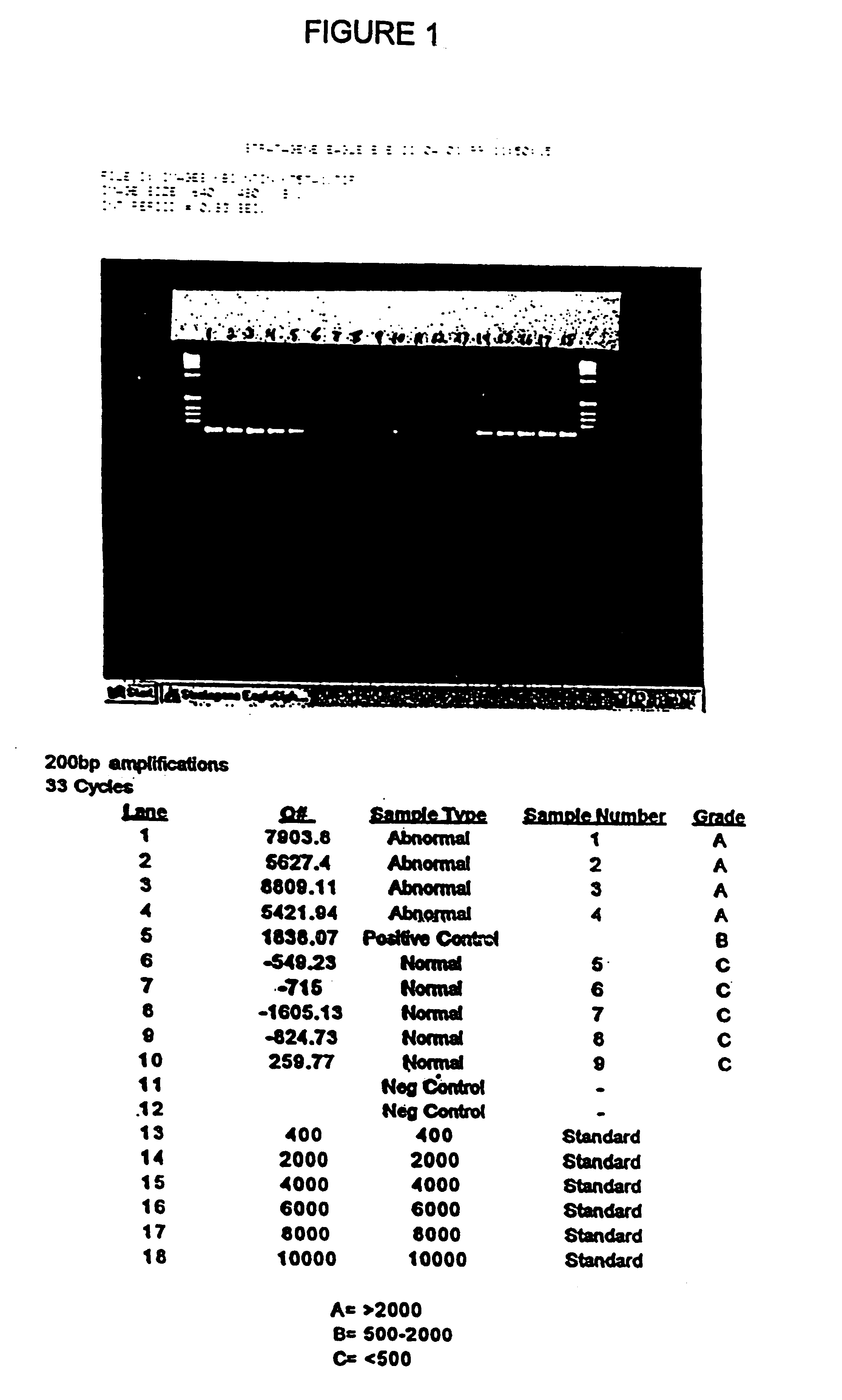
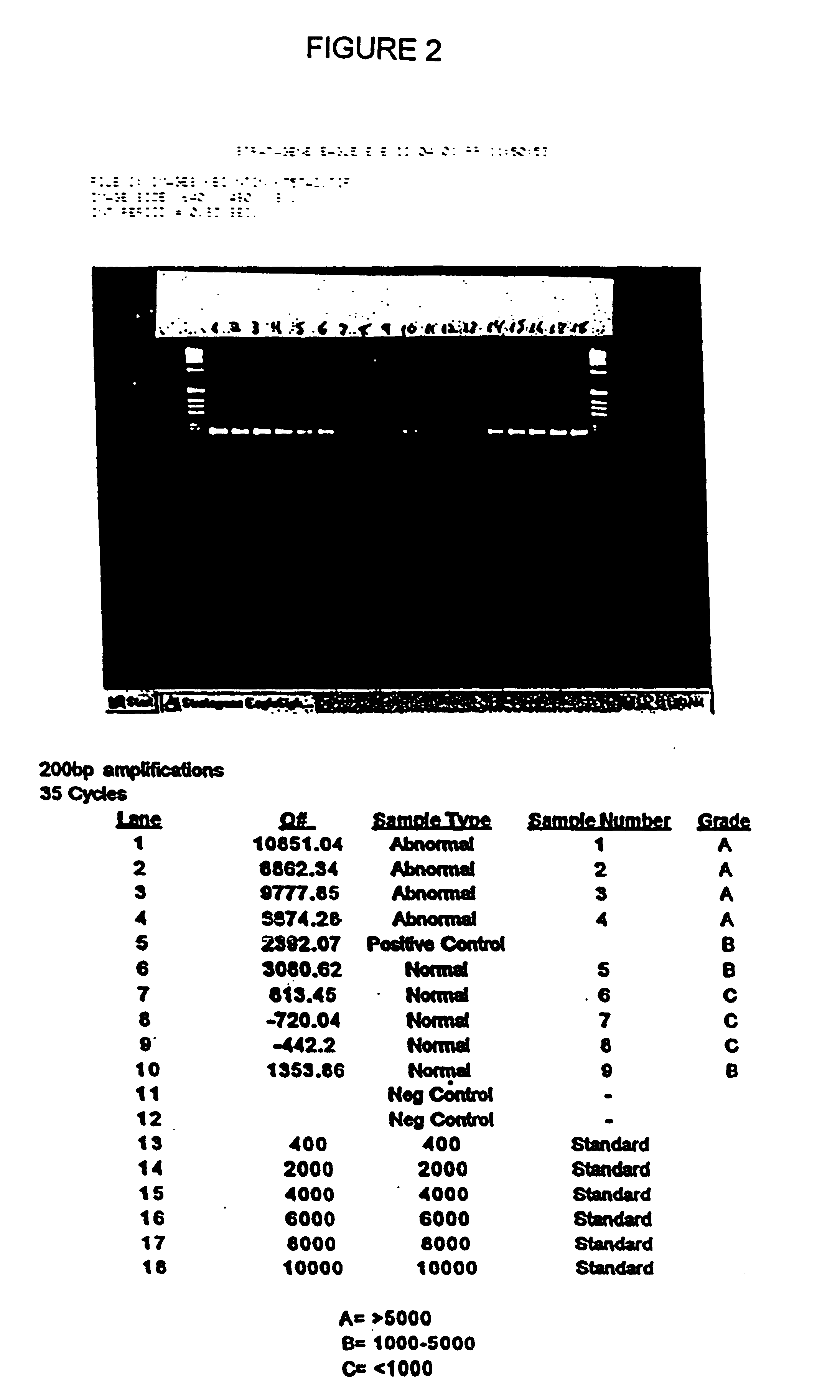
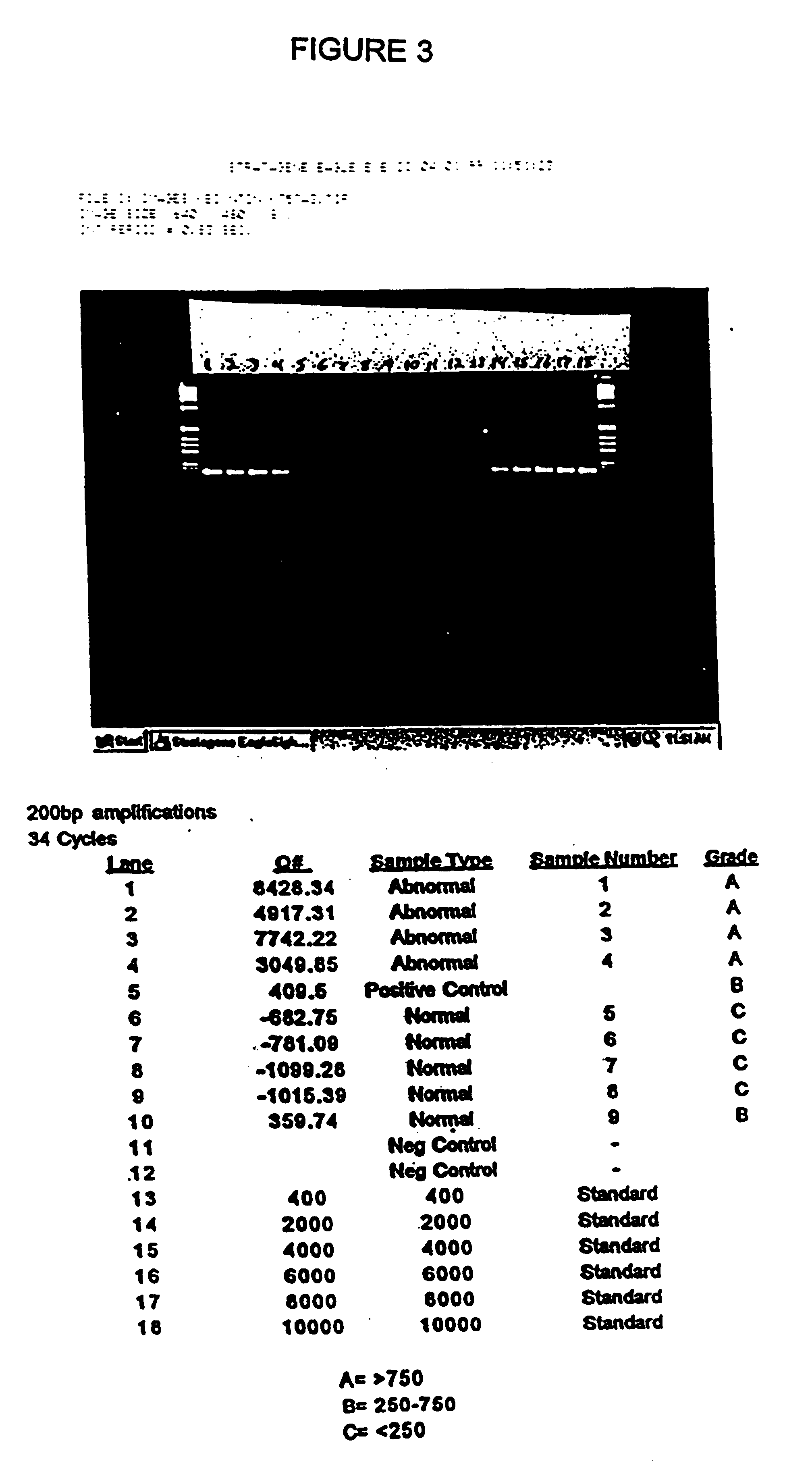
[0065]Stool samples were collected from 9 patients who presented with symptoms or a medical history that indicated that a colonoscopy should be performed. Each stool sample was frozen. Immediately after providing a stool sample, each patient was given a colonoscopy in order to determine the patient's disease status. Based upon the colonoscopy results, and subsequent histological analysis of biopsy samples taken during colonoscopy, individuals were placed into one of two groups: normal or abnormal. The abnormal group consisted of patients with cancer or with an adenoma of at least 1 cm in diameter. Based upon these results, 4 of the 9 patients were placed into the abnormal group.

[0066]The samples were screened by hybrid capturing human DNA, and determining the amount of amplifiable DNA having at least 200 base pairs. Each frozen stool specimen, weighing from 7-33 grams, was thawed and homogenized in 500 mM Tris, 16 mM EDTA, and 10 mM NaCl, pH 9.0 at a volume, to mass ratio of 3:1. Sa...

example 2

[0073]An experiment was conducted that was essentially identical to the one described above in Example 1, but forward and reverse primers were placed such that fragments of about 1.8 Kb and above were amplified.

[0074]DNA was prepared as described above. Forward and reverse primers were spaced so as to hybridize approximately 1.8 Kb apart on three different loci (Kras, exon 1, APC, exon 15, and p53 exon 5). Thirty-three rounds of amplification were performed, and the resulting DNA was placed on a 3% agarose gel. The results are shown in FIGS. 8-10. As shown in the Figures (which show results from three separate experiments to amplify and detect “long” product), samples from individuals having cancer or precancer produced large amounts of high-molecular weight (in this case 1.8 Kb and above) DNA; whereas samples from patients who did not have cancer or precancer produced no DNA in the range of about 1.8 Kb and higher. Thus, the presence of high-molecular weight DNA was indicative of t...

example 3

[0075]An experiment was conducted to determine the molecular weight profile of DNA from samples collected and prepared as part of a blind study on 30 patients who presented at the Mayo Clinic with suspected gastrointestinal disorders. Stool samples were obtained, and DNA was isolated as described above.

[0076]Prior to amplification, DNA was isolated from the samples by hybrid capture. Biotynilated probes against portions of the BRCA1, BRCA2, p53, APC genes were used.

[0077]The BRCA1probe was 5′GATTCTGMGAACCMCTTTGTCCTTMCTAGCTC173′ (SEQ ID NO: 8).

[0078]The BRCA2 probe was 5′CTAAGTTTGAATCCATGCTTGCTCTTCTTGATTATT3′ (SEQ ID NO 9).

[0079]The APC1 probe was 5′CAGATAGCCCTGGACAAACCATGCCACCAAGCAGAAG3′ (SEQ ID NO 10).

[0080]The p53 probe, hybridizing to a portion of exon 5, was 5′TACTCCCCTGCCCTCAACAAGATGTTTTGCCAACTGG3′ (SEQ ID NO:4).

[0081]The APC2 probe was 5′GAAGTTCCTGGATTTTCTGTTGCTGGATGGTAGTTGC3′ (SEQ ID NO 11).

[0082]A 300 μl aliquot of sample was placed in 300 μl of 6 M guanidine isothiocyanate ...


PUM

PropertyMeasurementUnit
diameteraaaaaaaaaa
mass ratioaaaaaaaaaa
lengthaaaaaaaaaa

Abstract

The present invention provides methods for detecting disease by analysis of a patient sample to determine the integrity of nucleic acids in the sample.

Description

RELATED APPLICATIONS[0001]This application claims priority to and the benefit of U.S. Ser. No. 60 / 169,457, filed Dec. 7, 1999, the entire disclosure of which is incorporated herein by reference.BACKGROUND OF THE INVENTION[0002]Many diseases are associated with genomic instability. That is, a disruption in genomic stability, such as a mutation, has been linked to the onset or progression of disease. Accordingly, various aspects of genomic instability have been proposed as reliable markers for disease. For example, mutations in the BRCA genes have been proposed as markers for breast cancer, and mutations in the p53 cell cycle regulator gene have been associated with numerous cancers, especially colorectal cancer. It has been suggested that specific mutations might be a basis for molecular screening assays for the early stages of certain types of cancer. See, e.g., Sidransky, et al., Science, 256: 102-105 (1992).[0003]The search for genomic disease markers has been especially intense i...

Claims


Application Information

Patent Timeline
Patent Type & Authority Patents(United States)
IPC IPC(8): C12Q1/68C12N15/09
CPCC12Q1/6886C12Q2600/156
Inventor SHUBER, ANTHONY P.
Owner ESOTERIX GENETIC LAB
Why Eureka
Try Eureka