Method and system for imaging the dynamics of scattering medium

a scattering medium and dynamics technology, applied in the field of scattering medium imaging, can solve the problems of high computing power to cost ratio, excessive branching of vessels, and highly disorganized tissues, and achieve the effects of reducing the need for undue system calibration, enhancing resolution and contrast, and enhancing stability

Inactive Publication Date: 2005-08-30
THE RES FOUND OF STATE UNIV OF NEW YORK
View PDF6 Cites 20 Cited by
  • Summary
  • Abstract
  • Description
  • Claims
  • Application Information

AI Technical Summary

Benefits of technology

[0029]The present invention satisfies these needs by providing a method for (1) enhancing resolution and contrast of various dynamic features of the medium being imaged, (2) imaging the dynamic properties of the target medium, and (3) generating images of the dynamic properties using techniques that reduce the need for undue system calibration and produce more stable solutions with the reconstruction algorithm.

Problems solved by technology

These techniques permit the use of types of energy and wavelengths, such as near infrared light energy, that are not suitable for projection imaging techniques, such as x-ray imaging.
Additionally, since a significant computational effort may be required for both image reconstruction and data analysis, the technology particularly gains from the exponential growth in the ratio of computing power to cost.
Ultrastructurally, these tissues are highly disorganized and exhibit functional abnormalities.
There is excessive branching of the vessels, including significant arterio-venous shunting as well as blind vascular endings.
Although these attributes of disease tissue are well appreciated, the availability of a suitable detection methodology able to take full advantage of these characteristics has been notably lacking.
These methods however are insensitive to hemoglobin states and only indirectly provide measures of altered blood flow.
The latter is well accomplished, in the case of larger vessels, using Doppler ultrasound, and for near-surface microvessels by laser Doppler measurements, but each is insensitive to variations in tissue blood volume or blood oxygenation.
Ultrasound measurements are also limited by their inability to penetrate bone.
First, known methods and systems provide images having low contrast and resolution.
Second, these methods and systems do not image the dynamic properties of highly scattering media.
Third, these methods and systems require accurate calibration and are susceptible to errors.
In addition, the physics of light transport in highly scattering media, such as tissue, imposes further practical constraints that relate to the method adopted for data analysis.
At issue is the required accuracy of assumptions made in order to generate predicted detector values, especially those adopted for the initial estimate.
Small errors in the initial guess of optical properties of the reference medium can lead to large errors in the computed detector values.
One consequence of this can be the severe corruption of the information content of the data vectors leading to artifact-laden images.
The net effect of these limitations can render solutions to image recovery problems of this type overly sensitive to the influence of experimental noise (i.e., ill-conditioned), provide nonunique solutions (ill-posed) or both.
It is also well appreciated that, in general, there are no simple or well-defined methods that can be universally applied to overcome the noted limitations.
For instance, it is known that heart rate variability is chaotic.
Presently, the capacity to monitor dynamic behavior in vascular structures is limited principally to near surface measures using laser Doppler methods.
However, these measures are insensitive to the activity of the microvasculature, do not provide for full cross-sectional views, and are not sensitive to the dynamics of hemoglobin states.
Although optical methods, such as Laser Doppler, pulse oximetry, photoplethysmography and the like, can be used to monitor dynamic states of the vasulature, none are capable of providing such measures in the form of a cross-sectional image, especially in the case of large tissue structures.
Moreover, known optical methods for cross-sectional imaging of the properties of a scattering media have not been used to derive dynamic measures of these states, and are plagued with a host of technical limitations, such as low contrast, low resolution images, prolonged computing times, excessive sensitivity to errors in initial estimates.
These limitations render it unlikely that useful information regarding dynamic states could be derived from these known methods.

Method used


Image

  • Method and system for imaging the dynamics of scattering medium
  • Method and system for imaging the dynamics of scattering medium
  • Method and system for imaging the dynamics of scattering medium

Examples


Embodiment Construction

Introduction

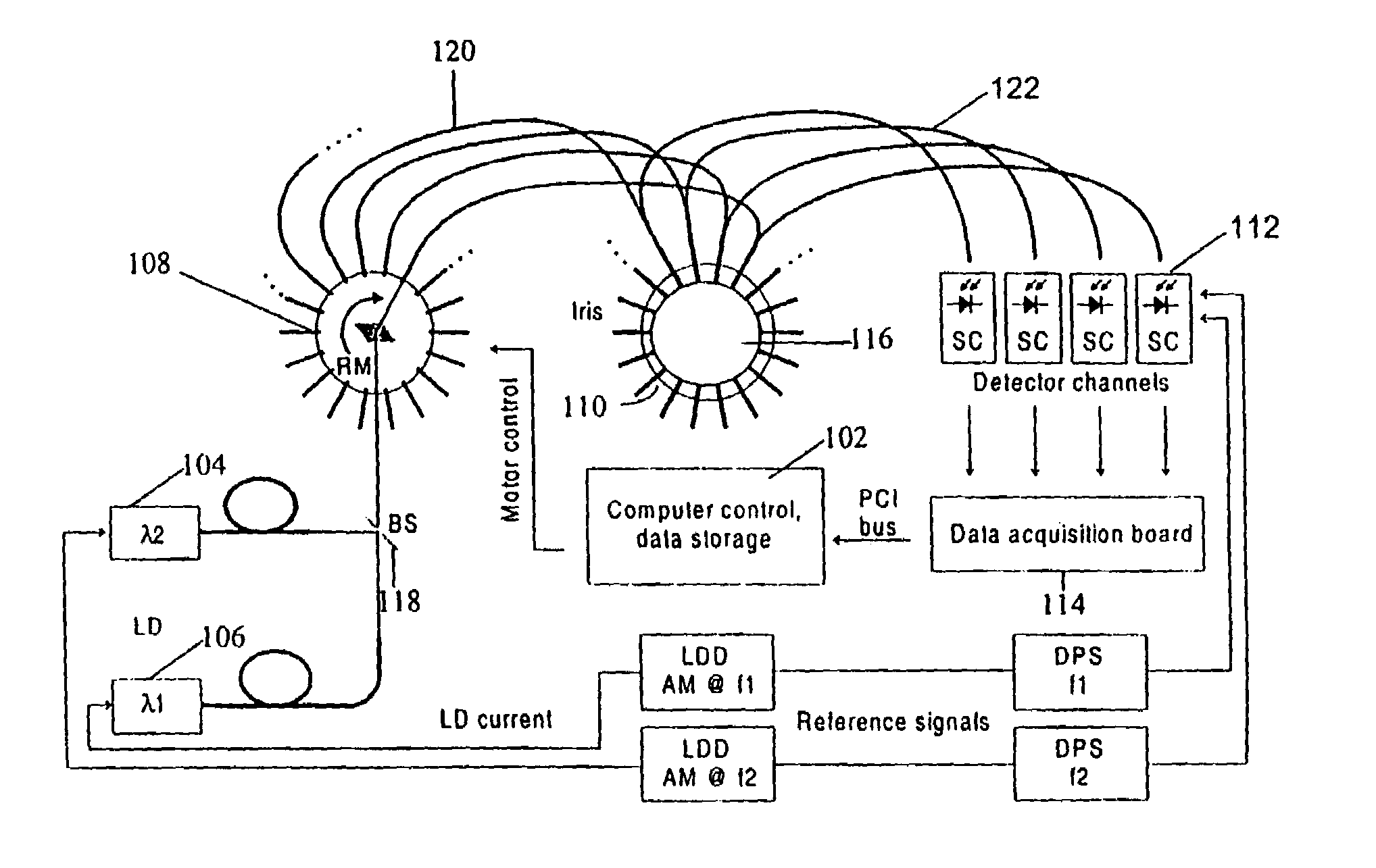
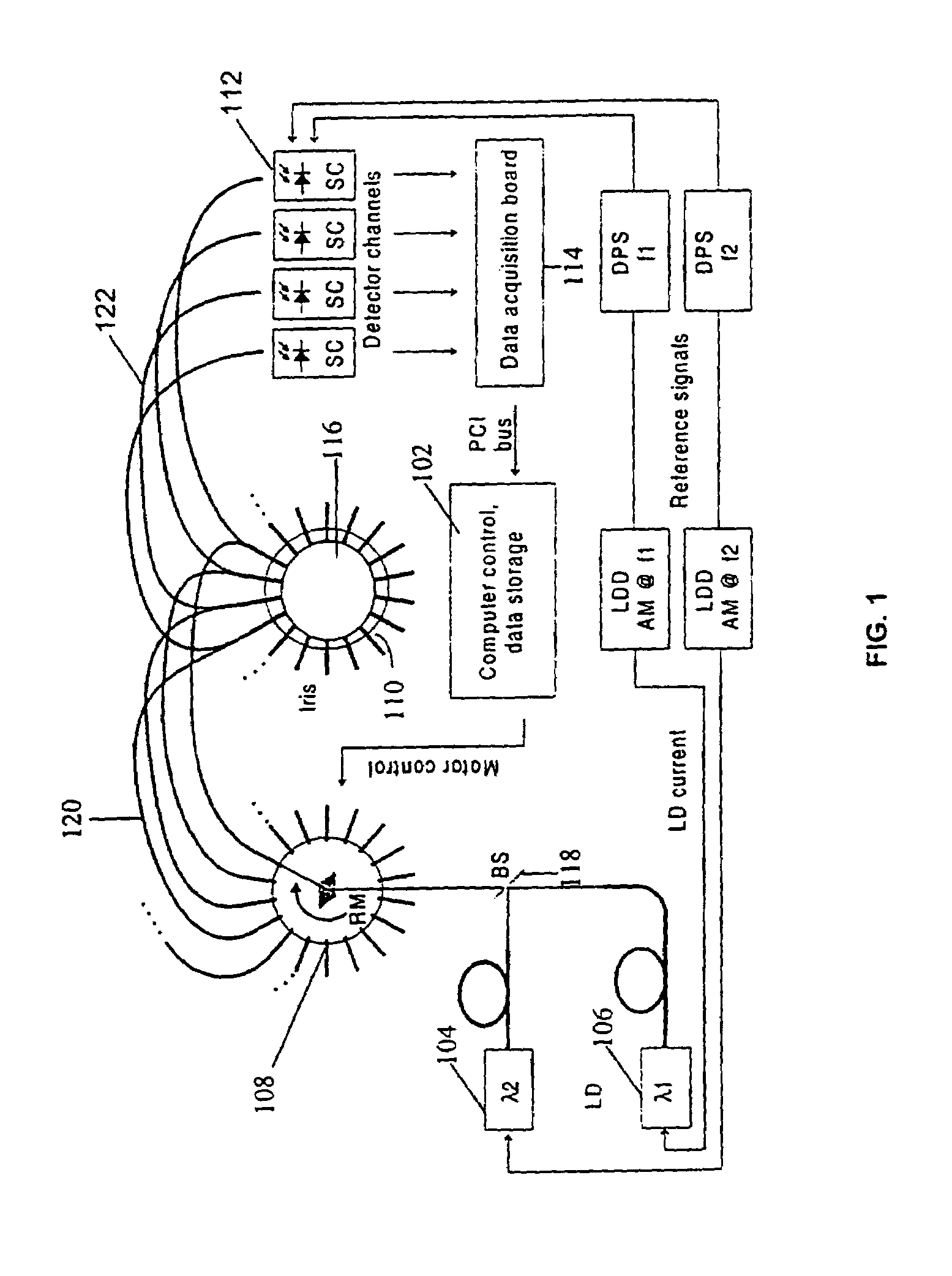
[0059]The present invention is built upon known basic approaches to tomographic imaging of a scattering medium, by enabling the investigation of the spatio temporal dynamics of a target medium. This enhancement represents a fundamentally new imaging modality with broad based applications such as in industrial processes involving preparation of powdered mixtures, imaging subsurface turbulence in murky waters and throughout clinical medicine both as a highly sensitive diagnostic imaging tool, as well as for use in treatment planning, therapeutic monitoring and follow-up.

[0060]For example, the present invention recognizes that the occurrence of dynamic behavior in human tissue lends the collected data and reconstructed images to evaluation using time series analysis methods, including pattern recognition schemes. Dynamic behavior is known to occur in vascular structures in response to cardiac, respiratory, vasomotor and local metabolic influences. Because optical tomography...


PUM

PropertyMeasurementUnit
diameteraaaaaaaaaa
modulation frequencyaaaaaaaaaa
modulation frequenciesaaaaaaaaaa

Abstract

A method and system for imaging the dynamics of a scattering medium (116) is provided. The method and system generates contrast and resolution enhanced images of dynamic properties of a medium having a temporal signature by using time series analysis methods on a time series of collected data or time series of images to extract and isolate dynamic properties of the medium (116).

Description

[0001]This application claims the benefit under 35 U.S.C. §120 of prior U.S. Provisional Patent Application Ser. No. 60 / 153,926 filed Sep. 14, 1999, entitled DYNAMIC TOMOGRAPHY IN A SCATTERING MEDIUM and 60 / 154,099 filed Sep. 15, 1999, entitled DYNAMIC TOMOGRAPHY IN A SCATTERING MEDIUM.[0002]This application is related to copending application Ser. No. PCT / US00 / 25155, filed on the same date as this application, entitled “SYSTEM AND METHOD FOR TOMOGRAPHIC IMAGING OF DYNAMIC PROPERTIES OF A SCATTERING MEDIUM” by inventors R. Barbour and C. Schmitz and is hereby incorporated by reference (hereinafter the “Barbour 4147PC1 application”). The counterpart U.S. patent application is application Ser. No. 10 / 088,254 filed Mar. 14, 2002.[0003]This application is also related to copending application Ser. No. PCT / US00 / 25156, filed on the same date as this application, entitled “IMAGING OF SCATTERING MEDIA USING RELATIVE DETECTOR VALUES” by inventor R. Barbour and is hereby incorporated by refer...

Claims


Application Information

Patent Timeline
Patent Type & Authority Patents(United States)
IPC IPC(8): A61B6/00G01N21/47
CPCG01N21/4795
Inventor BARBOUR, RANDALL L.
Owner THE RES FOUND OF STATE UNIV OF NEW YORK
Why Eureka
Try Eureka