Endoscope with a single image pick-up element for fluorescent and normal-light images

an endoscope and fluorescent light technology, applied in the field of endoscope devices, endoscopes and image processing devices for endoscopes, can solve the problems of high cost of endoscopes, inconvenient replacement of rotational filters, and insufficient thickness of insertable parts, so as to achieve finer and cost-reducing effects

Inactive Publication Date: 2005-11-01
OLYMPUS CORP
View PDF14 Cites 76 Cited by
  • Summary
  • Abstract
  • Description
  • Claims
  • Application Information

AI Technical Summary

Benefits of technology

[0025]It is an object of the present invention to provide an endoscope device (and an endoscope) whereby both a fluorescence image and a normal-light image can be captured by a single image pickup element, the insertable part can be made finer, and costs can also be reduced.
[0026]It is another object of the present invention to provide an endoscope device whereby excitation light of a wavelength suited to the endoscope is irradiated, in accordance with an endoscope actually connected, and both a fluorescence image and a normal-light image of good quality can be obtained.
[0029]Moreover, the endoscope according to the present invention is an endoscope for capturing a fluorescence image, by transmitting excitation light and irradiating same onto a subject by means of an illumination optical system, and receiving light by means of a single image pickup element, via an excitation light shielding filter which shields excitation light reflected by the subject, characterized in that an excitation light shielding filter is disposed in front of the image pickup element, the excitation light shielding filter having the characteristic whereby it shields excitation light of a part of the wavelength band of the blue wavelength band, while transmitting blue light in the wavelength band apart from the wavelength band of the excitation light, as well as transmitting green and red light wavelength bands; whereby it is possible to achieve capturing of the fluorescence image; and color capturing in the visible light region, on the basis of color component image in the green and red wavelength bands in the case of illumination by red, green and blue light, and of blue color component image in the case of illumination of blue light in a wavelength band shielding the wavelength of the excitation light.

Problems solved by technology

Therefore, such conventional endoscopes have drawbacks that the insertable parts are too thick, the endoscopes are expensive due to the necessity of providing two image pickup elements, and the like.
Therefore, in order to change the wavelength, it is necessary to replace the rotational filter, and if endoscopic examination is to be performed whilst changing the wavelength, then the laborious task of replacing the rotational filter is required, and time is also required for the endoscopic examination.

Method used


Image

  • Endoscope with a single image pick-up element for fluorescent and normal-light images
  • Endoscope with a single image pick-up element for fluorescent and normal-light images
  • Endoscope with a single image pick-up element for fluorescent and normal-light images

Examples


first embodiment

[0066](First Embodiment)

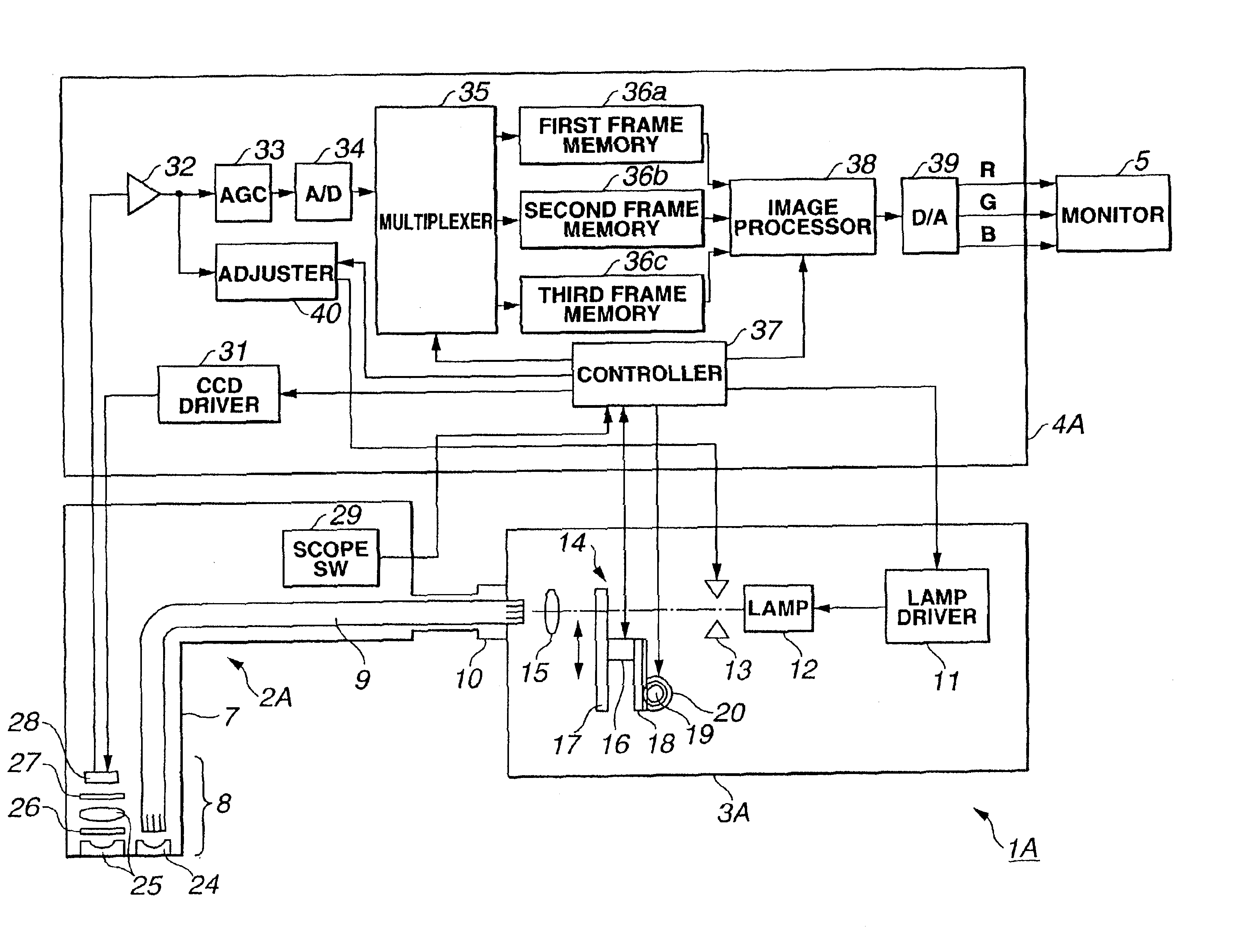
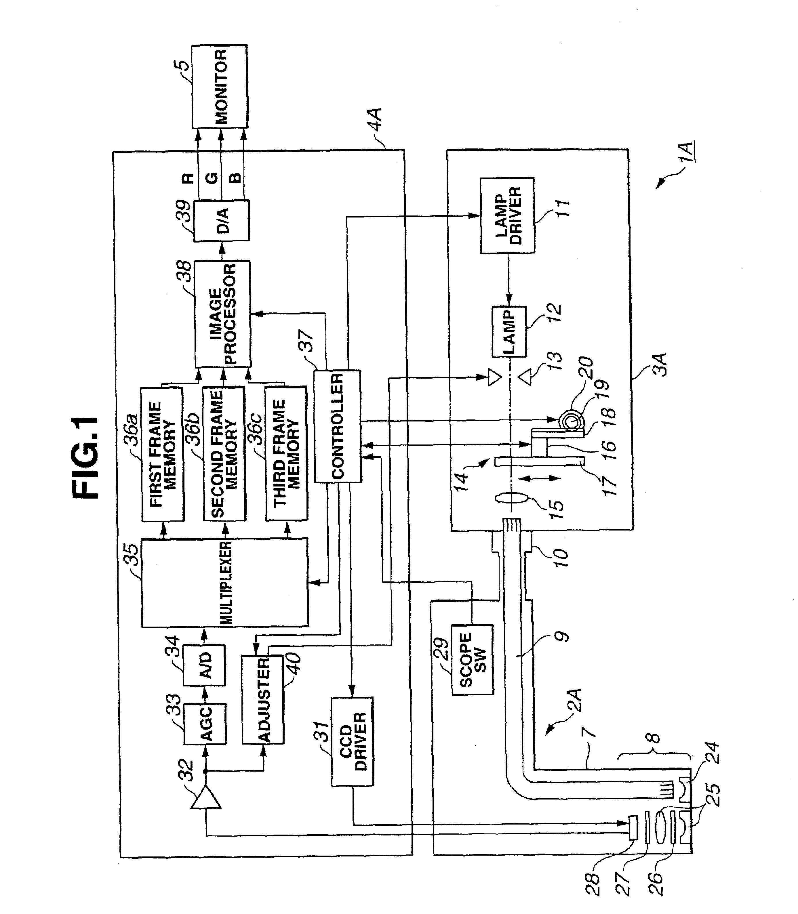
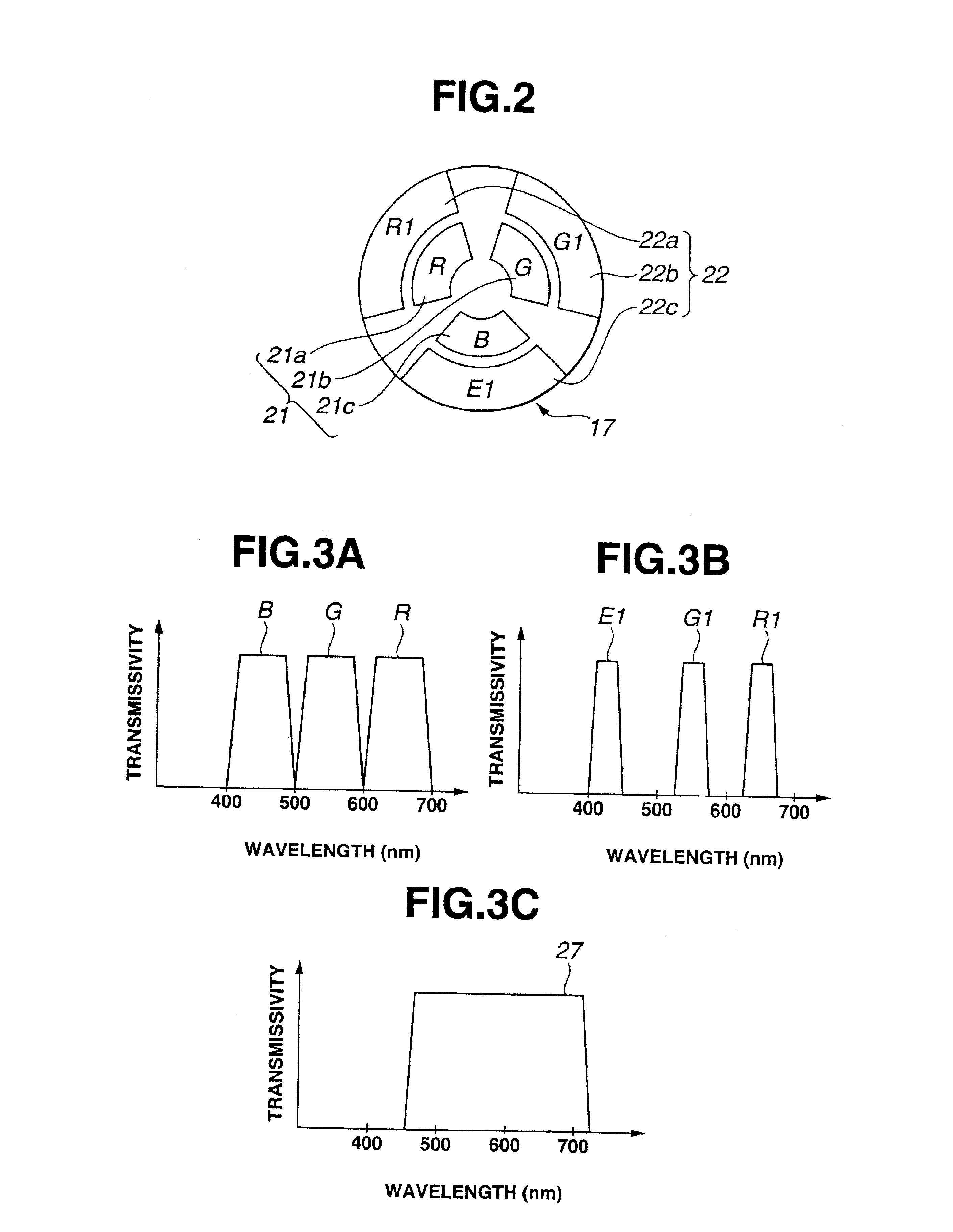
[0067]FIG. 1 to FIG. 13B relate to a first embodiment of the present invention: FIG. 1 shows the general composition of an endoscope device according to a first embodiment; FIG. 2 shows the composition of a switchable filter provided with a normal-light observation filter and a fluorescence observation filter; FIGS. 3A to 3C show transmission characteristic with respect to wavelength for a normal-light observation filter, fluorescence observation filter and excitation light shielding filter; FIGS. 4A and 4B show light intensity characteristic with respect to wavelength for light received by a CCD, when a white subject is observed in a normal-light observation mode and when skin is observed in a fluorescence observation mode; FIGS. 5A and 5B show fluorescence intensity and absorption characteristic in a case where normal tissue and cancerous tissue are observed in a fluorescence observation mode; FIGS. 6A to 6E show an operational diagram for normal-light obse...

second embodiment

[0176](Second Embodiment)

[0177]FIG. 14 to FIG. 20 relate to a second embodiment of the present invention; FIG. 14 is a general compositional diagram of an endoscope device comprising second embodiment; FIG. 15 is a chart showing the characteristic of light absorption with respect to wavelength of the haemoglobin contained in human tissue; FIG. 16 is a circuit block diagram showing the composition of the image processing circuit in FIG. 14; FIG. 17 is a graph showing the relationship between gain adjustment and haemoglobin concentration; FIG. 18 is a circuit block diagram showing a modification of the image processing circuit in FIG. 16; FIG. 19 is a chart showing transmission characteristic of an excitation light shielding filter which transmits the 500 to 700 nm wavelength band; and FIG. 20 is a chart showing the characteristic of light intensity received by the CCD with respect to wavelength, in normal-light mode, in the case of the excitation light shielding filter in FIG. 19.

[01...

third embodiment

[0218](Third Embodiment)

[0219]FIG. 21 is a circuit block diagram showing the composition of an image processing circuit according to a third embodiment of the present invention.

[0220]The third embodiment is constituted in such a manner that, in addition to the composition of the second embodiment, the B channel gain adjustment is corrected by generating a blue signal in accordance with the 1% to 4% haemoglobin concentration. The remaining composition is the same as the first embodiment and further description thereof is omitted. Similar constituent parts are assigned same reference numerals.

[0221]More specifically, as shown in FIG. 21, an image processing circuit 38C according to the second embodiment comprises a color element calculating section 61 for calculating the amount of haemoglobin by using the B′ light restricted to 470 to 500 nm and the R light (or the R light and G light), a parameter setting section 53c for setting parameters suited to the mode, on the basis of the amou...


PUM


Abstract

In a light source unit, a switchable filter section in which an RGB filter and a fluorescence observation filter can be shifted into the light path is disposed in front of a lamp. When fluorescence image mode is selected, excitation light in a part of the wavelength band of the blue wavelength band is supplied to an electronic endoscope, and the excitation light which is reflected by the subject is shielded by an excitation light shielding filter situated in front of a CCD, whereby a fluorescence image can be obtained. On the other hand, if normal-light image mode is selected, then R, G, B light is supplied sequentially, and even under illumination of B light, the color component image in the wavelength bands which are not shielded by the excitation light shielding filter are captured, thereby yielding a normal-light image also, and hence making it possible to capture both a fluorescence image and a normal-light image by means of a single image pickup element.

Description

[0001]This Application claims benefit of Japanese Patent Application No. 2001-146755 filed in Japan on May 16, 2001, No. 2001-302788 filed in Japan on Sep. 28, 2001, the contents of which are incorporated by this reference.BACKGROUND OF THE INVENTION[0002]1. Field of the Invention[0003]The present invention relates to an endoscope device, an endoscope and an image processing device for an endoscope, whereby normal white light images and fluorescence images can be observed.[0004]2. Description of the Related Art[0005]In recent years, endoscopes have come to be used widely in medical fields and industrial fields. Moreover, in medical fields, endoscope devices providing fluorescence images have also been achieved, in addition to endoscope devices providing standard images based on normal white light. As prior art examples of endoscope devices providing fluorescence images, the following devices have been disclosed, for example.[0006](a) U.S. Pat. No. 5,827,190[0007]This U.S. Patent dis...

Claims


Application Information

Patent Timeline
Patent Type & Authority Patents(United States)
IPC IPC(8): A61B1/04A61B1/045A61B5/00
CPCA61B1/00009A61B1/00059A61B1/00186A61B1/043A61B1/045A61B1/0638A61B1/0646A61B5/0071A61B1/0669A61B5/0084H04N2005/2255Y10S600/921A61B1/0655H04N23/555
Inventor UENO, HITOSHIOZAWA, TAKESHINAKAMURA, KAZUNARIHIRAO, ISAMIIBE, HIROSHIYOSHIMITSU, KOICHIMORIZANE, YUICHIKANEKO, MAMORUAKIMOTO, SHUNYATERAKUBO, MASAKIIMAIZUMI, KATSUICHIDOGUCHI, NOBUYUKITAKEHANA, SAKAE
Owner OLYMPUS CORP
Why Eureka
Try Eureka