Method and apparatus for targeting localized electroporation

a localized electroporation and electroporation technology, applied in biochemistry apparatus, biochemistry apparatus and processes, applications, etc., can solve the problems of inability to effectively target electroporation, lack of reproducibility, and previous electroporation efforts

Inactive Publication Date: 2005-12-20
INNOVATION & DEV CORP
View PDF7 Cites 7 Cited by
  • Summary
  • Abstract
  • Description
  • Claims
  • Application Information

AI Technical Summary

Benefits of technology

[0020]FIG. 2. The effect of varying pulse frequency on mean number of avian embryo cells transfected. Six, one-second trains were administered and pulse length was adjusted to provide equal power output. Electrodes were approximately the same size and 2–5 embryos were analysed per condition. Bars indicate standard errors.
[0021]FIG. 3. The effect of train number and polarity on mean number of avian embryo cells transfected. Trains were one second long and alternating train polarity is indicated by 3+3. All tests were performed at 500 pulses per second with a pulse duration of 1 ms. Electrodes were approximately the same size and 2–5 embryos were analysed per condition. Bars indicate standard errors.

Problems solved by technology

However, previous electroporation efforts have had a number of limitations, including the inability to effectively target the electroporation, lack of reproducibility of results, and exposure of non-targeted cells to potentially damaging current.
Specifically, the electroporation apparatuses of the prior art are generally paddle-like pairs of electrodes, having inherent limitations as to how small a target area would be electroporated and lacking integral means for introducing the foreign matter (commonly DNA) into the cells.
Accordingly, the prior art teaches unnecessary electroporation of (and thereby potential attendant injury to) cells outside the target area.
Moreover, methods of electroporation in the prior art have proved inefficient and unreliable in localising to any degree the transfection events, and cannot target a specific region of an organ or tissue.
Truly localised electroporation could neither be accomplished with prior methods, nor was it contemplated.
Prior art methods of in ovo electroporation have involved subjecting the entire embryo to potentially damaging current and the effects have been difficult to reproduce.
Cells and tissues subjected to these prior art methods would be not be uniformly electroporated, and as a result only random transfection events would occur within the selected areas.
Introduction of foreign compounds other than genetic material by electroporation has likewise suffered from a high degree of unpredictability, due to the inefficiency and unreliability of prior art methods of electroporation.

Method used


Image

  • Method and apparatus for targeting localized electroporation
  • Method and apparatus for targeting localized electroporation
  • Method and apparatus for targeting localized electroporation

Examples


example 1

Electroporation of Avian Embryos

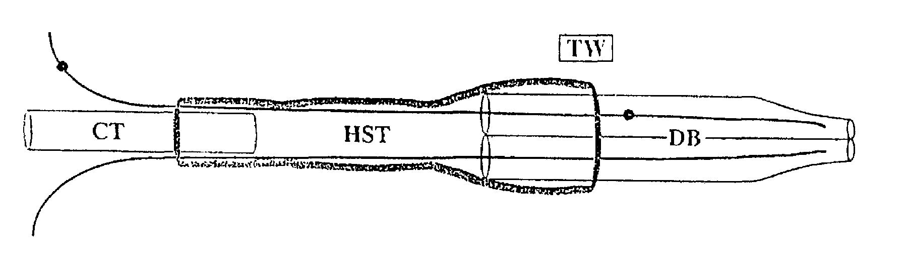
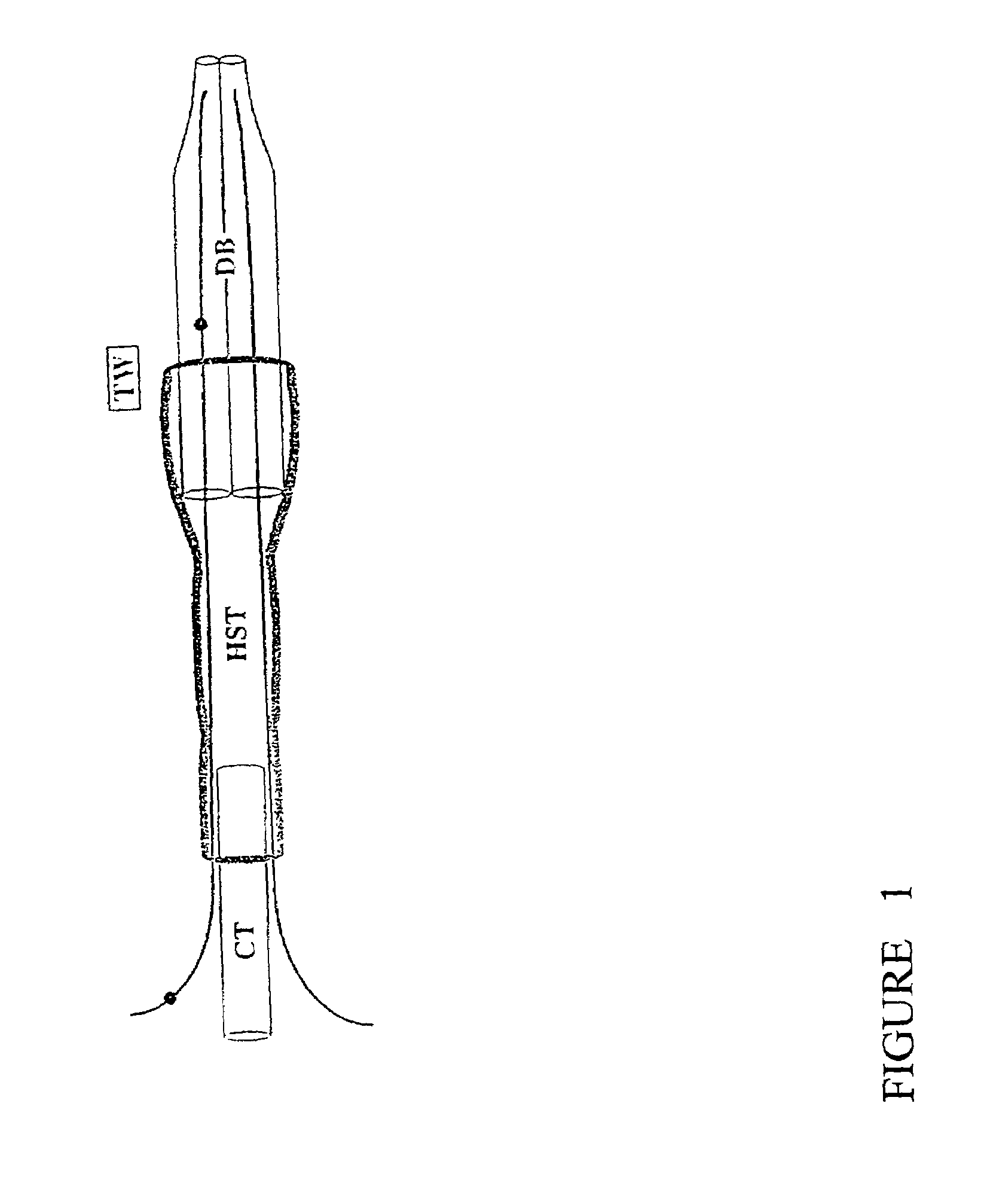
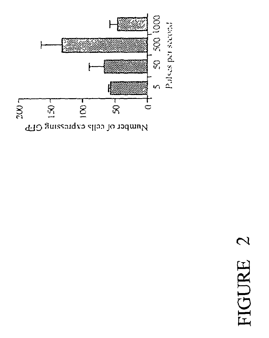
[0025]An electroporation apparatus, back-filled with DNA containing solution and driven by a conventional neurophysiological stimulator, was used to transfect plasmids containing green-fluorescent protein (GFP) into avian embryos. The electroporation apparatus was made from a 1.2 mm×0.6 mm, 4 inch, double-barrelled glass capillary tube (catalog #6070, A-M Systems) which was drawn on a vertical pipette puller (Model 700C, David Kopf Instruments), broken back to an inside diameter of 200–250 μm, and forged. Tungsten wire (catalog #7960, A-M Systems). coated with Teflon as an insulator and with the Teflon removed 5 mm from the ends, was inserted in each barrel to within 1 mm of the tip. A capillary tube mounting shaft was attached with heat-shrink tubing and sealed with epoxy (FIG. 1). This apparatus was connected with polyethylene tubing to a screw-drive syringe, and was held in a micromanipulator. The tungsten wire leads were connected to the poles of ...

example 2

In Vivo Mammalian Electroporation

[0032]The electroporation apparatus was made from a 1.2 mm×0.68 mm, 4 inch, double-barreled borosilicate glass capillary tube (catalog #6350, A-M Systems) which was drawn on a vertical pipette puller (Model 700C, David Kopf Instruments), broken back to and ground to a bevel edge with a Narashige EG-4 electrode grinder. Teflon-coated tungsten wire (catalog #7960, A-M Systems), with the Teflon removed 5 mm from the ends, was inserted in each barrel to within 1 mm of the tip. A capillary tube mounting shaft (catalog #6260, A-M Systems) was attached with heat-shrink tubing and sealed with hotmelt glue. The apparatus was held in a micromanipulator and connected with polyethylene tubing to a screw-drive syringe. The tungsten wire leads were connected to the poles of a BioRad Gene Pulser II with RF Module.

[0033]Plasmid (eGFP N1, Clontech) was diluted to 250 ng / μl in 0.85 M NaCl. Female Balb / C mice were restrained in a tube restraining device and their tails...


PUM

PropertyMeasurementUnit
diameteraaaaaaaaaa
diameteraaaaaaaaaa
Depthaaaaaaaaaa

Abstract

The invention comprises an apparatus and a method of effecting localized electroporation in a relatively small target area and for introducing foreign matter into cellular material in which the target area is located. The cellular material may be in vitro, in ovo or in vivo.

Description

[0001]This is a continuation of International Application No. PCT / CA00 / 01561, filed Dec. 22, 2000, which claims the benefit of U.S. Provisional Application No. 60 / 171,444, filed Dec. 22, 1999, each of which applications is incorporated herein by reference.FIELD OF THE INVENTION[0002]This invention relates to the fields of introduction of foreign compounds into cells by electroporation.BACKGROUND OF THE INVENTION[0003]Current transgenic methods include viral techniques, lipofection, microparticle bombardment, and paddle electroporation (Leber et. al 1996; Methods Cell Biol. 51: 161–83; Muramatsu et. al 1997; Bioc Biophys Res Comm. 23: 376–80). Electroporation is a versatile and popular method of transfecting cells by briefly subjecting them to an electric field, which forms pores in the lipid bilayer, and allows entry of compounds into the cytoplasm (Lurquin 1997; Mol Biotechnol. 7 (1): 5–35). Electroporation overcomes the disadvantages of viral techniques, such as small insert size ...

Claims


Application Information

Patent Timeline
Patent Type & Authority Patents(United States)
IPC IPC(8): A61N1/32C12M3/00C12N15/87
CPCA61N1/0424A61N1/325C12M35/02C12N15/87A61N1/042
Inventor BURKE, ROBERT D.ATKINS, ROSS L.WANG, DIANA
Owner INNOVATION & DEV CORP
Why Eureka
Try Eureka