
Systems and methods for preparing autologous fibrin glue

a technology of fibrin glue and autologous, which is applied in the field of systems and methods for preparing autologous fibrin glue, can solve the problems of no “ready to use” kit available on the market, serious risks for the receiver of fibrin glue, and possible viral contamination

Inactive Publication Date: 2005-12-27
CASCADE MEDICAL ENTERPRISES
View PDF84 Cites 168 Cited by
  • Summary
  • Abstract
  • Description
  • Claims
  • Application Information

AI Technical Summary

Benefits of technology

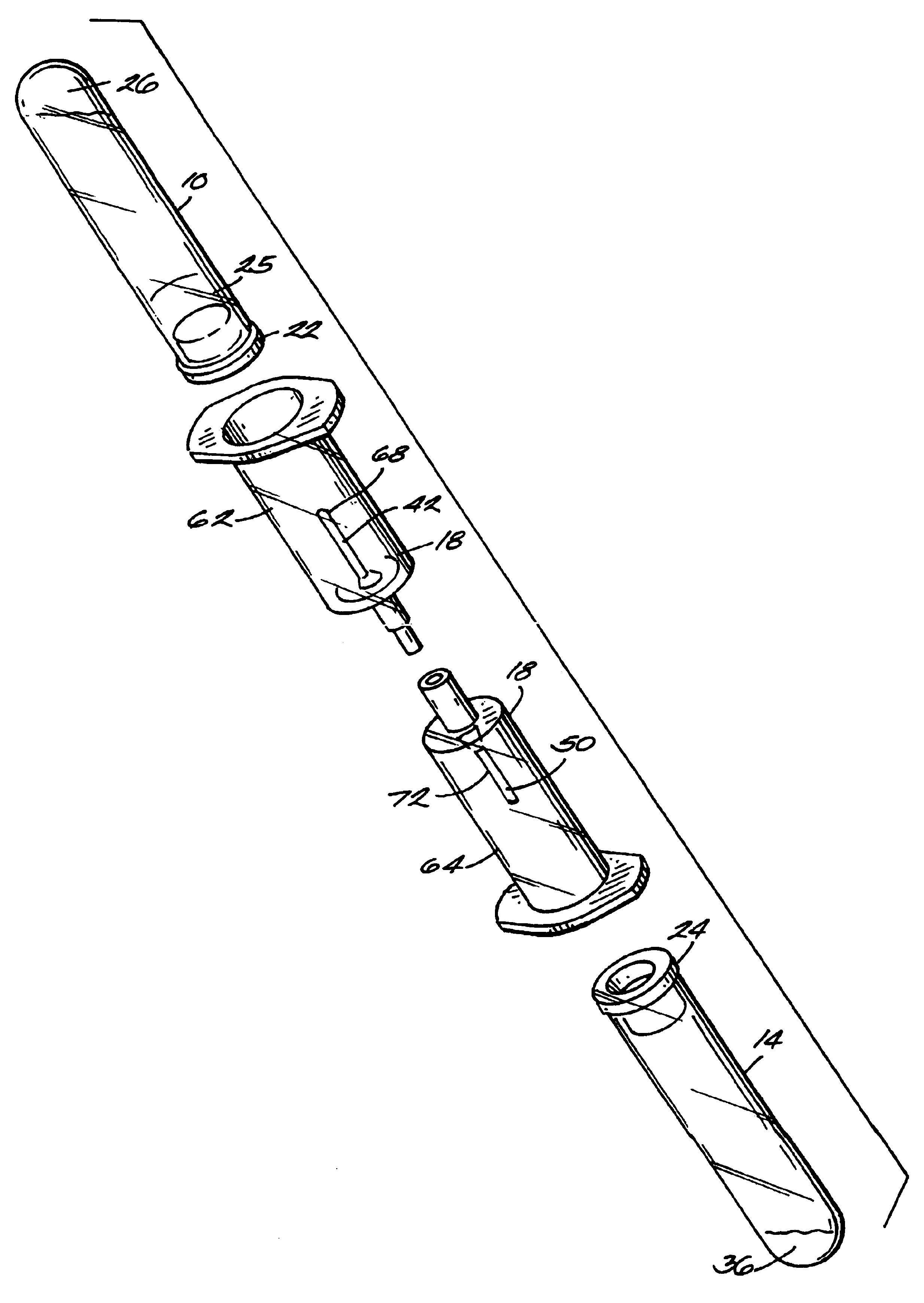
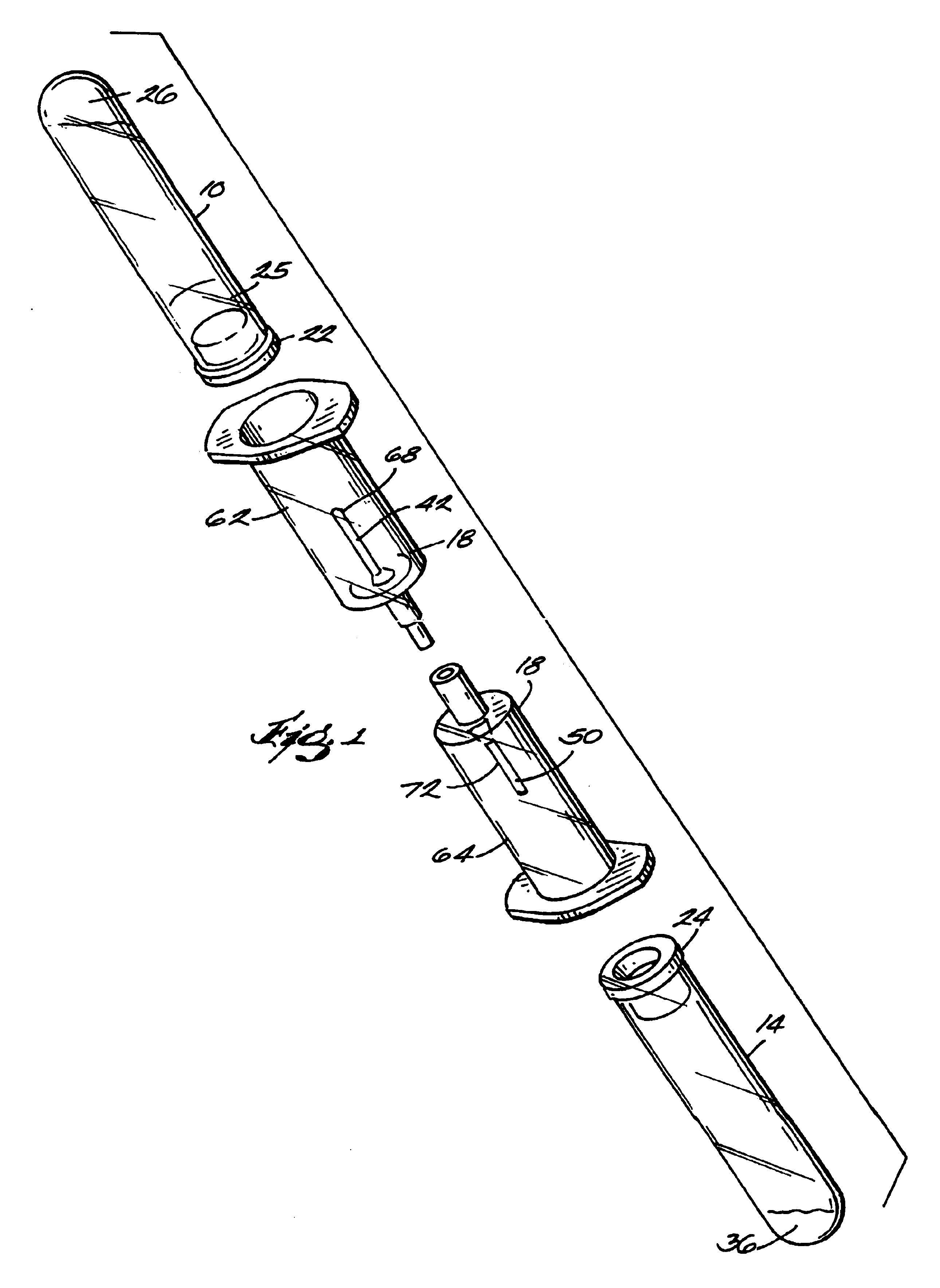
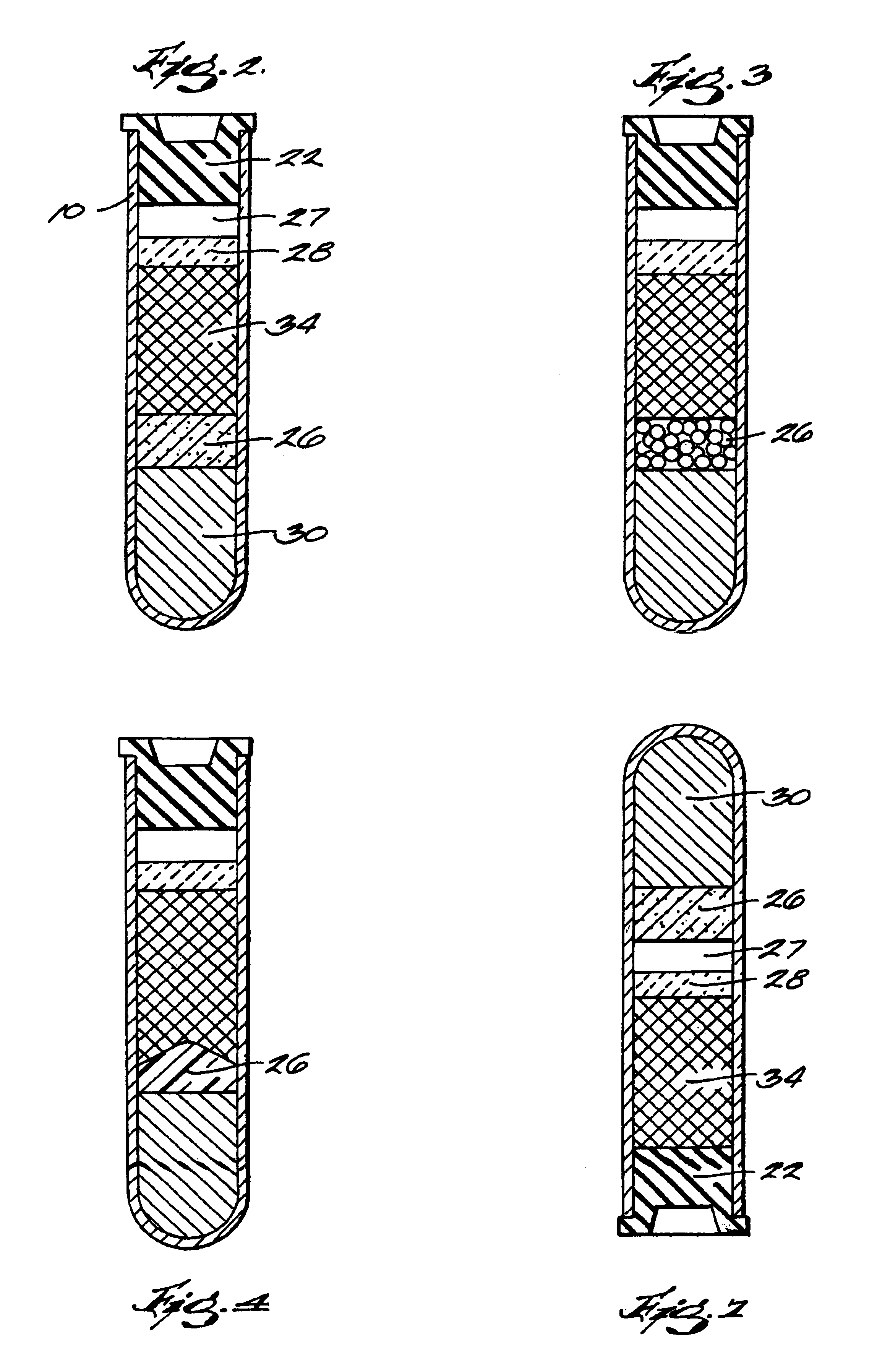
[0032]In another aspect, the invention provides another system for preparing a solid-fibrin web suitable for regenerating tissue in a living organism. The system comprises a sealed primary collection device having an interior and containing a separation medium. The primary collection device is capable of having blood drawn into the interior, and the separation medium is capable of separating plasma f

Problems solved by technology

Such known kits involve the use of material of human or animal origin, which, owing to its origin, could result in possible viral contamination and in serious risks for the receiver of the fibrin glue.
Several procedures have already been described for obtaining extemporary autologous fibrin glue, but no “ready to use” kit is available on the market although some relevant references can be found in the patent literature.
These activators are of human or animal nature and therefore still involve the risk of rejection and / or viral infections for the patient.
The method disclosed in this reference requires a long time for obtaining the plasma concentrate necessary for the subsequent preparation of autologous fibrin glue and the apparatus is expensive and not disposable.
The method does not disclose using a calcium-coagulation activator, and requires a pre-concentration step.
This practice, however, exposes the sample to environmental contaminants.
Frequently the high-density, undesirable, lower-fraction red blood cells contaminate the aspirated sample.
To avoid this problem, the pipette is frequently maintained a safe distance from the meniscus (i.e. the separator between the plasma and red blood cells), thereby resulting in an incomplete transfer of the sample.
The incomplete transfer of the desirable fraction results in lower than optimum volume yield and non-stoichiometric ratios of the sample reagents and those in the second container.
This second condition can be a serious source of performance variation of the product.

Method used


Image

  • Systems and methods for preparing autologous fibrin glue
  • Systems and methods for preparing autologous fibrin glue
  • Systems and methods for preparing autologous fibrin glue

Examples


example 1

[0049]In a 5 ml glass container for antibiotics, being sealable under vacuum, made of transparent white glass, inert and 1 mm thick were introduced 100 mg of tranexamic acid, acting as fibrin stabilizer. The synthetic tranexamic acid, being more than 98% pure, is put on the market by the American company Sigma Inc. Separately, a 1M CaCl2 solution was prepared, by weighing on a precision balance 147.0 g of CaCl2.2H2O (>99%pure), from the same American company Sigma Inc.

[0050]This salt was dissolved in exactly 1 liter of ultrapure nonpyrogenic distilled water, for a few minutes at room temperature, under frequent stirring. By using a precision piston dispenser, having a dispensing precision of ±5% (Eppendorf like), 80 μL of the activator solution were introduced in the glass container. In this step, at the same time as the dispensing, a filtering was carried out by using a 0.22μm Millpore sterilizing filter, while carefully preventing possible contamination from powders or filaments o...

example 2

[0051]10 ml of venous blood were drawn from a patient according to the provisions of the qualitative standards for clinical analysis, e.g. by using VACUTAINER® sterile test-tubes by Becton-Dickinson, added with a 0.106 M sodium citrate solution. For this purpose also test-tubes added with disodium or dipotassium ethylenediaminetetraacetate can be used.

[0052]The sample was carefully kept sterile during the blood drawing. Finally, the sample was gently shaken for wholly mixing the components, thereby ensuring the anticoagulating action of sodium citrate. The test-tube was then introduced in a suitable centrifuge, while carefully balancing the rotor weight in order to prevent the same centrifuge to be damaged. Once the lid is sealed, the sample was centrifuged at 3500 rpm for 15 minutes, thereby separating the red cells (being thicker) from the citrated plasma (supernatant). In this case the plasma yield, mainly depending upon the characteristics of the donor blood, was as high as 55%....

example 3

[0053]About 18 ml of venous blood were drawn from a normotype 49 years-old patient by using 5 ml sodium citrate VACUTAINER® test-tubes by Becton-Dickinson, taking care to shake gently just after the drawing of the sample. The so taken blood was immediately subjected to centrifugation (15 min. at 2500 rpm) to separate the plasma. The plasma (12 ml) was carefully transferred into two 10 ml test-tubes, containing 120 μL of CaCl2 (10 g / 100 ml) each, which had been prepared as described in Example 1, but without using tranexamic acid. After mixing the plasma with the activator, the test-tubes were centrifuged for 30 min. at 3000 rpm, finally obtaining two massive fibrin samples which were inserted, with all sterility precautions, within 2-3 hours from preparation, in the large vesicular mandibular cavity resulting from extraction of impacted left canine and right second incisor, as well as from abscission of the cyst present in the central area of the incisor teeth. Finally the gingival ...


PUM

PropertyMeasurementUnit
Pressureaaaaaaaaaa
Viscosityaaaaaaaaaa
Densityaaaaaaaaaa

Abstract

The invention provides a system for preparing an autologous solid-fibrin web suitable for regenerating tissue in a living organism. The system includes a sealed primary container containing a separation medium and a low-density high-viscosity liquid. The separation medium separates red blood cells from plasma when the container contains blood and is centrifuged, and the primary container has a first pressure. The system further includes a sealed secondary container containing a calcium-coagulation activator. The secondary container has a second pressure that is less than the first pressure. The system also comprises a transfer device including a cannula having a first end and a second end. The first and second ends puncture the sealed primary and secondary containers in order to provide fluid communication between the first and second containers. The low-density high-viscosity liquid of the primary container blocks flow through the cannula upon entering therein.

Description

CROSS-REFERENCE TO RELATED APPLICATIONS[0001]This application is a continuation-in-part of and claims priority to U.S. application No. 09 / 446,729 filed on Mar. 3, 2000 now U.S. Pat. No. 6,368,298, which is a 35 U.S.C. § 371 application of and claims priority to international application no. PCT / 1T98 / 00173 filed Jun. 24, 1998, which claims priority to Italian application no. M197A001490 filed Jun. 24, 1997. This application claims priority to each of the applications mentioned above.BACKGROUND OF THE INVENTION[0002]The present invention relates to systems, kits and methods for preparing a solid-fibrin web or autologous fibrin glue.[0003]Fibrin glue is known to be a haemoderivative largely that is used as a topical surgical adhesive or an haemostatic agent. Several kits are available on the market that contain concentrated fibrinogen from donors, associated to a proteic activator of human or animal origin, such as thrombin or batroxobin, for obtaining heterologous fibrin glue.[0004]Su...

Claims


Application Information

Patent Timeline
IPC IPC(8): A61L27/00A61L24/10A61L31/00
CPCA61B17/00491A61J1/2089A61L24/106A61L24/00A61B2017/00495A61C5/064A61J1/062A61J2001/2013A61J2001/2065A61J1/2065A61J1/2086A61J1/2013A61J1/201A61C5/64A61P7/04B01D21/262B01D33/15
Inventor BERETTAGRIPPI, NICHOLAS A.
Owner CASCADE MEDICAL ENTERPRISES
Learn More