Unitized electrode with three-dimensional multi-site, multi-modal capabilities for detection and control of brain state changes

a brain state and multi-site technology, applied in the field of medical devices, can solve the problems of poor spatial resolution, electrodes/devices not currently available, and current methodology, and achieve the effects of improving recording and control purposes, increasing the information content of signals, and reducing nois

Active Publication Date: 2006-02-28
FLINT HILLS SCI L L C
View PDF10 Cites 352 Cited by
  • Summary
  • Abstract
  • Description
  • Claims
  • Application Information

AI Technical Summary

Benefits of technology

[0034]The principal objects and advantages of the present invention include: providing a mechanism that ensures proper continuous close contact of constant degree between the cortex, both exposed and unexposed, and recording devices thereby considerably increasing the information content of the signal and decreasing noise for improved recording and control purposes; providing such a mechanism that minimizes displacement and flutter or flap of the recording contacts or surfaces thereby limiting recording and/or control signal degradation and loss of target acquisition; providing such a mechanism that considerably increases the spatial and temporal resolution by providing three-dimensional information to thereby allow for more precise and timely localization of the site of origin and time of onset of brain state changes; providing such a mechanism that allows more precise, timely and efficacious ...

Problems solved by technology

While recording of electrical activity directly from the cortical surface provides more information than scalp recordings, the current methodology, techniques and devices/probes have important limitations that must be overcome if further advances are to be made in the field of neurosciences in general and of epilepsy in particular.
Accomplishing this important objective, requires electrodes/devices not currently available since existing intracranial electrodes, either subdural or depth, have several significant limitations, which limitations as hereinafter described also apply to electrical stimulation of the cortex.
Poor Spatial Resolution
Furthermore, even if the diameter of depth electrodes were to be reduced, their ability to localize and track spatio-temporal spread (due to the fact that they would provide only one-dimensional information) would be impaired.
Also the possibility of radial migration of these type of electrodes (with consequent loss of the desired target) would remain unaddressed.
As a result, prior art recording devices and methods are unable to:(i) detect, from the surface without contamination or distortion, electrical fields generated below the cortical surface (or intracortically) especially from the deeper layers where generation of electrical signals is most active.
Moreover, signals recorded from the surface are highly filtered, which results in shape- and phase-distortions.
As a result, signals acquired with prior art devices provide limited and poor quality information about cortical dynamics;(ii) detect changes which may be close to the recording device but which project only within a very small solid angle;(iii) precisely localize the critical or primary region from where physiological or pathological activity of interest is generated because it is transmitted with delay, phase distortion, and loss of amplitude and information content to the top of a gyrus.
These limitations are due, in part, to the fact that the amplitude of an electrical signal decays as a function of the distance from where it is generated; in other words, the electrical potential at a point behaves according to V˜q/r where voltage, V, is proportional to charge, q, and inversely proportional ro r, which is the distance between the generator and the recording electrode.
In other words, electrical activity along those walls, which constitute a large portion of the gyrus, is either undetected or inadequately recorded at the surface.
The inability to resolve potentially discrete but relevant changes in BEA with high spatial accuracy negatively impacts scientific progress and compromises clinical care in the important areas of epilepsy, neurology, psychiatry, and mapping of cognitive or other functions of the brain; and(iv) extract three-dimensional information since electrical activity is generated and also integrat...

Method used


Image

  • Unitized electrode with three-dimensional multi-site, multi-modal capabilities for detection and control of brain state changes
  • Unitized electrode with three-dimensional multi-site, multi-modal capabilities for detection and control of brain state changes
  • Unitized electrode with three-dimensional multi-site, multi-modal capabilities for detection and control of brain state changes

Examples


first modified embodiment

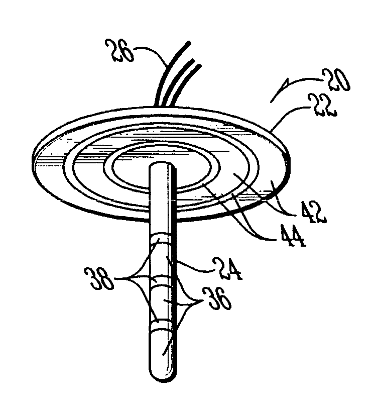
[0059]the unitized electrode with three-dimensional capabilities for detection and control of brain state changes, designated by the numeral 60, is depicted in FIG. 7. The first modified embodiment 60 includes a hollow body mechanism 61 having one or more retractable electrode devices 62 with distal ends 63 that can be selectively pushed out into the cortex, wherein the body mechanism 61 is secured to a disk portion 64 similar to that hereinbefore described for electrode 20. Disk portion 64 provides supporting and stabilizing structure for the first modified embodiment 60 but can also have one or more recording and stimulating contact surfaces on a lower surface 65 thereof, as hereinbefore described. While the body mechanism 61 is being inserted into a fold of the gyrus, as indicated in FIG. 7, the distal ends 63 of the retractable electrode devices 62 are retained within the body mechanism 61. Once the body mechanism 61 is suitably placed inter-gyrally as desired, i.e., in between ...

third modified embodiment

[0064]the unitized electrode with three-dimensional capabilities for detection and control of brain state changes, designated by the numeral 92 is shown, in FIG. 9. The third modified embodiment 92 includes a plate portion 93 and a plurality of shaft portions 94. As hereinbefore described, each shaft portion 94 may have a plurality of recording and / or stimulating electrode surfaces 95 separated by insulating material 96, as indicated on only one of the shaft portions 94 for simplification of illustration. Each of the recording and / or stimulating electrode surfaces 95 is independently connected to a different one of the conductors 97. In addition, the lower surface 98 of the plate portion 93 may have a single recording and / or stimulating contact surface connected in communication with a separate one of the conductors 97. Application of the third modified embodiment is substantially similar to that indicated in FIG. 6C for electrode 50. The multiple shaft portions 94 allow a fur...


PUM


Abstract

An electrode with three-dimensional capabilities for detection and control of brain state changes of a subject. The electrode includes a disk portion having an upper surface and a lower surface, and a shaft portion secured to and extending perpendicularly outwardly from the lower surface of the disk portion; the shaft portion having an outer surface. The disk portion and shaft portion may include one or more recording or stimulating contact surfaces structured to operatively interact with the brain of a subject. Insulating material isolates each of the recording or stimulating contact surfaces from each other. At least one conductor operatively and separately connect each of the recording or stimulating contact surfaces in communication with external apparatus. The disk portion and shaft portion are structured relative to each other to operatively provide support and anchoring for each other while providing three-dimensional capabilities for detection and control of brain state changes of a subject. Modified embodiments include insertible/retractable electrode wires, both contained in channels and sheathed in axially displaceable cannulae; activating mechanisms for inserting/retracting the electrode wires and/or cannulae; and multiple shaft portions.

Description

CROSS REFERENCE TO RELATED APPLICATIONS[0001]This application is a continuation-in-part of Provisional Patent Application No. 60 / 397,511 filed Jul. 20, 2002 and entitled “Self-anchoring, Unitized, Mesoelectrode with Multisite, Multimodal Dual (Recording and Control) Capabilities and a Method for Contingent Random, Nonrandom, Turbulent, or Nonturbulent Stimulation for Seizure Blockage.”BACKGROUND OF THE INVENTION[0002]1. Field of the Invention[0003]This invention relates generally to medical apparatus and, more specifically but without limitation, to an electrode apparatus for interacting with the brain of a subject.[0004]2. Description of Related Art[0005]Brain electrical activity (BEA) is a readily accessible and reliable index of brain state and function. It allows distinction between both normal states, such as wakefulness or sleep and its different substates, such as NREM and REM, and abnormal states, such as the ictal and inter-ictal substates of an epileptic brain. BEA is also...

Claims


Application Information

Patent Timeline
IPC IPC(8): A61B5/042A61N1/05
CPCA61B5/0478A61N1/0539A61N1/0531A61N1/36017A61B5/291A61B5/293
Inventor OSORIO, IVANBHAVARAJU, NARESH C.
Owner FLINT HILLS SCI L L C
Why Eureka
Try Eureka