Method for the quantitative detection of nucleic acids
a nucleic acid and quantitative technology, applied in the field of quantitative detection of nucleic acids, can solve the problems of long time and additional costs of amplified product detection steps, limited strategy, and unspecific pcr products generated signals, etc., to achieve enhanced sensitivity, accuracy and precision, and improve the efficiency of techniques
- Summary
- Abstract
- Description
- Claims
- Application Information
AI Technical Summary
Benefits of technology
Problems solved by technology
Method used
Image
Examples
example 1
Selection of the HHV-6, HHV-7, HHV-8, HIV 35s CAMV and Mycobacterial sequences.
[0040]The U67 HHV-6 region (A variant, GeneBank Accession N°. X83413) and the 26 HHV-8 orf region (Chang et al., Science 266, 1865-1869, 1994) the HHV-7 region (Gene Bank AF037218), 35sCAMV (Gene Bank AF140604), RegX-SenX (Gene Bank MTY 13628) and 156110 (Gene Bank X17848), and the HIV region mapping between the LTR and gag region from nucleotide 684 to 810 using the sequence HXB2CG-Accession number K03455 (Gene Bank) are reported in Table 1 (primers and probe of the target nucleic acid and calibrator probe). The HHV-6 sequences (primers and probe for the target, calibrator's primers and probe) are reported in Table 2.
[0041]The probe sequences of the calibrators (HHV-7, HHV-8, HIV-1, CAMV, Myc. T.) and the calibrator's primers (HHV-6) were designed randomizing the probe region of the standard, while maintaining:[0042]1. the same base composition (G+C / A+T) of the standard[0043]2. an identical Tm (calculate...
example 2
HHV-6 Standard (target nucleic acid) and Calibrator cloning and preparation.
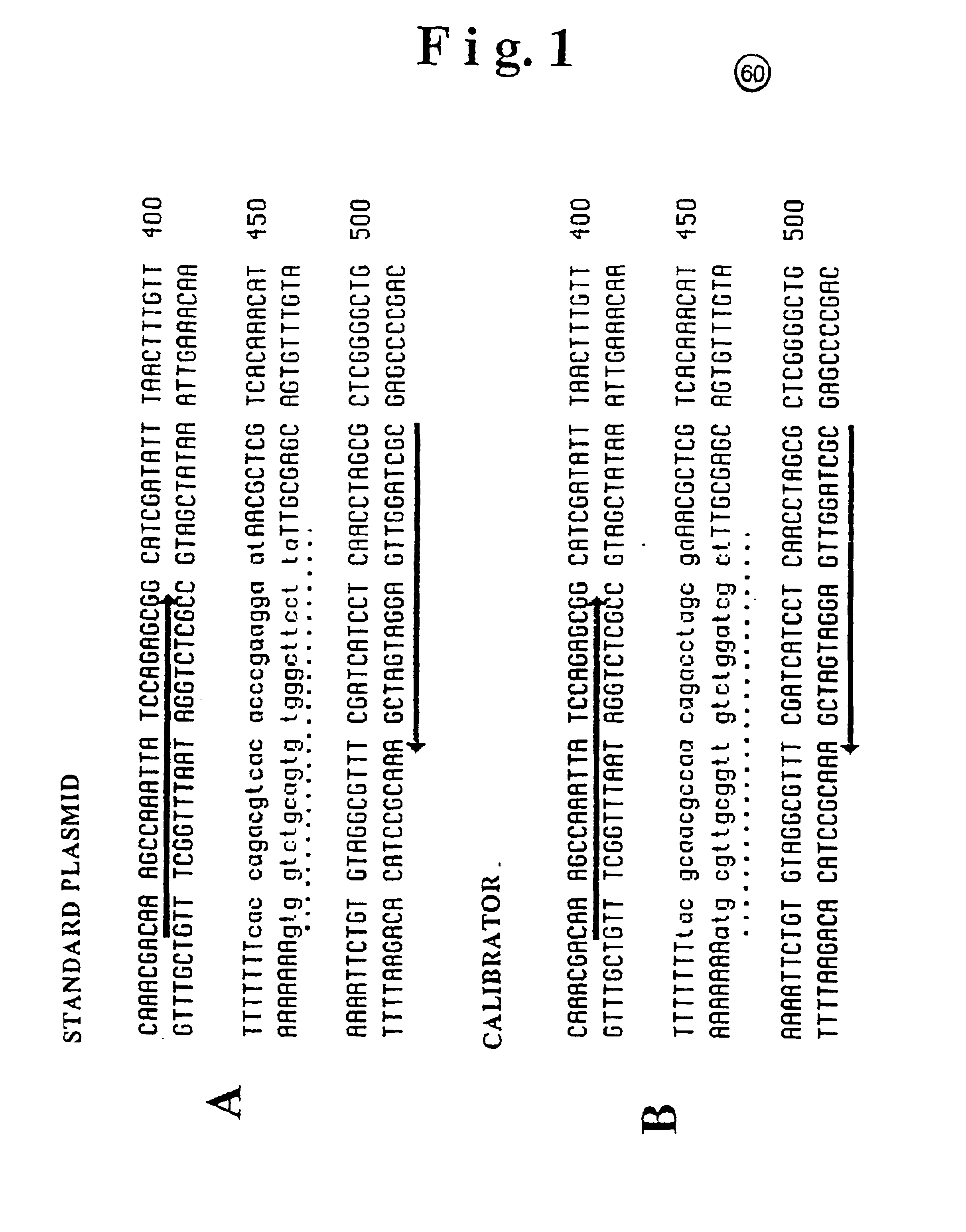
[0048]The fragments used for the standard and calibrator DNA construction in the HHV-6 virus detection system are schematically represented in FIG. 1 (1-A and 1-B respectively).
[0049]The Standard fragment sequence was obtained by amplification of the viral DNA from HHV-6 GS strain and subsequent cloning into pCRII plasmid vector (Invitrogen). The calibrator fragment (133 bp) was chemically synthesized by an Oligosynthetizer (Perkin Elmer), and then cloned in the same vector as above.
[0050]After cloning, both fragments were completely sequenced in order to identify: i) the identity (co-linearity) of the standard fragment with the original viral DNA, ii) the identity of the calibrator fragment with the artificially designed sequence.
[0051]As shown in FIG. 1 the arrows, oriented in the transcription direction, indicate the oligonucleotide sequence employed as primers. The dotted line identifies in both construc...
example 3
HHV-6 calibrator / standard system validation
[0052]Absence of cross-hybridization
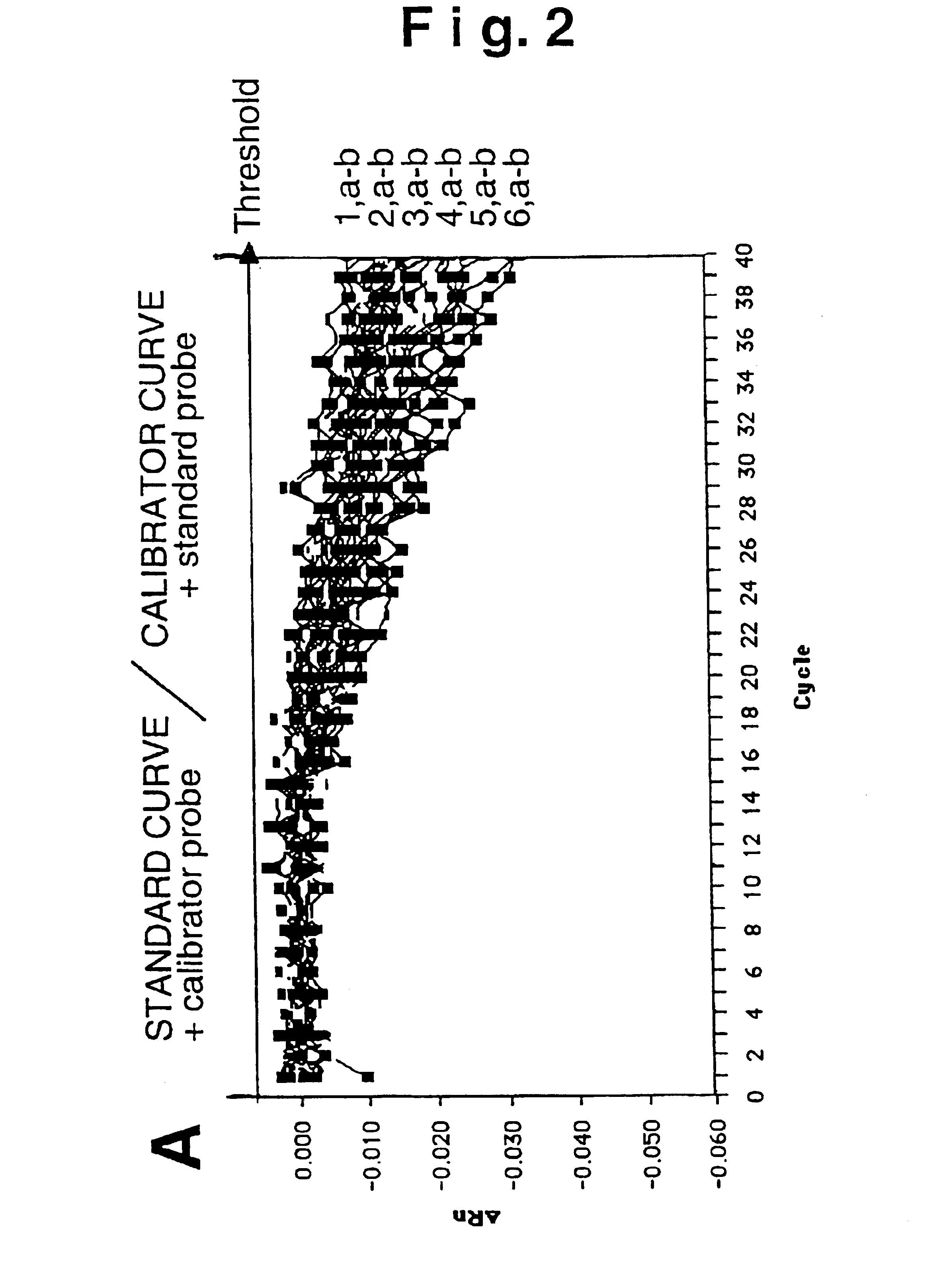
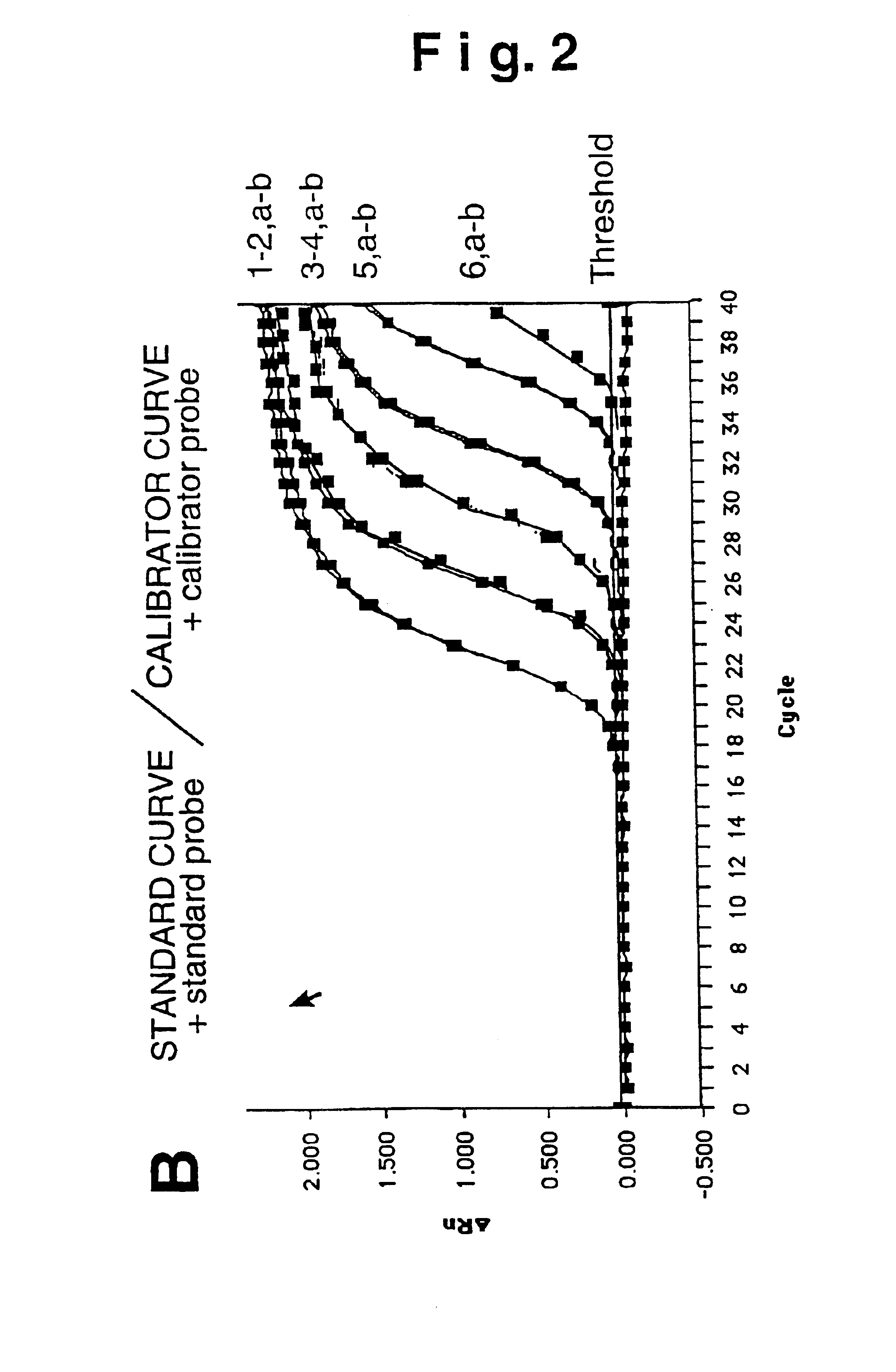
[0053]We thus experimentally verified the absence of spurious signals from cross-hybridization. Increasing concentrations of the standard and calibrator fragment (from 101 to 106 plasmid copies per PCR reaction) were measured using the homologue probe (standard probe for the standard DNA, calibrator probe for the calibrator DNA) or the heterologous probe (standard probe for the calibrator DNA, calibrator probe for the standard DNA). FIG. 2B shows the detection of the various template amounts employing the probe homologous to the fragment to measure, where: a) indicates the standard amplification curve and, b) the calibrator curve. Detection takes place for both constructs with overlapping kinetics (curves 1-5, a, b) furthermore indicating that the template amplification is proportional to the measured fluorescence signal. The use of heterologous probe (FIG. 2A) for both templates does not generate a signa...
PUM
| Property | Measurement | Unit |
|---|---|---|
| Tm | aaaaa | aaaaa |
| Tm | aaaaa | aaaaa |
| Tm | aaaaa | aaaaa |
Abstract
Description
Claims
Application Information
 - R&D
- Intellectual Property
- Life Sciences
- Materials
- Tech Scout
- Unparalleled Data Quality
- Higher Quality Content
- 60% Fewer Hallucinations
Browse by: Latest US Patents, China's latest patents, Technical Efficacy Thesaurus, Application Domain, Technology Topic, Popular Technical Reports.
© 2025 PatSnap. All rights reserved.Legal|Privacy policy|Modern Slavery Act Transparency Statement|Sitemap|About US| Contact US: help@patsnap.com


