
Methods for reducing distal embolization

a distal embolization and distal catheter technology, applied in the field of improved methods for reducing distal embolization, can solve the problems of serious and permanent injury, death, and reduced blood carrying capacity of the vessel, and achieve the effect of reducing distal embolization

Inactive Publication Date: 2006-04-25
MEDTRONIC VASCULAR INC
View PDF162 Cites 201 Cited by
  • Summary
  • Abstract
  • Description
  • Claims
  • Application Information

AI Technical Summary

Benefits of technology

[0012]The aspiration method disclosed herein can be used in any vessel of the body where the pressure is at least 0.2 psi at any time during the diastolic / systolic cycle of the heart, and, preferably, is about 1.2 psi, and is capable of providing a flow rate of at least 5 cc / minute when not occluded. Thus, although the pressure within any vessel may fall below 0.2 psi during relaxation between heartbeats, so long as the pressure created by the heartbeat rises to at least 0.2 psi, the pressure within the vessel will be sufficient.
[0014]In a preferred embodiment of the method of the present invention, a guide catheter is first introduced into the patient's vasculature through an incision made in the femoral artery in the groin and is used to guide the insertion of other catheters and devices to the desired site. A guidewire is then advanced until its distal end reaches a site proximal to the occlusion. Fluoroscopy is typically used to guide the guidewire and other devices to the desired location within the patient. The devices are frequently marked with radiopaque markings to facilitate visualization of the insertion and positioning of the devices within the patient's vasculature.
[0015]In contrast to previous methods, in one embodiment the guidewire does not cross the occlusion prior to aspiration with an aspiration catheter or other suitable catheter which can provide aspiration pressure. Instead, an aspiration catheter is inserted over the guidewire and the two are concomitantly moved past the occlusion, proximal to distal, while aspirating, to prevent material which dislodges or breaks off the occlusion from entering the vasculature and producing distal emboli.
[0016]A guidewire having an occlusive device on its distal end is preferably used in the present method. The method can be effectively carried out, however, using a number of guidewires or catheters that perform the function of occluding the vessel and allowing for the slidable insertion of various other catheters and devices. A standard guidewire can also be used to initially cross the occlusion, and then exchanged with a guidewire having an occlusive device at its distal end. When the standard guidewire is used, it may also be delivered with the aspiration catheter so that both are moved past the occlusion while aspirating. The occlusive device should be capable of preventing the migration of particles and debris from the working area, either through total or partial occlusion of the vessel. The occlusion of the vessel need not be complete.
[0018]After the distal end of the guidewire having an occlusive device is delivered past the site of the occlusion and aspiration is complete, the occlusive device is activated, preventing particles and debris from travelling downstream. Therapy can then be performed to remove or reduce the occlusion. A therapy catheter can be used if desired. The therapy catheter can be any of a number of devices, including a balloon catheter used to perform angioplasty, a catheter which delivers a stent, a catheter for delivering enzymes, chemicals, or drugs to dissolve and treat the occlusion, an atherectomy device, or a laser or ultrasound device used to ablate the occlusion. Alternatively, the therapy catheter can be eliminated and use of the guide catheter or the aspiration catheter alone can be used to aspirate the occlusion. This method is especially useful to remove emboli from the coronary arteries following acute myocardial infarction, because the aspiration catheter can be made small enough to enter the coronary arteries.
[0020]The method described herein provides an improved method for reducing distal embolization during treatment of emboli, thrombi and other types of occlusions in the human arterial or venous system, and is especially useful following myocardial infarction.

Problems solved by technology

Human blood vessels often become occluded or completely blocked by plaque, thrombi, emboli or other substances, which reduces the blood carrying capacity of the vessel.
Should the blockage occur at a critical location in the circulation, serious and permanent injury, or death, can occur.
Damage to or malfunction of the heart is caused by narrowing or blockage of the coronary arteries (atherosclerosis) that supply blood to the heart.
The coronary arteries are first narrowed and may eventually be completely blocked by plaque, and may further be complicated by the formation of thrombi (blood clots) on the roughened surfaces of the plaques.
AMI can result from atherosclerosis, especially from an occlusive or near occlusive thrombus overlying or adjacent to the atherosclerotic plaque, leading to death of portions of the heart muscle.
Thrombi and emboli also often result from myocardial infarction, and these clots can block the coronary arteries, or can migrate further downstream, causing additional complications.
It can be difficult, however, to treat plaque deposits and thrombi in the coronary arteries, because the coronary arteries are small, which makes accessing them with commonly used catheters difficult.
Each of these methods is not without the risk of embolism caused by the dislodgment of the blocking material which then moves downstream.
In addition, the size of the blocked vessel may limit percutaneous access to the vessel.

Method used


Image

  • Methods for reducing distal embolization
  • Methods for reducing distal embolization
  • Methods for reducing distal embolization

Examples


Embodiment Construction

[0075]The present invention involves a method for advancing a guidewire into position in the vasculature of a patient prior to therapy on plaque, a thrombus, emboli, or other occluding substance. The preferred devices used during the method will be described first, followed by a description of the preferred method of use.

I. Overview of Occlusion System

A. Syringe Assembly

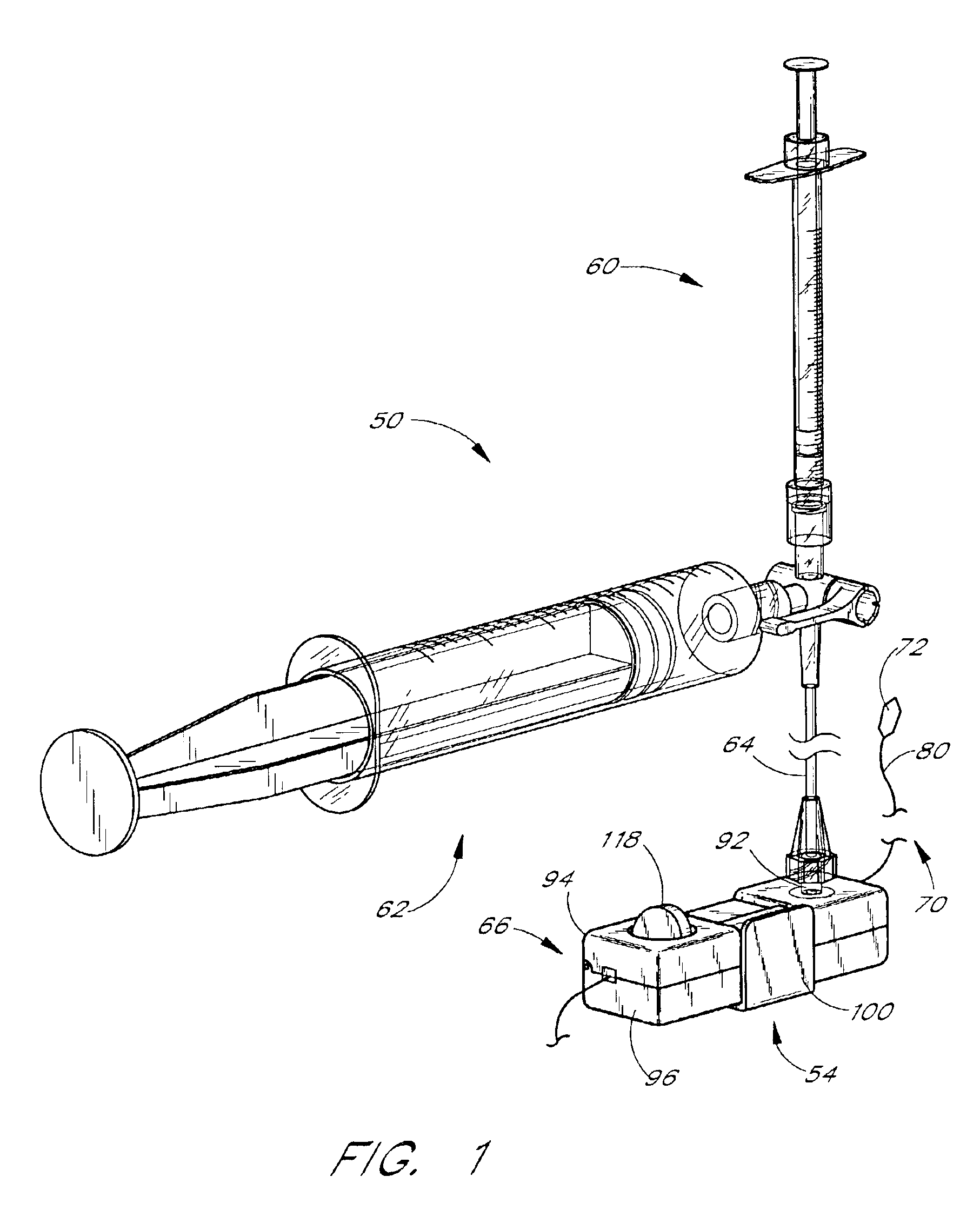
[0076]The preferred embodiments of the present invention may comprise or be used in conjunction with a syringe assembly such as that generally illustrated in FIG. 1. Also shown in FIG. 1 is an illustrative connection of the syringe assembly 50 to an occlusion balloon guidewire catheter 70 utilizing an inflation adapter 54. The syringe assembly 50, comprising the inflation syringe 60 and a larger capacity or reservoir syringe 62, is attached via tubing 64 to the inflation adapter 54 within which a low profile catheter valve 66 and the balloon catheter 70 are engaged during use.

[0077]The catheter valve 66, described in...


PUM


Abstract

A method for reducing distal embolization during insertion of devices to treat plaque, thrombi and other intravascular occlusions. Using a guide catheter, a guidewire is delivered to a site proximal to the lesion. An aspiration catheter is then introduced over the guidewire, and the two are moved together in a distal to proximal direction across the occlusion. The aspiration catheter is then moved in a proximal to distal direction while continuously aspirating, which prevents embolization of any particles which may be created during the crossing of the guidewire.

Description

CROSS REFERENCE TO RELATED APPLICATIONS[0001]The present application is a continuation of application Ser. No. 09 / 438,030, filed Nov. 10, 1999, now U.S. Pat. No. 6,652,480, which is a continuation-in-part of application Ser. No. 09 / 049,857, filed Mar. 27, 1998, now U.S. Pat. No. 6,135,991, which is a continuation-in-part of application Ser. No. 08 / 813,807, filed Mar. 6, 1997, now abandoned.BACKGROUND OF THE INVENTION[0002]1. Field of the Invention[0003]The present invention relates generally to an improved method for reducing distal embolization during treatment of emboli, thrombi and other types of occlusions in the human arterial or venous system. The method is particularly well suited for use when treating stenoses or occlusions within saphenous vein grafts, coronary arteries, cerebral arteries and similar vessels.[0004]2. Description of the Related Art[0005]Human blood vessels often become occluded or completely blocked by plaque, thrombi, emboli or other substances, which reduc...

Claims


Application Information

Patent Timeline
IPC IPC(8): A61M31/00A61M29/00A61B17/22A61B18/24A61M29/02
CPCA61B17/22A61M25/104A61B18/245A61B2017/22067A61B2017/22084A61M2025/1093A61M2025/09183A61M2025/1015A61M2025/1052A61M2025/109
Inventor IMRAN, MIR A.
Owner MEDTRONIC VASCULAR INC
Learn More