
Electrochemical biosensor by screen printing and method of fabricating same

a biosensor and electrochemical technology, applied in the field of electrochemical biosensors, can solve the problems of inability to effectively and efficiently control the process of affixing two insulating substrates with electrodes thereon is complicated and expensive, and the speed of filtering is still not considered ideal, etc., to achieve the effect of simple structure, less volume and rapid sample inflow

Active Publication Date: 2006-11-21
GENERAL LIFE BIOTECHNOLGOY
View PDF21 Cites 135 Cited by
  • Summary
  • Abstract
  • Description
  • Claims
  • Application Information

AI Technical Summary

Benefits of technology

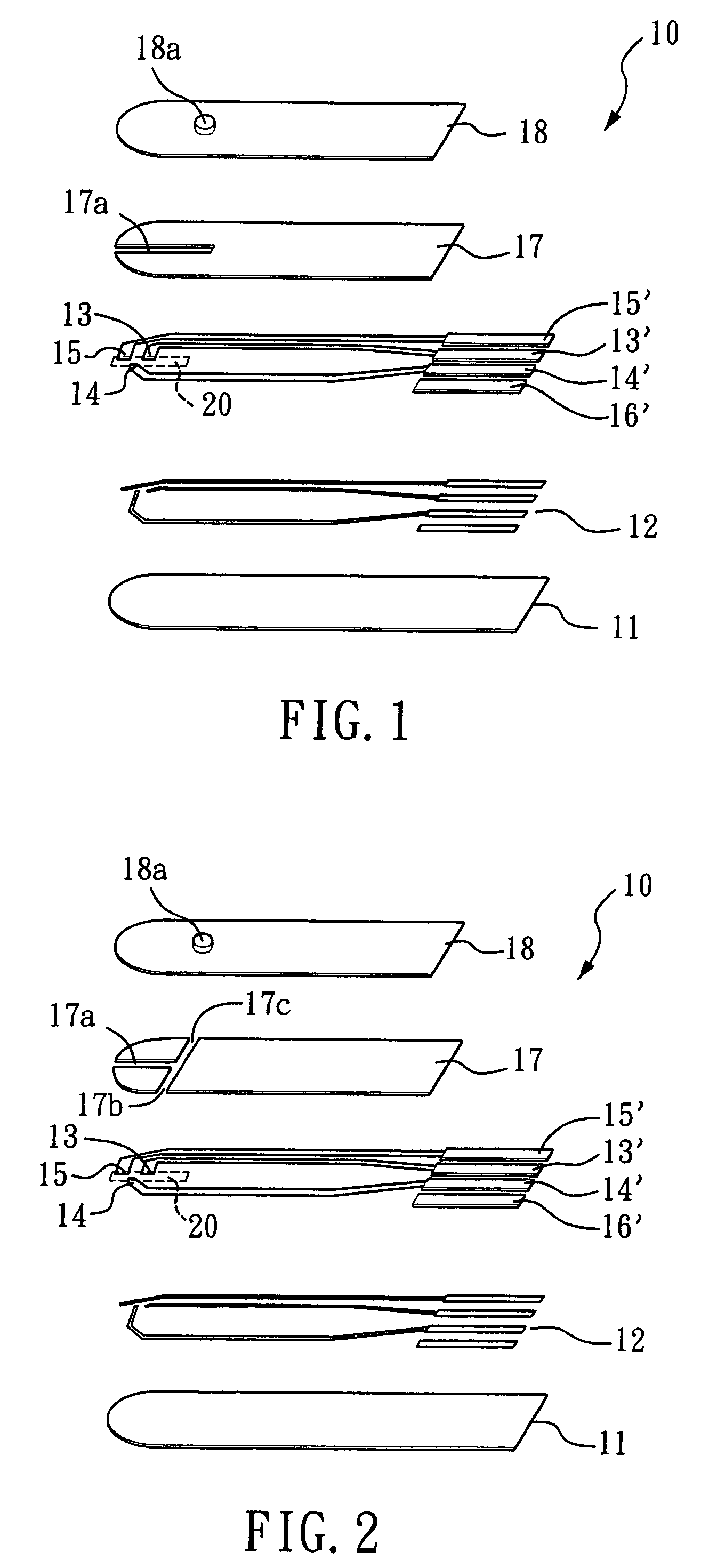
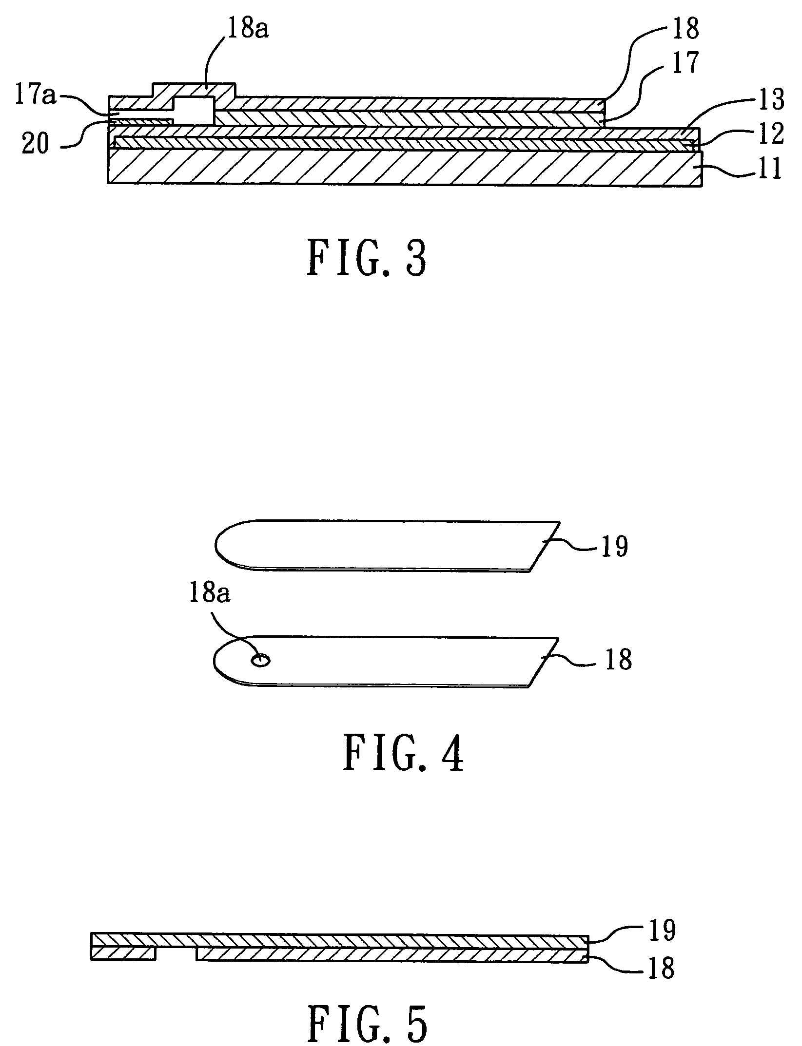
"The present invention provides a biosensor with a fast sample inflow and less volume. The biosensor has a simple structure and does not require a mesh, which prevents contamination. Only 0.5-0.8 μl of sample is required and analysis can be completed in about 5-10 seconds. The biosensor is formed by screen printing and includes an electrode layer with three electrodes (working, reference, and auxiliary). An active reaction layer containing reactant, reaction catalyst, mediator, wetting agent, and surfactant is spread on the surface of the electrode layer. A sample inflow channel is used to introduce sample solution into the electrode area and the active reaction layer by siphon or capillary. The biosensor can analyze samples using electrochemical potentiometric or amperometric methods. The biosensor also has an upwardly extended close chamber that can control sample volume and effectively position the sample."

Problems solved by technology

Hence, they are still not considered ideal although the amounts of blood sample and measuring time they require are much less than those conventional colorimetric method does.
Such sensors can be fabricated by photolithography or screen printing but processes of affixing two insulating substrates with an electrode thereon are very complicated and expensive.
However, the amount of the sample introduced and the speed of filtering cannot be effectively and efficiently controlled.
However, the processes of fabricating such sensors are very complex and extremely costly.
Such technique requires a larger amount of sample and processing such sensors introduces numerous electroplating process steps.
Such application is similar to that developed by Abbott, which utilizes mesh for the inflow of sample and is thus more costly, is also restricted to the amount of sample required.
By this way, the volume of sample flowing into the channel cannot be controlled and the sample is likely to float a vent opening, which causes contamination.
With this structure, sample fluid may float the vent opening when the size of which is too small.
However, as the size of sensors is getting smaller, it is likely to touch the vent opening by hand which causes outflow of sample fluid and thus contamination.

Method used


Image

  • Electrochemical biosensor by screen printing and method of fabricating same
  • Electrochemical biosensor by screen printing and method of fabricating same
  • Electrochemical biosensor by screen printing and method of fabricating same

Examples


example 1

Fabrication of Glucose Sensor by Screen Printing

[0028]A layer of electrically conductive silver paste is formed on a polyporpylene synthetic substrate 11. by 300 mesh screen printing, which is dried and heated for 30 minutes at 50° C., and three electrodes (working electrode 13, reference electrode 14 and auxiliary electrode 15) are printed by carbon paste thereon. The substrate 11 is again heated for 15 minutes at 90° C. and printed by insulating gel, which is subsequently dried and hardened under ultraviolet light to form an insulating layer with an inflow reaction are 17c, 17b and 17c (for sensors with air vents). Reaction reagents of 2–6 μl, containing 0.5–3 units of glucose oxidase, 0.1–1% of polyvinyl alcohol, pH 4.0–9.0 and 10 14 mM potassium phosphate as buffer solution, 10–100 mN potassium chloride, 0.05–0.5% of dimethylferrocene, 0.005%–0.2% tween−20, 0.005%–0.2% of sufynol and 011%–1.0% of carboxymethyl cellulose are spread on the recessed inflow channel area 17a. The sub...

example 2

Standard Glucose Solution and Whole Blood Measurement

[0029]Standard potassium phosphate buffer solution (pH 7.4) is disposed containing glucose with a concentration of 0–400 mg / dl. The sample solution is measured by an electrochemical device (CHInstrument Co. 650A) in conjunction with a sensor according to Example 1 under a measuring potential of 100 mV for 8 seconds. The volume of sample solution is 3 μl for every measurement and the volume of sample solution introduced into the sensor for every measurement is less than 3 μl. The measuring results are listed in Table 1:

[0030]

TABLE 1Results of standard glucose measurementsGlucose Concentration (mg / dl)Charge (μ coulomb) 00.690 251.532 502.9521005.2482007.4004009.577

[0031]Whole blood sample can also be measured by sensors according to the present invention. Table 2 shows results of by measuring fresh vain whole blood sample with glucose additive with a measuring potential of 100 mV and volume of 2 μl.

[0032]

TABLE 2Results of whole bloo...

example 3

Measurements of Blood Sugar with Varied Volume of Whole Blood

[0033]Electrode sensors according to Example 1 are employed, which provide whole blood samples with different volume required in the present invention. Vein whole blood samples are mixed with standard glucose solution, which in turn form solutions with a concentration of 300 mg / dl.

[0034]The method of measurements is to provide whole blood samples with different volume and supply samples by siphon under conditions set out in Example 2. As shown in FIG. 7, when the volume of a sample is insufficient (e.g., of less than 0.5 1), the concentration of glucose is low. Conversely, when the volume of a sample is above 0.8 1, the measured glucose concentration si near that in the sample solution, and the whole amount of the sample cannot be introduced into the sensor. That is, the more the volume of a sample is supplied, the more volume of the sample will be redundant, since inflow reaction channel is saturated with the sample and c...


PUM

PropertyMeasurementUnit
widthaaaaaaaaaa
widthaaaaaaaaaa
widthaaaaaaaaaa

Abstract

An electrochemical biosensor formed by screen printing and method of fabricating such biosensor is disclosed in the present invention. The biosensor can quickly absorb a sample to be measured therein, effectively control volume of the sample fed and “fill-and-position” the sample therein. The biosensor includes an electrode layer (electrode area) comprising two or three electrodes, which are a working electrode, a reference electrode and an auxiliary electrode (tri-electrode) on an insulating substrate. An active reaction layer containing reactant, reaction catalyst, mediator, wetting agent and surfactant is spread on the surface of the electrode layer. A sample inflow area is formed above the electrode area by adding an upper cover on top of a middle insulating layer with a U-shaped opening formed therein. Sample solution with a minute amount about 0.8 to 1 μl can be rapidly introduced into the electrode area and the active reaction layer via the inflow area by siphon or capillary, where the ingredient of the sample can be analysed by measuring reaction between the sample, reaction catalyst and mediator in the reaction layer using electrochemical potentiometric or amperometric method. An upwardly extended closed space formed within the upper cover above the electrode area adjacent to the front of conductive wires can be effectively used to control sample volume and “fill-and-position” the sample.

Description

BACKGROUND OF THE INVENTION[0001]1. Field of the Invention[0002]The present invention relates to an electrochemical biosensor formed by screen printing and a method of fabricating such biosensor.[0003]2. Description of the Related Art[0004]Recently, electrode sensors have been commercially utilized successfully for the fabrication of a variety of clinical measuring products, such as blood sugar, uric acid and cholesterol measuring devices, for their easy and low cost production processes and wide application of cheaper portable measuring devices. Taking the biggest and the most widely used blood sugar measuring device on market as an example, the leading manufacturers include Roche, Abbott, Bayer and Therasense and all of which fabricate blood sugar sensors by electrochemistry. The first generation of such sensors requires higher amount of blood sample (5–10 μl and above) and takes longer (30–60 seconds) to measure a sample. Hence, they are still not considered ideal although the am...

Claims


Application Information

Patent Timeline
Patent Type & Authority Patents(United States)
IPC IPC(8): G01N27/327G01N27/26G01N33/487
CPCG01N27/3272
Inventor SU, CHEIN-SHYONGCHANG, CHIA-WEIHUNG, MIAO-LINGCHENG, WEN-JINGWU, TAI-GUANG
Owner GENERAL LIFE BIOTECHNOLGOY
Learn More