Method and apparatus for multiple spectroscopy analysis by using nuclear magnetic resonance

a technology of nuclear magnetic resonance and multiple spectroscopy, applied in the field of multiple spectroscopy analysis, can solve the problems of large number of isotope labeled samples, difficult to obtain structural information, and affecting the skill of sample preparation, and achieves high binding activity

Inactive Publication Date: 2007-04-10
HITACHI LTD
View PDF9 Cites 1 Cited by
  • Summary
  • Abstract
  • Description
  • Claims
  • Application Information

AI Technical Summary

Benefits of technology

[0017]An object of the present invention is to provide a method and apparatus which can control and detect the interaction of protein in a mixed sample and measure the process of interaction.
[0028]According to the present invention, the interaction and binding of the specified amino residue having a group with high binding activity such as carbonyl in the side chain are selectively changed to make it possible to observe the binding of protein of high molecular weight to ligand and the interaction between proteins by a nuclear magnetic resonance signal. Since a wavelength variable THz oscillator is used, resonance oscillation of the entire protein and resonance in the specified structure are provided to make it possible to observe its molecular structure and chemical change by nuclear magnetic resonance. The energy of a THz electromagnetic wave applied is low and 0.4 kJ / mol when the frequency is 1 THz. The problem of the denaturation of protein which must be considered in an experiment such as Ramann spectroscopy using light above infrared rays can be reduced.

Problems solved by technology

Large numbers of isotope labeled samples are very expensive.
It is difficult to obtain structural information as a guide for developing more excellent ligand.
The difference in experience and skill among sample preparers is largely affected to increase the sample preparing cost.
Measurement of the process of interaction is very difficult.
In the experimental method of analyzing the response of the entire sample to an incident light of Ramann spectroscopy, selective observation of the specific portion is difficult.

Method used


Image

  • Method and apparatus for multiple spectroscopy analysis by using nuclear magnetic resonance
  • Method and apparatus for multiple spectroscopy analysis by using nuclear magnetic resonance
  • Method and apparatus for multiple spectroscopy analysis by using nuclear magnetic resonance

Examples


embodiment 1

[0044]Protein analysis of multiple measurement of THz electromagnetic wave application and NMR measurement will be described in detail.

[0045]Protein included in the sample 4 has about 20 kinds of amino acid residues which are bound by peptide bond. Some of the residues having carbonyl in the portion called a side chain not contributing to the peptide bond exist. Depending on an aqueous solution condition, the side chain carbonyl has a doublet state in which a proton is ionized and one unpaired spin is provided near an oxygen atom. FIG. 5 shows the structural formula of the carbonyl in which a proton is ionized. As indicated by the arrows, when the entire sample is placed under a strong uniform static magnetic field, the unpaired electron spin of the side chain carbonyl is arranged in parallel with the magnetic field.

[0046]When the unpaired spin produced in the carbonyl is placed under a strong static magnetic field, energy is different in the state that it is directed in the paralle...

embodiment 2

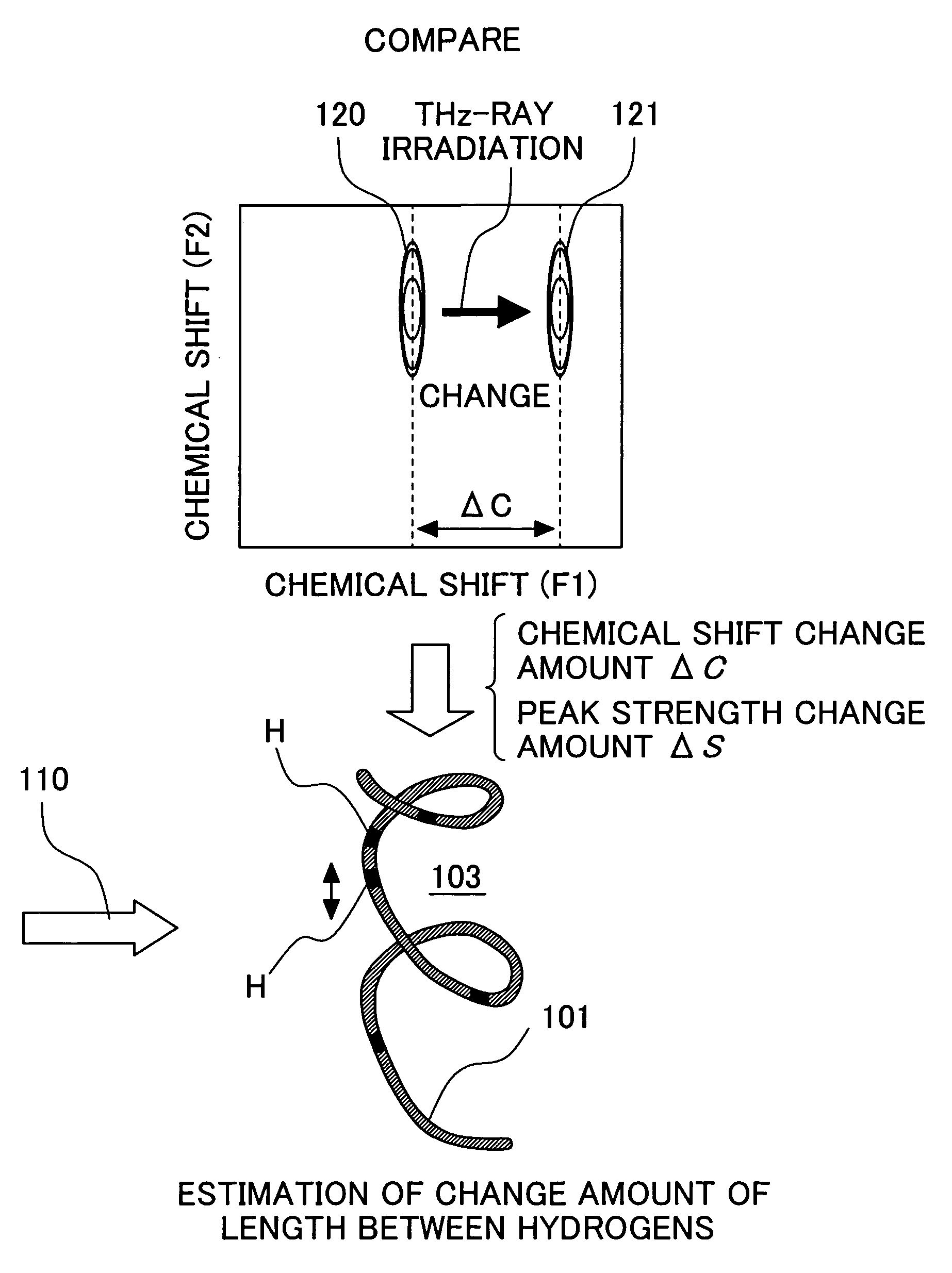
[0054]The electromagnetic wave application is combined with the nuclear magnetic resonance measurement to make it possible to observe the state change of various samples. The state change includes not only change brought about in the entire protein but also selective change focusing on the specified residue. The observation of the change in a protein structure brought about by electromagnetic wave application will be described below in detail.

[0055]FIG. 6 shows a target protein sample 103 obtained by subjecting protein 101 having aspartic acid or glutamic acid in the active center to isotope labeling 102 of aspartic acid or glutamic acid.

[0056]The binding of nuclear spins to be observed is determined. When comparing the change in chemical shift of an HN atom and an N atom, the 1H-15N-HMQC spectrum is used. When inspecting the strength of the interaction between hydrogens (that is, length between atoms), an NOESY spectrum is used.

[0057]FIG. 7 shows the procedure for observing the str...

embodiment 3

[0065]An observation example about the protein-ligand interaction using THz electromagnetic wave application will be described below in detail.

[0066]FIG. 9 shows a target protein sample 105 obtained by binding, to ligand 104, sample protein obtained by subjecting the protein 101 having aspartic acid or glutamic acid in the active center to the isotope labeling 102 of aspartic acid or glutamic acid. In this way, sample preparation is performed so as to perform selective isotope labeling to the sample protein and binding the ligand 104 thereto.

[0067]FIG. 10 shows the procedure for observing the protein-ligand interaction.

[0068](1) NMR measurement is performed by the sample 103 of target protein to obtain the spectrum 124-1 such as the 1H-15N-HMQC. The measurement is the same as measurement 1 described in Embodiment 2. The spectrum is a spectrum in the state that the target protein 103 is bound to none and is a spectrum as a reference of the following analysis. In particular, the peak ...


PUM

PropertyMeasurementUnit
frequencyaaaaaaaaaa
time constantaaaaaaaaaa
resonance frequencyaaaaaaaaaa

Abstract

An object of the present invention is to provide a method of controlling and observing binding of sample substances in an aqueous solution. An apparatus of the present invention has a unit applying a strong magnetic field to a sample and an oscillator applying an electromagnetic wave in a THz field onto the sample. A probe and an observing unit which can bring about nuclear magnetic resonance in the sample and detect it are arranged. An electric signal is sent to a THz-ray oscillator to apply an electromagnetic wave having a resonance frequency of unpaired electron spin of the sample onto the sample. An electric characteristic of the nuclear magnetic resonance is detected to observe a binding change in the sample.

Description

CLAIM OF PRIORITY[0001]The present invention claims priority from Japanese application JP 2003-395167 filed on Nov. 26, 2003, the content of which is hereby incorporated by reference into this application.FIELD OF THE INVENTION[0002]The present invention relates to judgment of the function and binding strength of molecules. More specifically, the present invention relates to a method and apparatus for judging the interaction of a protein-ligand complex and a protein-protein complex.BACKGROUND OF THE INVENTION[0003]In many applications, it is desirable to detect the interaction between substances in a sample. For instance, the detection of the interaction between protein and ligand, protein and glycoprotein, or proteins is very useful for diagnosis of diseases, studies on biotechnology, and development of agriculture and pharmaceuticals.[0004]Many methods for detecting the interaction between molecules have been developed. In the detection of the interaction of protein, SAR-by-NMR an...

Claims


Application Information

Patent Timeline
Patent Type & Authority Patents(United States)
IPC IPC(8): G01V3/00G01N22/00G01N24/00G01R33/32G01R33/46G01R33/465G01R33/60
CPCG01R33/46G01R33/60
Inventor KITAGAWA, ISAO
Owner HITACHI LTD
Why Eureka
Try Eureka