Non-invasive screening of skin diseases by visible/near-infrared spectroscopy

a spectroscopy and non-invasive technology, applied in the field of spectroscopy, can solve the problems of limited in-vivo diagnostic potential of mid-ir spectroscopy and difficult clinical diagnosis

Inactive Publication Date: 2007-10-09
NAT RES COUNCIL OF CANADA
View PDF26 Cites 45 Cited by
  • Summary
  • Abstract
  • Description
  • Claims
  • Application Information

AI Technical Summary

Benefits of technology

[0007]According to a first aspect of the invention, there is provided a method of diagnosing skin diseases comprising: providing a patient having a disease; emitting a beam of visible / near-IR light into a portion of the skin afflicted with the skin disease; collecting and analyzing reflected light from the beam, thereby producing a condition spectrum; emitting a beam of visible / near-IR light into a control skin portion of the patient which is not afflicted with the skin disease; collecting and analyzing reflected light from the beam, thereby producing a control spectrum; comparing the control spectrum and the condition spectrum; and identifying the skin disease based on said comparison.

Problems solved by technology

The clinical diagnosis is often difficult since many benign skin diseases resemble malignancies upon visual examination.
However, the diagnostic potential of mid-IR spectroscopy in-vivo is limited, since complete absorption of mid-IR light results with samples greater than 10-15 μm in thickness.

Method used


Image

  • Non-invasive screening of skin diseases by visible/near-infrared spectroscopy
  • Non-invasive screening of skin diseases by visible/near-infrared spectroscopy
  • Non-invasive screening of skin diseases by visible/near-infrared spectroscopy

Examples


example 1

ELECTION

[0038]A total of 195 cases were sampled from a study population of 153 (83 women and 70 men) referred to a dermatology clinic for definitive diagnosis of a skin disease, and for whom proper management necessitated a biopsy of their lesion(s). Upon decision by the dermatologist that a biopsy(ies) was required, the patient was referred to the study nurse and the spectrum was recorded. Subjects were excluded from the study if they: 1) were using any skin medication on the site of the lesion, 2) were presently undergoing radiotherapy or chemotherapy, 3) had either Type I or Type II diabetes (which may alter blood flow in the skin). Following an explanation and discussion of the study, informed consent was obtained. Ethical approval for this study was obtained from the Research Ethics Board of the National Research Council of Canada.

example ii

N OF SPECTRA

[0039]Spectra were recorded in the 400-2500 nm range in 2 nm steps using a commercial spectrometer (Foss NIRSystems Model 6500) equipped with a bifurcated visible / near-IR fiber optic probe with a 7 mm active area. Each reflectance spectrum was collected with a 10 nm slit width, and consisted of 32 scans, which were co-added to improve signal to noise. Prior to obtaining the readings, the subject's skin and the end of the probe were cleansed with 70% alcohol. The fiber optic probe was then positioned 0.5 mm from the measurement site by measuring with a micrometer. For all 195 cases, three (3) visible / near-IR spectra were taken from: 1) the lesion and 2) an area of normal appearing skin (the control site). Acquisition of each spectrum took 40 seconds.

[0040]After acquisition of visible / near-IR spectra, a biopsy of the lesion was taken. Biopsies were sent to the pathologist, and hematoxylin and eosin stained sections of formalin fixed, paraffin embedded slides were evaluated...

example iii

OCESSING AND ANALYSIS

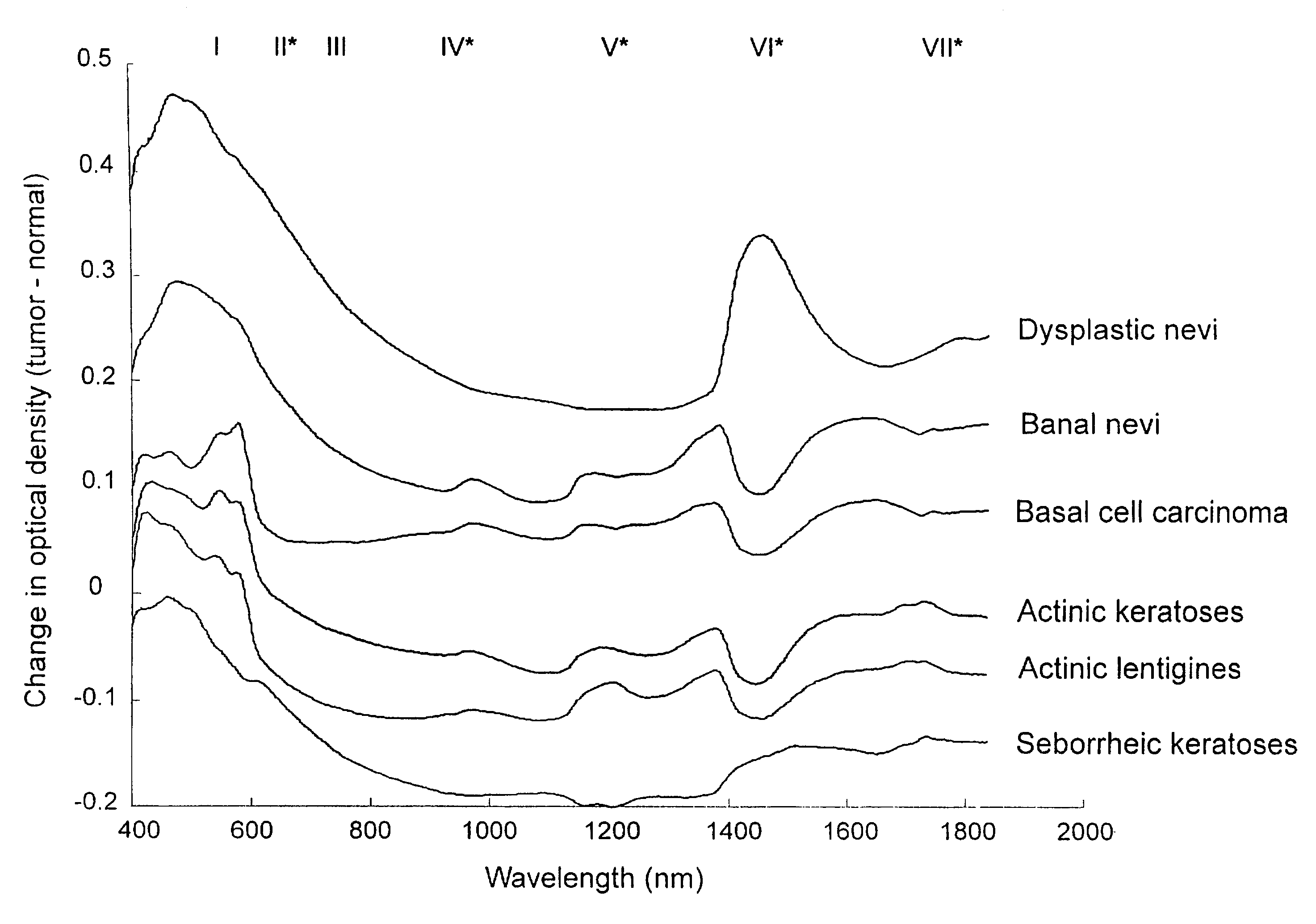
[0041]Significant noise was apparent in the 1850-2400 nm region due to the strong absorption of light by water in that spectral range. Prior to data analysis spectra were therefore truncated to 400-1840 nm, leaving a total of 720 data points per spectrum. Spectra were pooled according to the above 6 lesion categories. Spectra were pre-processed by normalizing to their total area and offset correcting.

[0042]The mean and standard deviation spectrum for each lesion category was generated by calculating the mean (+ / −SD) intensity at each of the 720 spectral data points for each category. Any spectrum that lay outside 2 standard deviations from the mean for each lesion group was removed from the study. It is interesting to note that in all instances spectra that lay outside 2 standard deviations were associated with patient movement as recorded by the study nurse. The remaining spectral database consisted of 94 (of 99) actinic keratosis spectra, 90 (of 96) BCC spectr...


PUM


Abstract

A non-invasive tool for skin disease diagnosis would be a useful clinical adjunct. The purpose of this study was to determine whether visible/near-infrared spectroscopy can be used to non-invasively characterize skin diseases. In-vivo visible- and near-infrared spectra (400-2500 nm) of skin neoplasms (actinic keratoses, basal cell carcinomata, banal common acquired melanocytic nevi, dysplastic melanocytic nevi, actinic lentigines and seborrheic keratoses) were collected by placing a fiber optic probe on the skin. Paired t-tests, repeated measures analysis of variance and linear discriminant analysis were used to determine whether significant spectral differences existed and whether spectra could be classified according to lesion type. Paired t-tests showed significant differences (p<0.05) between normal skin and skin lesions in several areas of the visible/near-infrared spectrum. In addition, significant differences were found between the lesion groups by analysis of variance. Linear discriminant analysis classified spectra from benign lesions compared to pre-malignant or malignant lesions with high accuracy. Visible/near-infrared spectroscopy is a promising non-invasive technique for the screening of skin diseases.

Description

PRIOR APPLICATION INFORMATION[0001]This application is a national phase application of PCT application CA00 / 01187, filed Oct. 5, 2000 which claims benefit of U.S. Provisional Application 60 / 157,857, filed Oct. 6, 1999.FIELD OF THE INVENTION[0002]The present invention relates generally to the field of spectroscopy. More specifically, the present invention relates to a method for non-invasively diagnosing skin diseases using visible and near-infrared spectroscopy.BACKGROUND OF THE INVENTION[0003]Skin cancer is the most common human cancer. In 1999, it is estimated that there will be 70000 new cases of skin cancer in Canada (Canadian Cancer Statistics: Toronto: National Cancer Institute of Canada, 1999) and more than 1 million new cases in the United States. The clinical diagnosis is often difficult since many benign skin diseases resemble malignancies upon visual examination. As a consequence, histopathological analysis of skin biopsies remains the standard for confirmation of a diagn...

Claims


Application Information

Patent Timeline
Patent Type & Authority Patents(United States)
IPC IPC(8): A61B5/00
CPCA61B5/0075A61B5/444A61B5/445G01N21/31G01N2201/129G01N21/359G01N2021/3155A61B5/7264
Inventor MCINTOSH, LAURA M.JACKSON, MICHAELMANTSCH, II, HENRYMANSFIELD, JAMES R.CROWSON, A. NEILTOOLE, JOHN W. P.
Owner NAT RES COUNCIL OF CANADA
Why Eureka
Try Eureka