
Production of "biological carriers" for induction of immune responses and inhibition of viral replication

a technology of immune response and viral replication, applied in the field of antigen presentation, can solve the problems of cumbersome, laborious, time-consuming, and expensive current procedures

Inactive Publication Date: 2009-08-25
JDM TECH +1
View PDF4 Cites 6 Cited by
  • Summary
  • Abstract
  • Description
  • Claims
  • Application Information

AI Technical Summary

Benefits of technology

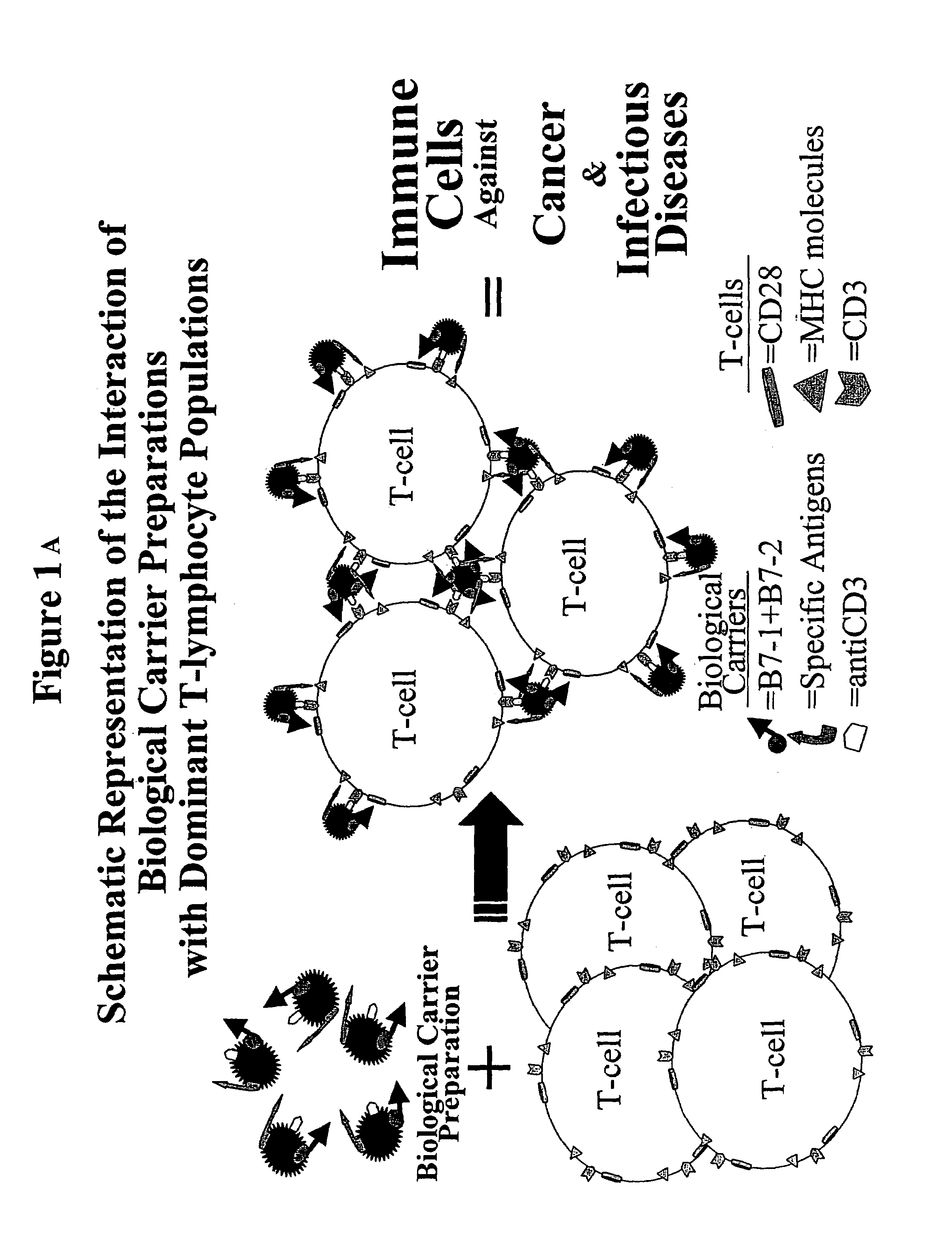
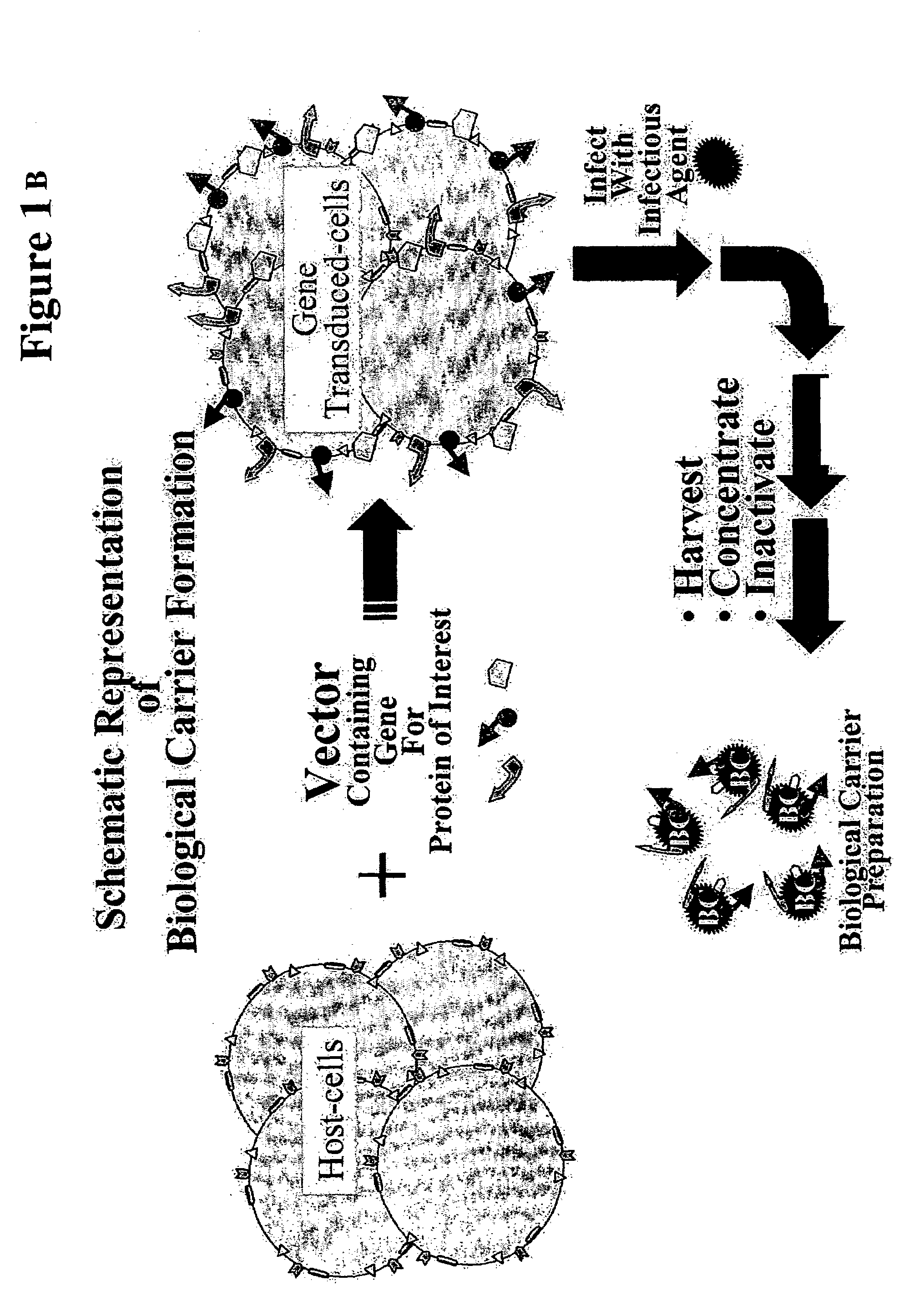
[0010]Viruses are ideal candidates for utilization as non-professional antigen presenting carriers. The invention uses viruses not for their infectious abilities (all preparations will be inactivated), but as a scaffold that contain specific antigens and co-stimulatory molecules to induce immune responses. The major advantage of this approach is the ease of production of potential therapeutic and / or vaccine doses. At the end of the virus life cycle, large amounts of virions are released from infected cells, reaching concentrations of 1012 virus particles per milliliter of culture fluid. Intrinsic to virus release, portion of the cell membrane are removed as the mature virus particle buds from the cell. The cells that support the productive viral infection can be genetically engineered to over-express surface molecules that would be carried with the virus particle as it is released from the cell. In addition, MHC class I and class II molecules containing viral peptides (due to the active viral infection) will decorate the virus particles, thereby presenting the antigens needed to stimulate the CD3 receptor on T-lymphocytes. These interactions supply one of the two signals required for T-cell stimulation. The over expression of co-stimulatory molecules by genetic engineering supplies the second signal, leading to antigen specific immune stimulation. These are distinct advantages over the use of cells (professional or non-professional) to present antigens to T-lymphocytes. The current procedure with professional antigen presenting cells (APCs) involves isolating cells from a patient, growing them in culture, and transplanting them back into that same person, a process that takes weeks. With non-professional APCs, a genetic engineering step (where co-stimulatory and / or MHC molecules would be introduced into the cell) would be added to the process. Even if the cells can be implanted allogeneically, current procedures are cumbersome, labor intensive, time consuming, and expensive. The present invention simplifies the process, thereby making immunotherapy potentially available world-wide in the area of infectious diseases.
[0013]In another aspect, the invention provides a method of activating or priming a naïve T-lymphocyte to respond to viral antigens, by contacting the T-lymphocyte with a Biological Carrier preparation containing either viral-specific antigen(s) fragments or with non-specific immune activators in the proper conformation for presentation. In addition, the Biological. Carrier may contain at least one co-stimulatory molecule. This T-cell priming can allow for the induction and expansion of not only effector cells but also long-lasting memory T-lymphocytes against a specific virus.

Problems solved by technology

Even if the cells can be implanted allogeneically, current procedures are cumbersome, labor intensive, time consuming, and expensive.

Method used


Image

  • Production of "biological carriers" for induction of immune responses and inhibition of viral replication
  • Production of "biological carriers" for induction of immune responses and inhibition of viral replication
  • Production of "biological carriers" for induction of immune responses and inhibition of viral replication

Examples


example 1

Requirement for Host Cell Modification(S) for Biological Carrier-Dependent Lymphocyte Stimulation

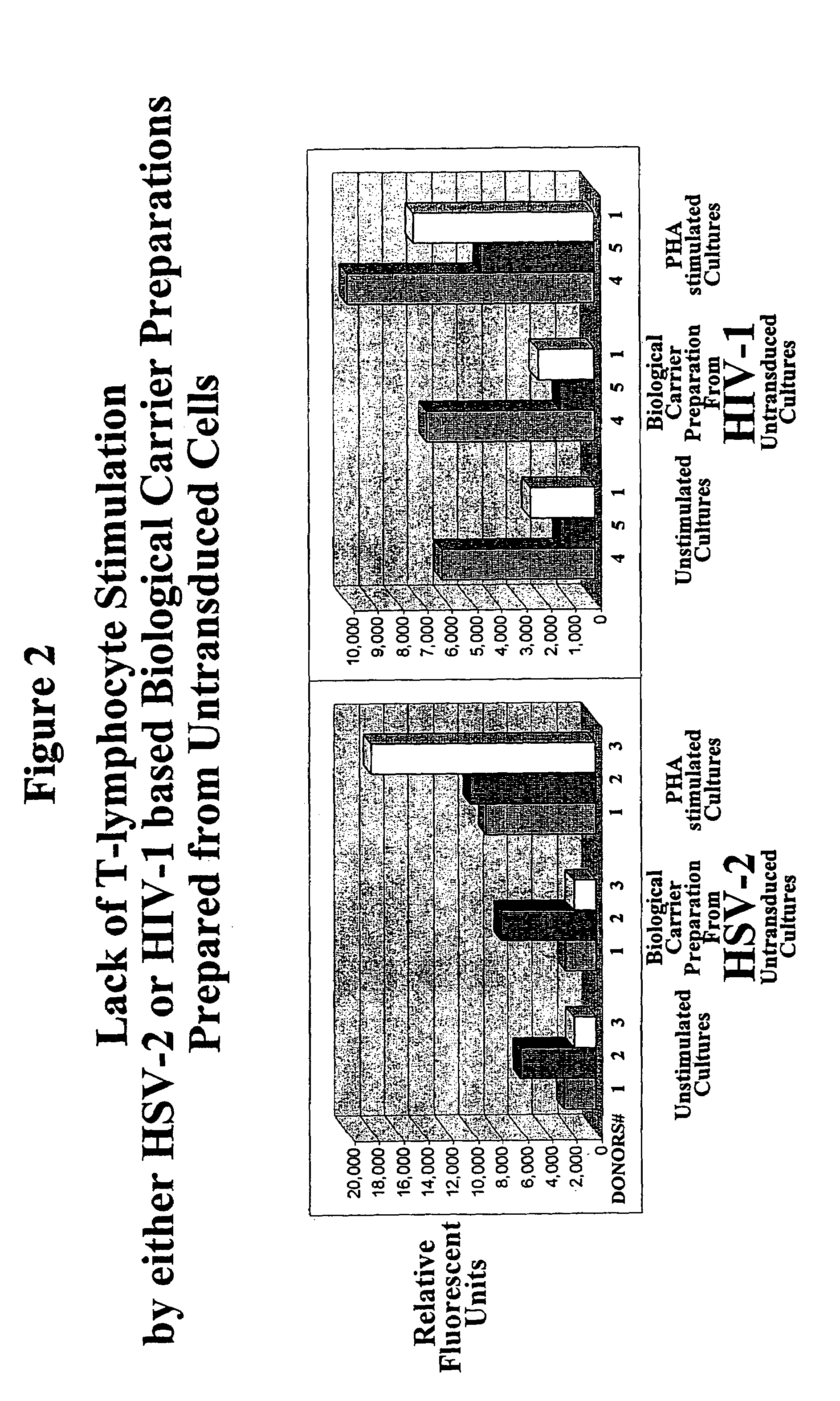
[0037]The principle of this invention is demonstrated by proliferation experiments comparing the degree of stimulation of biological carrier preparations obtained from untransduced, anti-CD3, B7-1+B7-2, and anti-CD3&B7-1+B7-2 transduced cultures.

[0038]Elutriated lymphocytes that were treated for six day with a preparation of biological carriers obtained from untransduced cultures showed a degree of proliferation (lymphocyte stimulation) similar to unstimulated (un-treated) cells (FIG. 2). Proliferation was measured by AlamarBlue™ assay. The assay is designed to measure quantitatively the proliferation of human cells by the incorporation of an oxidation-reduction indicator that fluoresces in response to chemical reduction of growth medium resulting from cell growth. The lack of biological carrier dependent stimulation when biological carriers were prepared from untransduced cultures was i...

example 2

Biological Carrier Preparations Retain Their Biological Activity After Lyophilization and Storage at Room Temperature

[0039]The principle of this invention is further demonstrated by retention of cellular proliferating biological activity when biological carrier preparations were lyophilized and stored at ambient temperatures.

[0040]The ability of native harvested culture fluid from HSV-2 based biological carriers from anti-CD3, B7-1+B7-2, and anti-CD3&B7-1+B7-2 transduced cells were compared to aliquots of the same sample fluids after lyophilization with respect to the ability of the preparations to stimulate T-lymphocytes in culture. In addition to lyophilization, the lyophilized material was stored at room temperature for two weeks before testing biological activity. After thirteen days in culture with Donor #6 T-lymphocytes (Table 1), the lyophilized culture supernatant from the transduced host cells showed similar stimulation of proliferation to that observed with the native cult...

example 3

Biological Carrier Preparations Retain Their Biological Activity After Concentration

[0043]The principle of this invention is further demonstrated by retention of cellular proliferating biological activity when biological carrier preparations were concentrated.

[0044]The ability to stimulate T-lymphocytes with native harvested culture fluid from HSV-2 based biological carriers obtained from untransduced or anti-CD3&B7-1+B7-2 transduced cells were compared to the same supernatants concentrated by polyethylene glycol (PEG) precipitation. The addition of PEG to culture fluid results in the formation of a precipitate. Virus (HSV-2) infected harvested culture supernatants were centrifuged at 4,000 times the force of gravity for 10 minutes, removing large particulate material from the culture fluid. Polyethylene glycol was added to the clarified supernatant to 6% and after 4 to 16 hours of incubation at 4° C. a precipitate was collected by centrifugation. Following resuspension of the pelle...


PUM

PropertyMeasurementUnit
volumesaaaaaaaaaa
volumesaaaaaaaaaa
timeaaaaaaaaaa

Abstract

This application provides a method to form non-infectious Biological Carrier that may be used to deliver signals to cells either in vitro or in vivo. The Biological Carriers are inactivated virus particles that have been specifically modified to give biological properties different from the virus particles deriving from an unmodified host cell that (i) expresses at least one co-stimulatory molecule and (iia) at least one antigen that can initiate an immune response, and / or (iib) express surface molecules that suppress viral replication.

Description

PRIORITY DOCUMENTS[0001]This application is a 371 application of PCT / US02 / 04157, filed Feb. 12, 2002, which claims priority to U.S. Provisional application 60 / 268,066, filed Feb. 13, 2001.FIELD OF THE INVENTION[0002]This invention relates to the field of antigen presentation for the activation and resultant induction of antigen-specific immune responses and the inhibition of viral replication by the formation of “Biological Carriers.”BACKGROUND OF THE INVENTION[0003]In mammals, antigen-presenting cells (APCs) process foreign antigens. The processing of the antigen within the APC triggers an efficient immune response within the host. Antigens are degraded into peptide fragments and become bound to major histocompatibility complex (MHC) molecules that are expressed on the cell surface and are able to interact with other cells of the immune system. Dendritic cells, macrophages, and Kupffer cells in the liver are among the most commonly encountered type of APCs. These cells readily engu...

Claims


Application Information

Patent Timeline
Patent Type & Authority Patents(United States)
IPC IPC(8): A61K39/00A61K39/12A61K39/21C12N7/04C12N15/09C12P21/00A61K39/245A61K39/385C07K16/28
CPCA61K39/0011A61K39/245A61K39/385C07K16/2809C12N7/00A61K39/21C12N2810/855A61K2039/505A61K2039/5252A61K2039/5258A61K2039/55516A61K2039/57A61K2039/605C12N2710/16634C12N2740/16034A61K39/12
Inventor MOSCA, JOSEPH D.
Owner JDM TECH
Learn More