
Method and apparatus for determining conditions of biological tissues

a biological tissue and condition technology, applied in the field of biological tissue condition determination methods and apparatuses, can solve the problems of requiring bulky equipment and expensive installation, affecting the accuracy of titration, so as to facilitate home diagnosis and home titration, and facilitate auto-titration of positive pressure

Inactive Publication Date: 2010-05-04
ISONEA LTD
View PDF65 Cites 68 Cited by
  • Summary
  • Abstract
  • Description
  • Claims
  • Application Information

AI Technical Summary

Benefits of technology

"The present invention provides a method and apparatus for determining characteristics of biological tissue, particularly the upper airways, in a non-invasive and effective way. The method involves introducing a sound into the tissue and detecting it after it has travelled through the tissue. The detected sound is analyzed to determine its velocity and attenuation, which can be correlated to the characteristics of the tissue. The apparatus includes a sound generating device and a recording device for this purpose. The invention can be used to monitor airway patency and can be used in sleep apnea treatments. It is simple to use and does not require specialist skills or expensive equipment."

Problems solved by technology

These are generally expensive and involve some degree of risk which is usually associated with the use of X-rays, radioactive materials or gamma-ray emission.
Furthermore, these techniques are generally complicated and require equipment which is bulky and expensive to install and, in most cases, cannot be taken to the bedside to assess biological tissues in patients whose illness prevents them being moved.
However, the process requires sophisticated and sometimes expensive technology and cannot be used in tissues in which there is a substantial quantity of gas, such as the lung.
The vast majority of infants now survive initial acute respiratory illness, but lung injury associated with mechanical ventilation causes many infants to develop ‘chronic lung disease’.
However, trials of new strategies in mechanical ventilation which were expected to reduce barotrauma and / or exposure to oxygen have often had disappointingly little impact on the incidence of chronic lung disease (HIFI Study Group, 1989; Bernstein et al, 1996; Baumer, 2000).
Comparison of strategies of conventional mechanical ventilation in animals (Dreyfuss et al, 1985) have indicated that high lung volumes may be more damaging than high intrapulmonary pressures, and has led to the concept of ‘volutrauma’ due to over-inflation of the lung.
However, this technique fails to confer benefit if the average lung volume is low (HIFI Study Group, 1989), yet it appears to be successful if a normal volume is maintained (McCulloch et al, 1988; Gerstmann et al, 1996).
Attempts to maintain an optimal lung volume in the clinical setting are frustrated by a lack of suitable methods by which the degree of lung inflation can be monitored.
However, oxygen requirements may be influenced by factors other than lung volume (for example intra- or extracardiac right to left shunting), and the hazards of radiation exposure prevent radiological examination being performed with the frequency required.
Furthermore, while absolute lung gas volume can be measured using ‘gold-standard’ techniques of Nitrogen (N2) washout or Helium (He) dilution, these are impractical for routine clinical use.
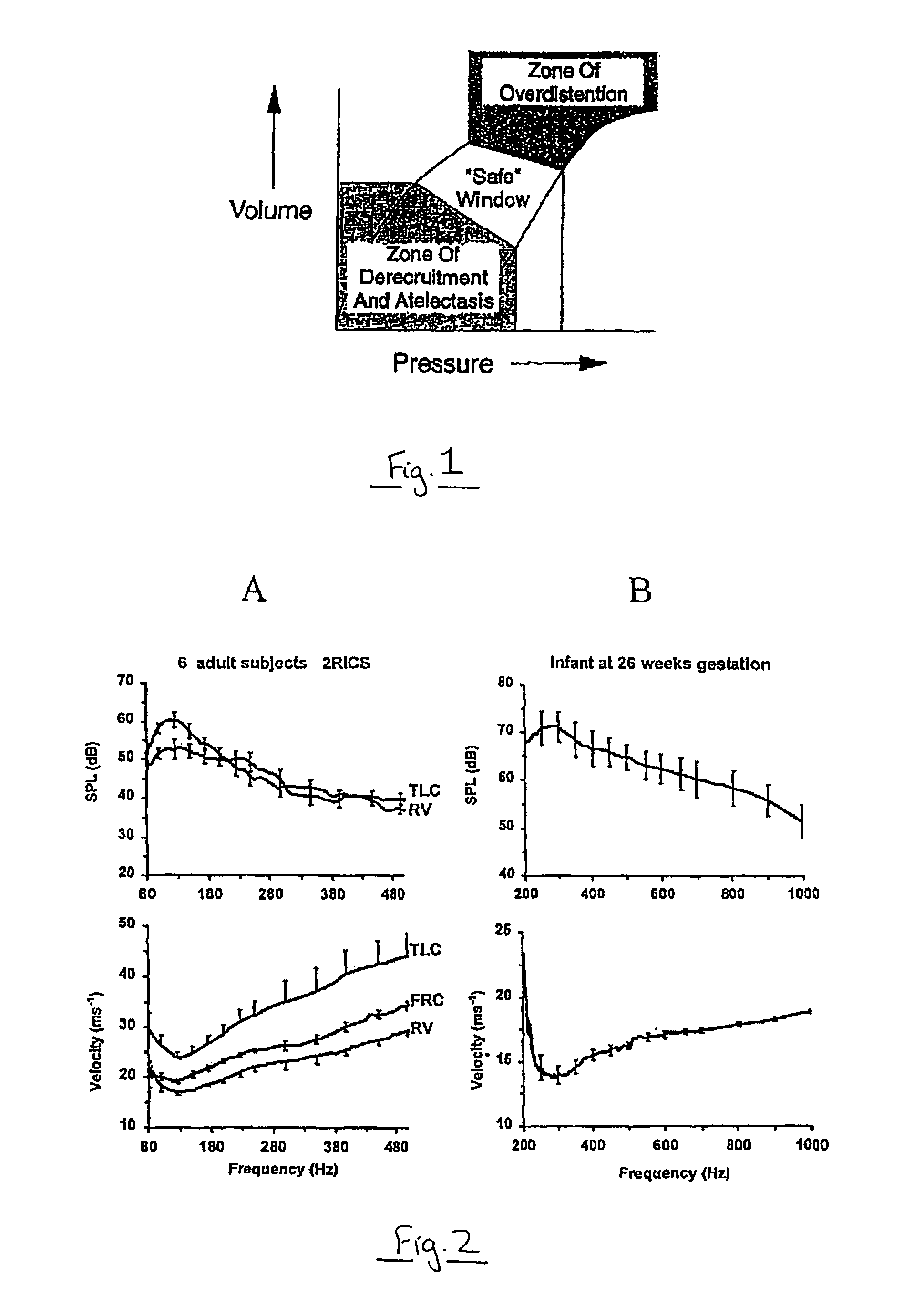
Even when lung volume is maintained in the “safe window”, changes in the lung condition may manifest due to the generally damaged or underdeveloped condition of the lung.
Fluid and blood may accumulate in the lung, posing additional threats to the patient.
However, in the sick newborn, the infant's small size, inability to co-operate and the presence of background noise greatly limits the value of such techniques.
Whilst determining and monitoring lung condition in newborn babies is difficult, determining lung condition in adults can be equally challenging, particularly if a patient is unconscious or unable to cooperate.
This places a further limitation on the presently available techniques for monitoring lung condition.
Sleep-disordered breathing is an increasingly recognised clinical problem, with severe obstructive sleep apnea affecting an estimated 5% of adults and 2% of infants.
In sleep-disordered breathing a reduction in airflow to the lungs may result in reduced blood oxygen levels (hypoxemia) and arousal from sleep.
This airflow reduction and arousal can occur many times a night, and can have a number of adverse effects, including excessive daytime sleepiness and an increased incidence of cardiovascular disease and stroke.
In obstructive sleep apnea, a patient's upper airway may be prone to collapse during sleep, thereby causing an obstruction and hypoxemia.
At present, the diagnosis of sleep-disordered breathing is difficult and is generally carried out by a polysomnography test, in which a number of physiological variables of a patient are monitored overnight in a sleep laboratory.
It is expensive, time-consuming and subject to inaccuracies.
Such methods are however too invasive for routine clinical use.

Method used


Image

  • Method and apparatus for determining conditions of biological tissues
  • Method and apparatus for determining conditions of biological tissues
  • Method and apparatus for determining conditions of biological tissues

Examples


example 1

Measurement of Lung Volume in Adults

[0247]In 5 healthy adult subjects, the velocity and attenuation of sound which was transmitted from one side of the chest to another, in a range of frequencies from 50-1000 Hz was measured at a number of defined positions on the chest. These measurements were taken while the lung volume was varied between Residual Volume (RV) and Total Lung Capacity (TLC). A reference position was established over the right upper zone of the chest. Using this position, a region in the frequency spectrum (around 100-125 Hz) where sound attenuation was much reduced and where the degree of attenuation was directly related to lung inflation (see FIG. 2A, upper panel) was found. The difference in attenuation between RV and TLC was approximately 7.5 dB and statistically significant (P=0.028). Further, it was found that sound velocity was low, averaging around 30 m / sec, and it showed a clear and strong sensitivity to the degree of lung inflation, being appreciably faster...

example 2

Measurement of Lung Density in Rabbits

[0250]Experiments were conducted in 1-2 kg New Zealand white rabbits. These animals were chosen for their similarity in size to the human newborn and their widespread use as a model of neonatal surfactant deficiency. Animals were anaesthetised with intravenous thiopentone, before performing a tracheostomy during which a 3 mm endotracheal tube was inserted into the airway to allow ventilation using a conventional neonatal ventilator (Bournes BP200). Maintenance anaesthesia was achieved with intravenous fentanyl. The chest was shaved and a microphone and transducer secured in various pre-defined positions, including a reference position over the right upper chest. The animal was then placed in a whole body plethysmograph to monitor absolute lung gas volume at intervals throughout the experiment. Tidal volume was monitored continuously with a pneumotachograph attached to the tracheostomy tube. The sound velocity and attenuation was determined at ea...

example 3

Measurement of Lung Inflation in Infants

[0259]To be a valuable clinical tool, measurements of sound velocity and attenuation must be sensitive to changes in lung inflation that are of a clinically relevant magnitude. A test of whether measurable changes in sound velocity and attenuation which occurred after clinical interventions which were confidently predicted alter the degree of lung inflation was conducted. It was found that clinical interventions which cause a significant change in lung inflation are associated with changes in sound transmission and velocity which are measurable using the present invention.


PUM


Abstract

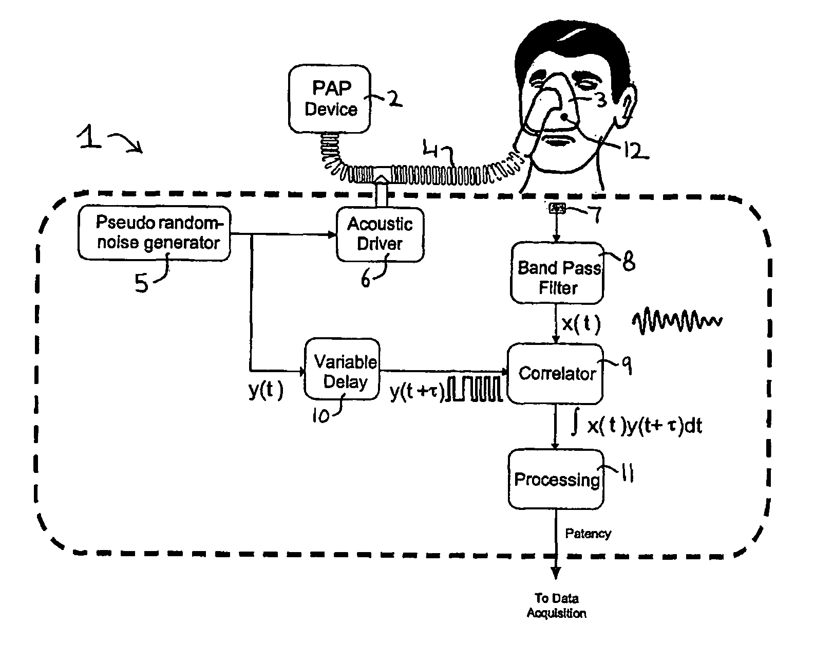
Airway monitoring apparatus 30 includes a driver 6 for applying sound in the audible frequency range to the airway of a subject and a detector 7 for monitoring the response of the airway, e.g. by detecting transmitted sound signal components and / or reflected sound signal components. Variations in the detected sound signal, e.g. energy, due to attenuation of the signal, give an indication of the state of the airway, e.g. airway patency, and can be used to monitor sleep-disordered breathing events, such as apnea and hypopnea, and to provide a breathing event index, e.g. AHI. The apparatus may provide servo-control for a respiratory assist device, such as a positive airway pressure device, so as to provide an appropriate pressure level setting. It may allow for home use of such devices and for home titration. It may also assist in discriminating central and obstructive breathing events, and in providing a measure of airway resistance.

Description

[0001]The present application is a continuation-in-part of U.S. Ser. No. 10 / 272,494 entitled “Method and Apparatus for Determining Conditions of Biological Tissues” and filed on 15 Oct. 2002 now U.S. Pat. No. 7,347,824, which is a continuation of International Patent Application No. PCT / AU01 / 00465 entitled “Method and Apparatus for Determining Conditions of Biological Tissues” filed on 20 Apr. 2001. The present application also claims the benefit of U.S. Provisional Patent Application No. 60 / 664,011 entitled “Apparatus, Systems and Methods for Monitoring Airway Patency” filed on 22 Mar. 2005. The contents of each of these applications are incorporated by reference in their entirety in the present application.[0002]The present invention relates to a method of determining characteristics of biological tissues in humans and animals. In particular, it relates to determining the characteristics of tissues such as the lungs and airways by introducing a sound to the tissue, and recording t...

Claims


Application Information

Patent Timeline
Patent Type & Authority Patents(United States)
IPC IPC(8): A61B5/08A61B8/08
CPCA61B5/08A61B5/085A61B5/4818A61B8/08A61M16/00A61B5/7257A61M2230/65A61M16/0666A61M2016/0039A61M2230/04A61M2230/205A61M16/0006A61M16/0051A61M16/026A61M2205/3375A61M2230/20
Inventor WILKINSON, MALCOLM HOWARDRAMSDEN, CLIVE ANDREWBERGER, PHILIP JOHNCAMILLERI, PETERWILSON, JACQUELINE ANNESAMUHEL, FRANK
Owner ISONEA LTD
Learn More