Human neutralizing antibodies against hemolytic uremic syndrome

a technology of hemolytic uremic syndrome and neutralizing antibodies, which is applied in the field of dose forms, can solve the problems of difficult development of effective treatments, permanent kidney damage, and adult susceptibleness, and achieve the effect of few adverse side effects

Inactive Publication Date: 2011-03-22
TRUSTEES OF TUFTS COLLEGE
View PDF13 Cites 7 Cited by
  • Summary
  • Abstract
  • Description
  • Claims
  • Application Information

AI Technical Summary

Problems solved by technology

Even as of 2007, there is no available treatment and antibiotics and anti-diarrheals may exacerbate the problem.
Although STEC infection occurs mainly in children, adults are also susceptible.
Bloody diarrhea usually occurs prior to systemic complications which can be either fatal, due to acute renal failure and serious neurological involvement, or lead to permanent kidney damage.
This has made development of effective treatments extremely difficult due to the general unreliability and lack of predictability of animal models for the human disease.

Method used


Image

  • Human neutralizing antibodies against hemolytic uremic syndrome
  • Human neutralizing antibodies against hemolytic uremic syndrome
  • Human neutralizing antibodies against hemolytic uremic syndrome

Examples


example 1

Development of a Gnotobiotic Piglet Model of Enterohemorrhagic Escherichia coli Infection

[0041]Materials and Methods:

[0042]Toxin Purification and Toxoid Production

[0043]Hydatid cysts isolated from sheep infected with Echinococcus granulosus contain material, identified as a glycoprotein, which has P1 blood group reactivity. The P1 glycoprotein's antigenic determinant was subsequently shown to consist of a trisaccharide, Galα-4Galβ1-4G1cNAc, identical to the nonreducing end of the P1 glycolipid on human erythrocytes. Shiga toxin, SLT-I and -II bind to terminal Galα-4Gal disaccharide of glycolipids and hence, the P1-glycolipid is a receptor for these toxins. The P1 glycoprotein in hydatid cyst fluid interacts directly with Shiga toxin and inhibits Shiga toxin binding and cytotoxicity to tissue culture cells. By covalently coupling the hydatid cyst glycoprotein to Sepharose 4B, a solid phase system for capturing toxin is generated. To purify SLT-I, C600(933J) is grown in low syncase me...

example 2

Construction of Monoclonal Antibodies by Creation of a Phage Display Library

[0059]Anti SLT-I and SLT-IT antibodies were generated by phage surface display technology as follows: In this approach, a library of heavy (VH-CH1) and Light (VL-CL) chain genes were generated in vitro. This library was cloned into an M13 surface display vector (pComb3 or its equivalent) and the resulting M13 phages, displaying anti SLT I and SLT II antibodies on their surface, were screened and selected by bio-panning.

[0060]Materials and Methods:

[0061]Enrichment of Lymphocytes Secreting Anti SLT I and Anti SLT II Antibodies

[0062]Lymphocytes secreting anti SLT-I and anti SLT-II antibodies are enriched according to Linton, et al. (Linton, et al., Cell, 59:1049-1059 (1989)). Purified lymphocytes are incubated for 45 minutes with 60 nM biotin-SLT-I or biotin-SLT-II toxin, washed twice, and then poured onto Petri dishes coated with streptavidin and blocked with bovine serum albumin, incubated for another 60 minu...

example 3

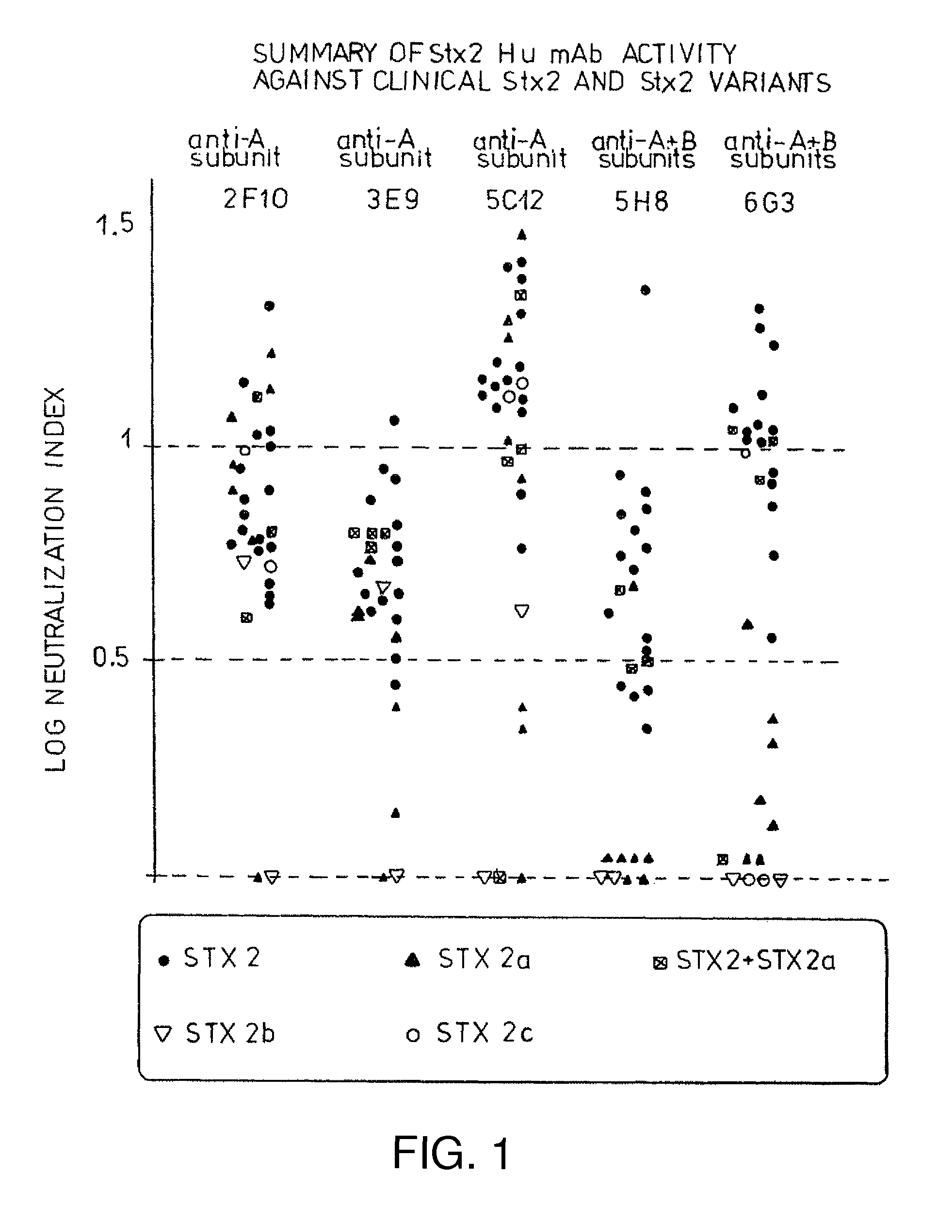
Preparation of Specific Human Monoclonal Antibodies Against Shiga-like Toxin II Using a Transgenic Mouse and Neutralization of this Toxin Using the Monoclonal Antibodies

[0074]Materials and Methods:

[0075]Isolation of Stx2

[0076]Stx2 was isolated, purified, and quantified as described in Donohue-Rolfe, et al., Infect. Immun., 57(12):3888-93 (1989). Briefly, Stx2 was isolated from E. coli strain C600 (933W) which bears the 933WJ bacteriophage that encodes Stx2. E. coli strain C600 (933W) was grown in Modified Syncase Broth at 37° C. with agitation in the presence of 200 ng / ml mitomycin C. Mitomycin C induces the 933J bacteriophage carrying the genes for Stx2. Stx2 in the culture supernatant was precipitated by the addition of 70% ammonium sulfate. The precipitate was dissolved in 10 mM Tris (pH 7.4) and dialyzed against the same buffer. The dialyzed and dissolved precipitate was applied to a Sepharose 4B affinity column containing P1 glycoprotein isolated from Echinococcus granuloses hy...


PUM

PropertyMeasurementUnit
concentrationaaaaaaaaaa
dissociation constantaaaaaaaaaa
weightaaaaaaaaaa

Abstract

Human and humanized monoclonal antibodies which binds specifically to subunit A of Shiga like toxin II have been developed which are effective to prevent or ameliorate one or more symptoms of HUS in a human. Effective dosages for treatment or prevention range from approximately 0.1 to 5.0 mg of antibody / kg of patient weight. The examples demonstrate the preferred dosage ranges based on the pig model, and what is being tested in phase I clinical trials. Antibodies are preferably transfused over a period of two hours, although this will depend on the patient and the disease state at the time of treatment. Preferred dosages for treatment of humans are between 0.1 mg / kg-5.0 mg / kg of 5C120, or an equivalent dosage of another antibody to subunit A of STX2. In the most preferred embodiments, dosages of 0.1 mg / kg, 0.5 mg / kg, or 5.0 mg / kg of 5C12 (low dose, anticipated therapeutic dose based on animal data and high dose) are administered.

Description

CROSS-REFERENCE TO RELATED APPLICATIONS[0001]This application is a continuation-in-part of U.S. Ser. No. 10 / 041,958, filed Jan. 7, 2002, which is a continuation-in-part of U.S. Ser. No. 09 / 302,125, filed Apr. 29, 1999, which is a division of U.S. Ser. No. 08 / 749,704, filed Nov. 1, 1996.GOVERNMENT SUPPORT[0002]The federal government may have certain rights in the invention since funding for the work described herein was provided in part by the NIH grants R01-AI41326 and R01-DK58993.FIELD OF THE INVENTION[0003]The present invention relates to dosage forms of human monoclonal antibodies capable of neutralizing Shiga or Shiga-like toxins which cause hemolytic uremic syndrome in humans, a process for the preparation of the new human monoclonal antibody dosage forms, and a method of administration thereof to a human to prevent or treat the development of hemolytic uremic syndrome.BACKGROUND OF THE INVENTION[0004]Since the first documented outbreaks in 1982, infections from enterohemorrhag...

Claims


Application Information

Patent Timeline
Patent Type & Authority Patents(United States)
IPC IPC(8): A61K39/00A61K39/395A61K39/40
CPCC07K16/1232A61K38/00A61P13/00
Inventor TZIPORI, SAULBALAKRISHNAN, RAMASWAMYDONOHUE-ROLFE, ARTHUR
Owner TRUSTEES OF TUFTS COLLEGE
Why Eureka
Try Eureka