Methods for the diagnosis of fetal abnormalities

a fetal abnormality and method technology, applied in the field of methods for the diagnosis of fetal abnormalities, can solve the problems of unreliable and accurate tests, many cancers continue to go undetected, and the amniocentesis and chorionic villus sampling (cvs) are potentially harmful to the mother and the fetus, so as to achieve reliable and accurate clinical diagnosis.

Active Publication Date: 2012-03-20
VERINATA HEALTH INC +2
View PDF543 Cites 165 Cited by
  • Summary
  • Abstract
  • Description
  • Claims
  • Application Information

AI Technical Summary

Benefits of technology

[0005]The methods of the present invention allow for enrichment of rare cell populations, particularly fetal cells or cancer cells, from peripheral blood samples which enrichment yields cell populations sufficient for reliable and accurate clinical diagnosis. The methods of the present invention also provide analysis of said enriched rare cell populations whereby said methods allow for detection, diagnosis and prognosis of conditions or diseases, in particular fetal abnormalities or cancer.

Problems solved by technology

However, the available methods today, amniocentesis and chorionic villus sampling (CVS) are potentially harmful to the mother and to the fetus.
For example, maternal serum alpha-fetoprotein, and levels of unconjugated estriol and human chorionic gonadotropin are used to identify a proportion of fetuses with Down's syndrome, however, these tests are not one hundred percent accurate.
Moreover, despite decades of advances in cancer diagnosis and therapy, many cancers continue to go undetected until late in their development.
However, fetal cells and cancer cells are rare as compared to the presence of other cells in the blood.
Enriching fetal cells from maternal peripheral blood and cancer cells from patient's blood is challenging, time intensive and any analysis derived there from is prone to error.

Method used


Image

  • Methods for the diagnosis of fetal abnormalities
  • Methods for the diagnosis of fetal abnormalities
  • Methods for the diagnosis of fetal abnormalities

Examples


example 1

Separation of Fetal Cord Blood

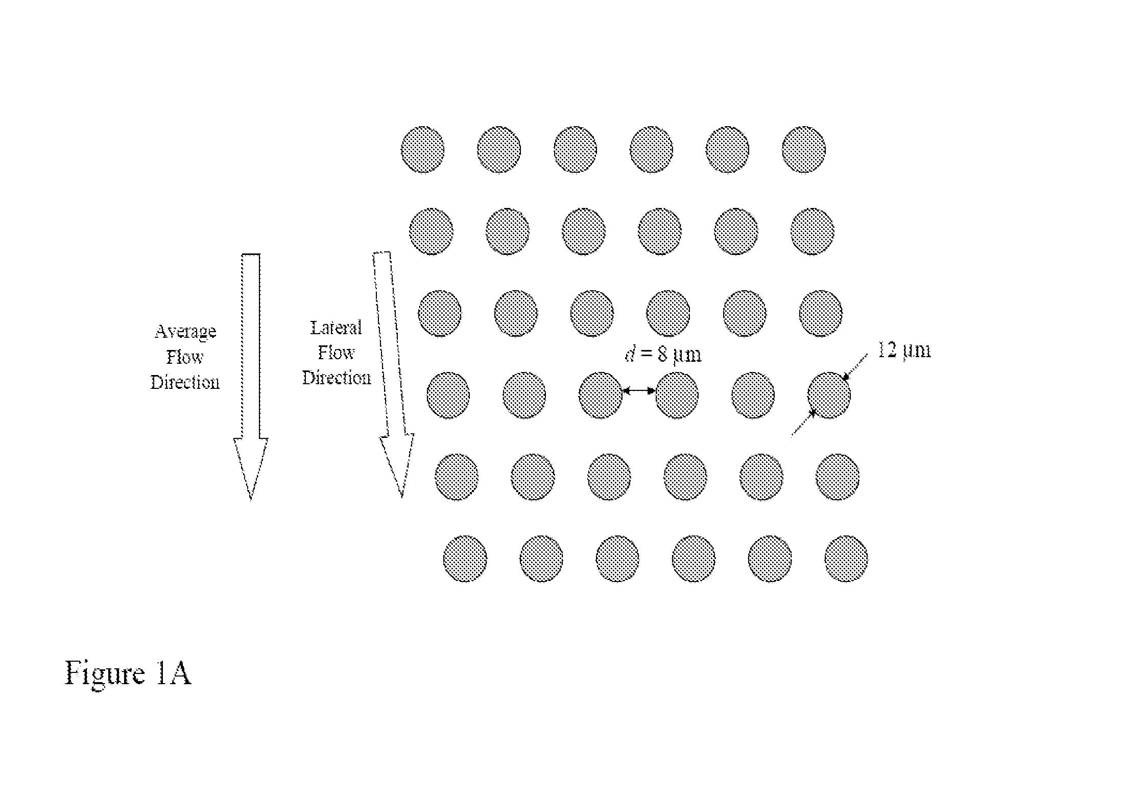
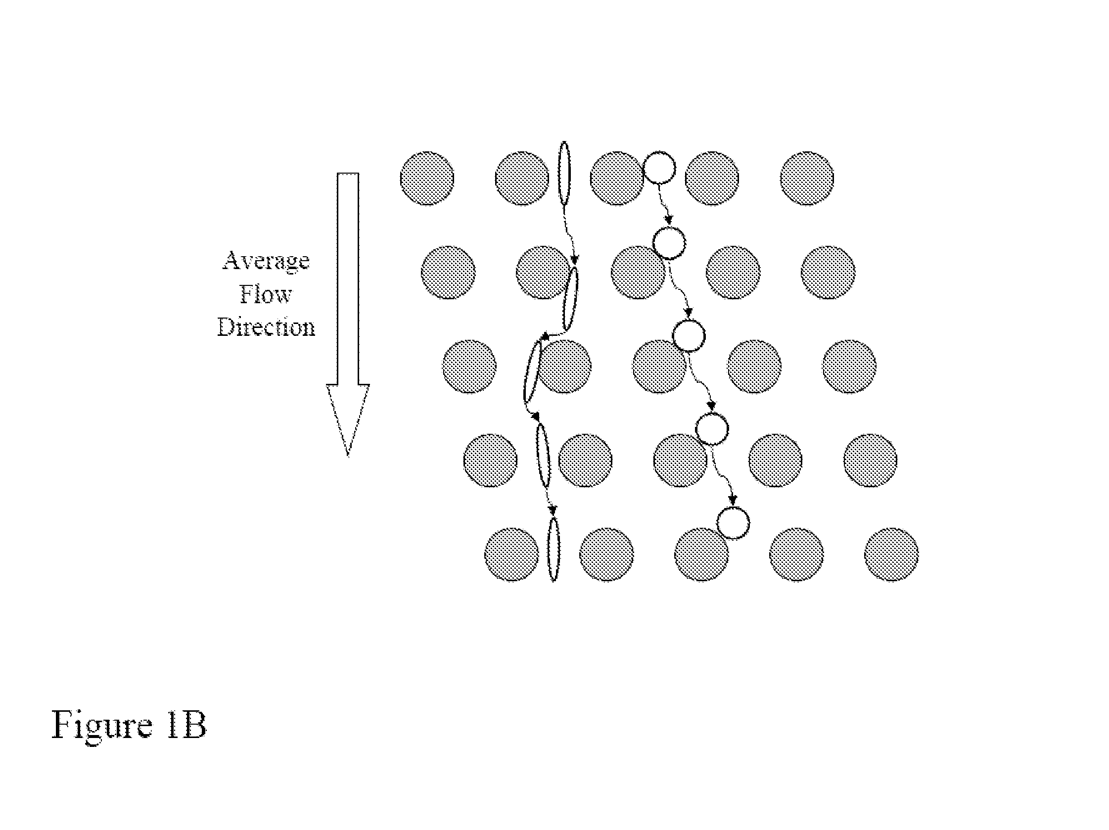
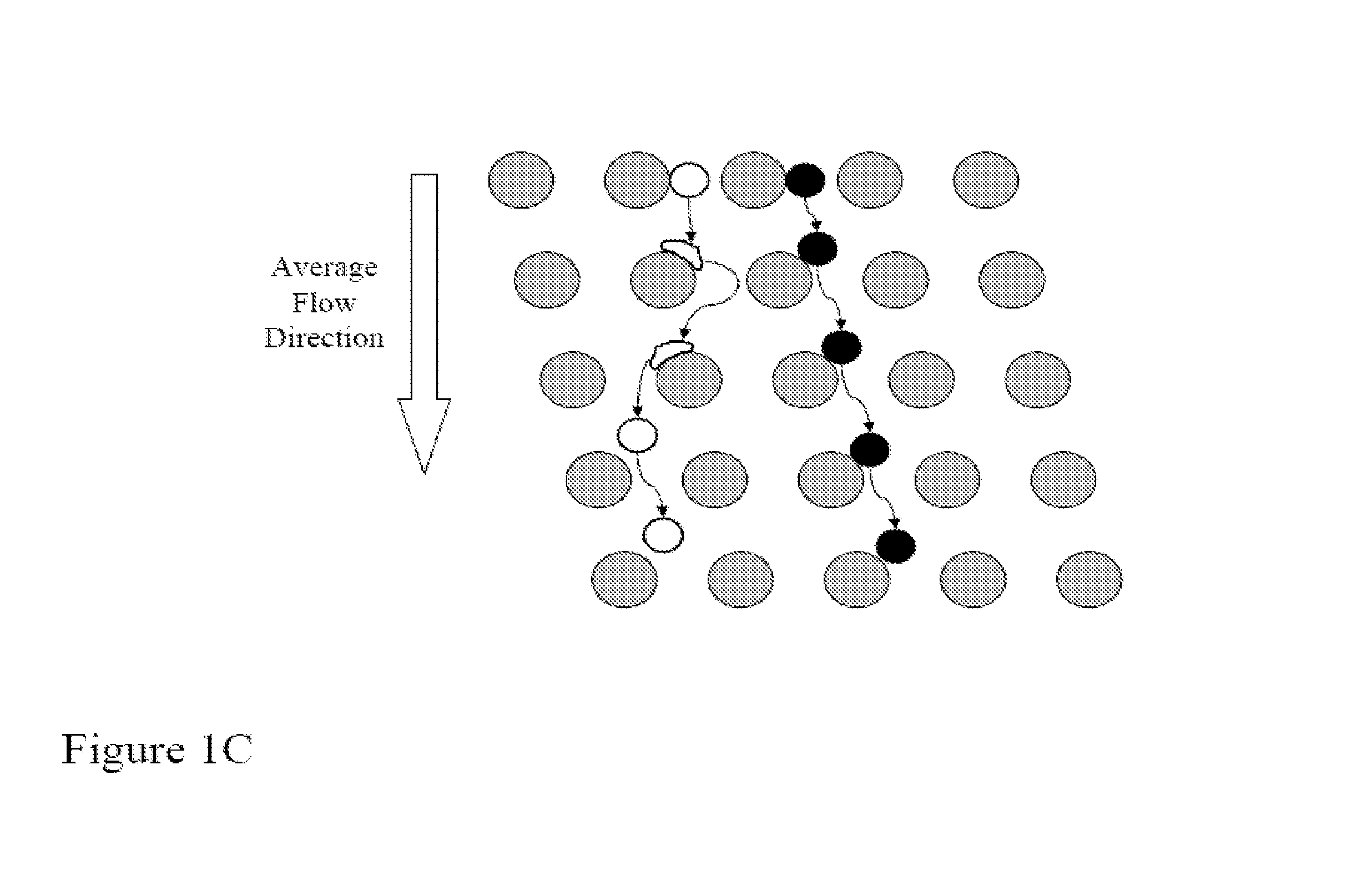
[0181]FIGS. 14A-14D shows a schematic of the device used to separate nucleated cells from fetal cord blood.

[0182]Dimensions: 100 mm×28 mm×1 mm

[0183]Array design: 3 stages, gap size 18, 12 and 8 μm for the first, second and third stage, respectively.

[0184]Device fabrication: The arrays and channels were fabricated in silicon using standard photolithography and deep silicon reactive etching techniques. The etch depth is 140 μm. Through holes for fluid access are made using KOH wet etching. The silicon substrate was sealed on the etched face to form enclosed fluidic channels using a blood compatible pressure sensitive adhesive (9795, 3M, St Paul, Minn.).

[0185]Device packaging: The device was mechanically mated to a plastic manifold with external fluidic reservoirs to deliver blood and buffer to the device and extract the generated fractions.

[0186]Device operation: An external pressure source was used to apply a pressure of 2.0 PSI to the buffer and blood r...

example 2

Isolation of Fetal Cells from Maternal Blood

[0190]The device and process described in detail in Example 1 were used in combination with immunomagnetic affinity enrichment techniques to demonstrate the feasibility of isolating fetal cells from maternal blood.

[0191]Experimental conditions: blood from consenting maternal donors carrying male fetuses was collected into K2EDTA vacutainers (366643, Becton Dickinson, Franklin Lakes, N.J.) immediately following elective termination of pregnancy. The undiluted blood was processed using the device described in Example 1 at room temperature and within 9 hrs of draw. Nucleated cells from the blood were separated from enucleated cells (red blood cells and platelets), and plasma delivered into a buffer stream of calcium and magnesium-free Dulbecco's Phosphate Buffered Saline (14190-144, Invitrogen, Carlsbad, Calif.) containing 1% Bovine Serum Albumin (BSA) (A8412-100ML, Sigma-Aldrich, St Louis, Mo.). Subsequently, the nucleated cell fraction was ...

example 3

Confirmation of the Presence of Male Fetal Cells in Enriched Samples.

[0194]Confirmation of the presence of a male fetal cell in an enriched sample is performed using qPCR with primers specific for DYZ, a marker repeated in high copy number on the Y chromosome. After enrichment of fnRBC by any of the methods described herein, the resulting enriched fnRBC are binned by dividing the sample into 100 PCR wells. Prior to binning, enriched samples may be screened by FISH to determine the presence of any fnRBC containing an aneuploidy of interest. Because of the low number of fnRBC in maternal blood, only a portion of the wells will contain a single fnRBC (the other wells are expected to be negative for fnRBC). The cells are fixed in 2% Paraformaldehyde and stored at 4° C. Cells in each bin are pelleted and resuspended in 5 μl PBS plus 1 μl 20 mg / ml Proteinase K (Sigma #P-2308). Cells are lysed by incubation at 65° C. for 60 minutes followed by inactivation of the Proteinase K by incubation...


PUM

PropertyMeasurementUnit
timeaaaaaaaaaa
timeaaaaaaaaaa
timeaaaaaaaaaa

Abstract

The present invention relates to methods for detecting, enriching, and analyzing rare cells that are present in the blood, e.g. fetal cells. The invention further features methods of analyzing rare cell(s) to determine the presence of an abnormality, disease or condition in a subject, e.g. a fetus by analyzing a cellular sample from the subject.

Description

CROSS-REFERENCE[0001]This application claims the benefit of U.S. Provisional Application Nos. 60 / 804,817, and 60 / 804,819 both filed Jun. 14, 2006 and U.S. Provisional Application No. 60 / 820,778 filed Jul. 28, 2006, which applications are incorporated herein by reference.BACKGROUND OF THE INVENTION[0002]Analysis of specific cells can give insight into a variety of diseases. These analyses can provide non-invasive tests for detection, diagnosis and prognosis of diseases, thereby eliminating the risk of invasive diagnosis. For instance, social developments have resulted in an increased number of prenatal tests. However, the available methods today, amniocentesis and chorionic villus sampling (CVS) are potentially harmful to the mother and to the fetus. The rate of miscarriage for pregnant women undergoing amniocentesis is increased by 0.5-1%, and that figure is slightly higher for CVS. Because of the inherent risks posed by amniocentesis and CVS, these procedures are offered primarily ...

Claims


Application Information

Patent Timeline
Patent Type & Authority Patents(United States)
IPC IPC(8): C12Q1/68C12P19/34C07H21/02C07H21/04
CPCG01N33/6893G01N1/30G01N2800/385C12Q1/6883C12Q2600/16C12Q2600/156C12Q2600/158
Inventor KAPUR, RAVITONER, MEHMETSTOUGHTON, ROLANDSHOEMAKER, DANIELDAVIS, RONALD W.
Owner VERINATA HEALTH INC
Why Eureka
Try Eureka