Method for repairing intervertebral discs

a technology of intervertebral discs and tissue defects, which is applied in the field of repairing tissue defects in intervertebral discs, can solve the problems of patient loss of bowl and bladder function, severe compression of the exiting nerve root, and strong and progressive pain that emanates from the compromised segment of the spine, etc., to achieve the effect of increasing the height of the disc space, preventing leakage, and increasing the pressure of the dis

Active Publication Date: 2013-01-22
PAUZA KEVIN
View PDF14 Cites 26 Cited by
  • Summary
  • Abstract
  • Description
  • Claims
  • Application Information

AI Technical Summary

Benefits of technology

[0013]The in-situ curable sealant material provides the surgeon with means for repairing the annulus fibrosus by injecting the in-situ curable composition into the disc so that the composition flows out into and at least partially seals fissures in the annulus to thereby delimit material from the nucleus pulposus from flowing through the fissures to the outside of the disc. The injection of the curable composition into the disc has been determined to be superior to the deposition of curable composition disclosed in U.S. Pat. No. 6,428,576, wherein curable composition was not injected into the disc such that composition flowed outward from the center of the disc to fill voids and fissures in the annulus. Thus, in accordance with this invention, by the superior process of injection disclosed herein, the annulus is at least partially sealed to thereby reduce or eliminate migration of nucleus pulposus cells into the epidural space, and to prevent, by sealing the annulus, diffusion of nucleus pulposus-derived cytokines and inflammatory mediators into the epidural space through the annulotomy site. The thus resulting prevention of contact between nucleus pulposus cells, and its cytokines or inflammatory mediators, with nerve roots after discectomy is another object of the invention and will assist to minimize nucleus pulposus-induced nerve root injury and nerve root pain. Likewise, the present invention contemplates injection of the curable composition in other spinal joints where a fibrous capsule surrounds a core.
[0018]In another broad respect, this invention is a process of injecting the curable composition into the disc such that the curable composition flows outward to repair a ruptured annulus fibrosus, where the nucleus pulposus contains a sufficient number of viable cells, or such cells are co-injected, that assist in the restoration of the load-bearing and viscoelastic properties of the defective intervertebral disc.
[0019]In another broad respect, this invention is an implant that minimizes removal of nucleus pulposus material during a discectomy intervention without having an elevated risk of recurrent disc hernia, where the implant has been formed by injecting the curable composition into the disc. Since the nucleus pulposus tissue in most disc hernia patients is viable and has regenerative potential, leaving as much nucleus pulposus tissue as possible in the disc space may be conducive to the gradually regeneration of the disc and restoration of its physiological functions.
[0021]Advantageously, the method and kit of this invention facilitate extended pain relief for patients with leaky disc syndrome, wherein for example nucleus pulposus leaks out of the disc through defects (e.g. tears or fissures) in the annulus fibrosus. The pressure monitor provides a heightened level of safety whereby the physician can measure pressure in real time so as to avoid over-pressurizing a disc being treated. Likewise, the physician can observe the pressure reading in conjunction with injection of the fibrin sealant to determine whether the disc is being sealed and whether sufficient fibrin sealant has been injected. In this way the physician can use the delivery device as a diagnostic tool to assess whether the disc is treatable.
[0025]In general, the curable composition of this invention is injected into the disc, the epidural space, the zygapophyseal joints (aka: facet joints), and / or sacroiliac joint. With respect to an injection of curable composition into a disc, an intra-discal injection serves to create a cured composition which seals the disc from leaking material from the nucleus into the area outside the disc. For example, the curable composition can be delivered by fluoroscopic transforminal lumber epidural or intra-discal injection, such as described in U.S. Pat. No. 6,468,527. For the treatment of back injuries such as these, the curable composition is injected into the nucleus pulposus, to fill any fissures or voids of the annulus fibrosus, to seal the bone end plates to the disc, increase pressure of the disc, and to increase the height of the disc space. In general, the curable composition is injected at a location near the defect in the annulus fibrosus. Typically the curable composition will flow into the fissures in the annulus fibrosus, and some may thus flow out of the intra-discal space. The injection may also serve to coat areas adjacent to the disc, directly on the nerve roots and surrounding areas which serve to protect those areas from the effects of the leaking nucleus material. Sealing the fissures and bone end plates halts the leakage of harmful chemicals from the disc environment and prevents the initiation of foreign-body reactions towards the damaged disc and nociceptors in the outer annulus of the disc by the immune system. Increasing the disc space relieves pressure on the nerve root. That is, as a result of the injection, an increase of the disc height occurs, which increases the spacing between lamina, and which in turn relieves pressure on the nerve roots on the lamina. For this application, supplementation of the curable composition with growth factors and a variety of biologically or pharmaceutically active agents, including tumor necrosis factor inhibitors (TNF-α inhibitors) may promote rehabilitation of the damaged tissues or the gradual replacement of the curable composition with healthy tissue and / or reduce inflammation in surrounding tissues.

Problems solved by technology

This usually leads to strong and progressive pain that emanates from the compromised segment of the spine.
This will typically result in severe compression of the traversing nerve root, the exiting nerve root, and the lateral aspect of the Thecal Sac.
Sequestration (aka: sequester, free-fragment) may be excruciatingly painful (back and leg pain—sciatica) and, if centrally located, may occasionally cause the patient to lose control of their bowl and bladder function, i.e., Cauda Equina Syndrome, which is considered a medical emergency.
Without repair, a considerable risk of post-discectomy complications, such as a re-herniation of the disc, will remain.
However, it has been shown that severe post-discectomy complications may occur in about 6-16% of all surgical interventions.
Both events may result in post-discectomy complications such as persistent nerve root pain and / or irritation of nocioceptors in the outer ⅓ of the disc annulus.

Method used


Image

  • Method for repairing intervertebral discs
  • Method for repairing intervertebral discs
  • Method for repairing intervertebral discs

Examples


example 1

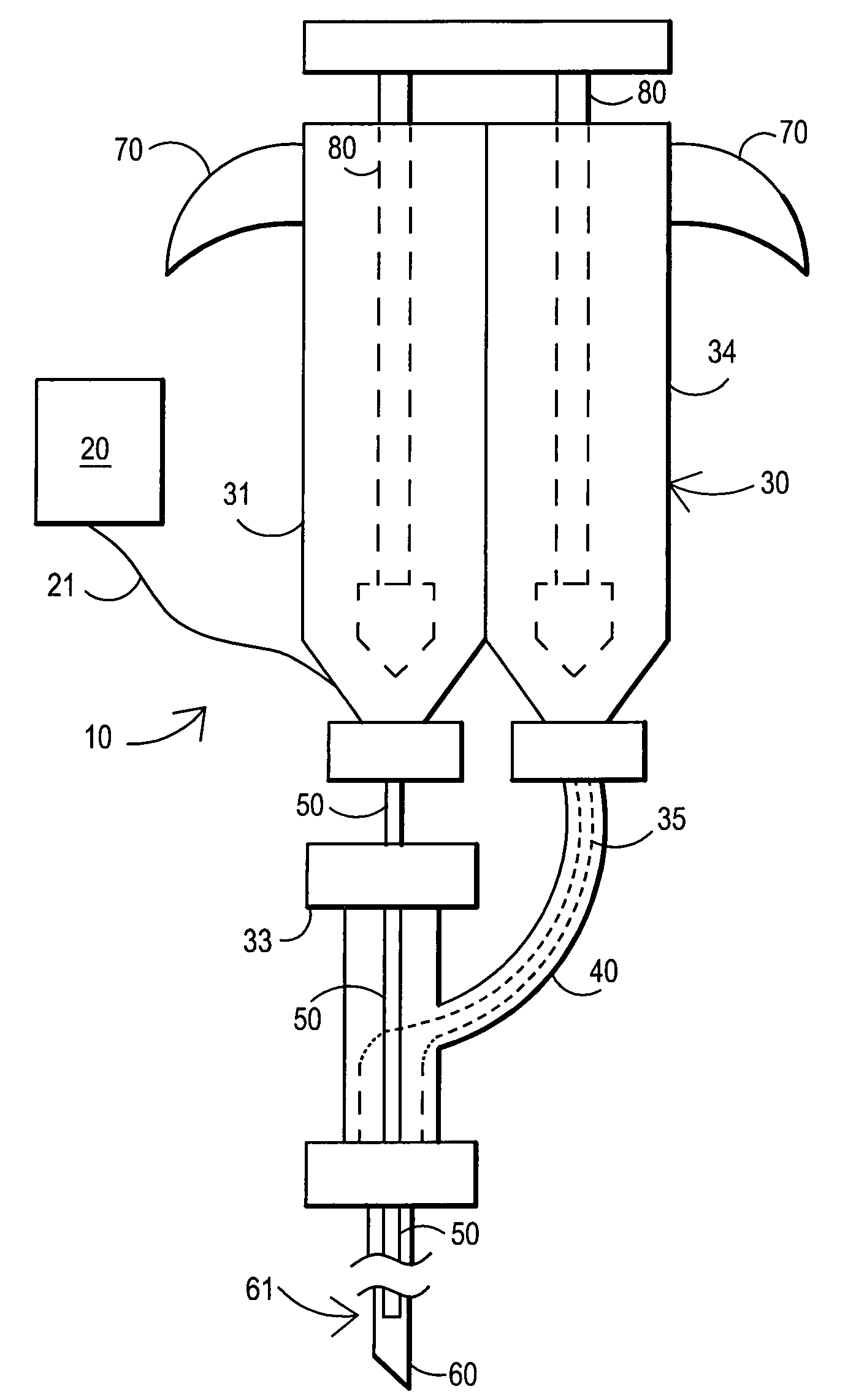
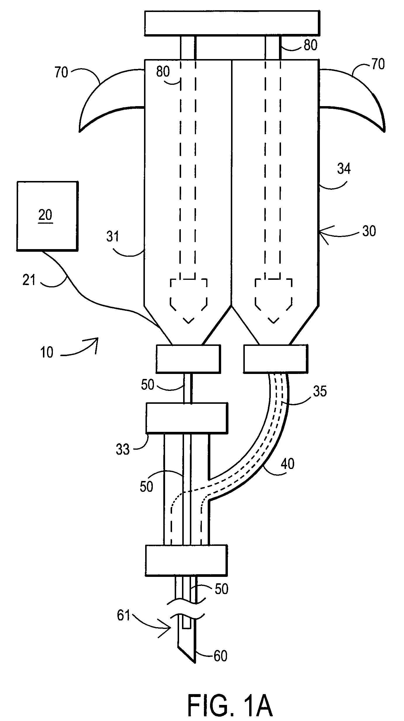
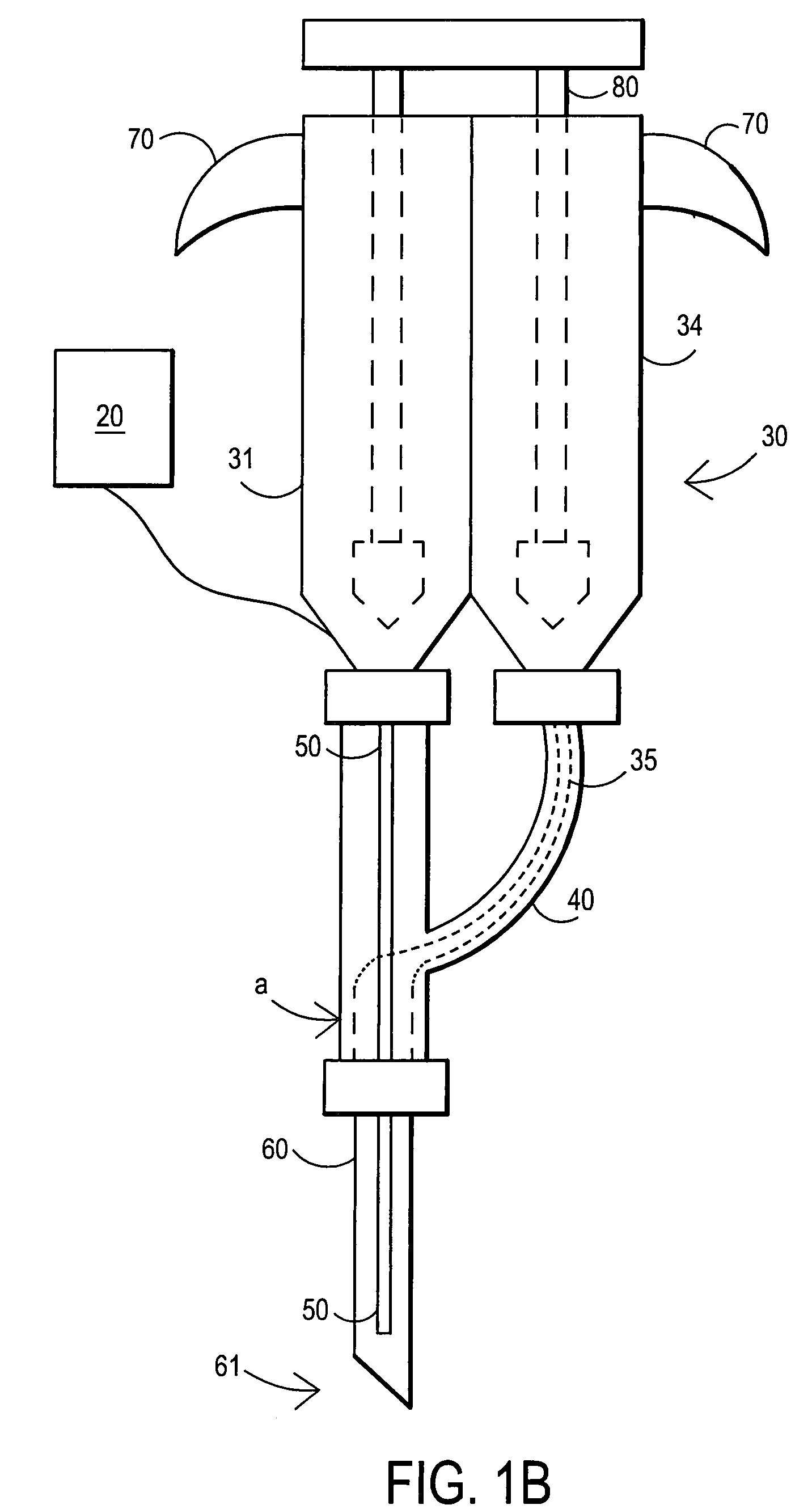
[0126]A 105 mg / ml (10.5% W / V) aqueous solution of fibrillar collagen in 0.05 M sodium bicarbonate buffer and 0.15 M sodium chloride is adjusted to pH 9.5. 2.5 ml of this biomaterial solution (solution A) is aspirated into a dual chamber polypropylene cartridge through one of the two extrusion flanges of the cartridge. 2.5 ml of a solution of difunctionally activated N-succinimidyl carbonate PEG (DSC-PEG, MW 3600) in 0.005 M sodium carbonate / bicarbonate buffer and 0.15 M sodium chloride at pH 6.0 and in a 1 to 10 molar ratio of collagen (solution A) to DSG-PEG (solution B) is filled into the second chamber of the cartridge.

[0127]Both chambers of the cartridge are closed by attaching a spiral mixer nozzle (3.2 mm inner diameter, 6.2 cm length, 0.38 ml void volume) onto the dual extrusion flanges. The cartridge is placed into a manual application instrument that allows for a reproducible and volume controlled extrusion of the bio-material in increments of 0.5 ml per step. A blunt tip a...

example 2

[0129]A 380 mg / ml (38% w / v) aqueous solution of human serum albumin (MW 68000) in 0.1 M sodium bicarbonate buffer and 0.15 M sodium chloride is adjusted to pH 8.2 (solution A buffered protein solution). A 200 mg / ml (25% w / v) aqueous solution of difunctionally activated N-succinimidyl propionate PEG (DSP-PEG, MW 3400) in 0.01 M sodium carbonate / bicarbonate buffer at pH 6.0 is prepared as solution B (cross linking agent). Solutions A and B are placed in the dual chamber cartridge and injected as described in Example 1. In this Example 2, the application of the sealant without delay is particularly important because of the short curing time of this type of sealant (2-3 minutes).

example 3

[0130]180 mg / ml (18% w / v) of PEG tetraacrylate (MW 8200) is dissolved in a buffer of 0.02 M sodium phosphate at pH 7.4 and 0.15 M sodium chloride. Ammonium persulfate (0.01 M) and sodium bisulfite (0.005 M) are added to the solution that now represents the polymerizable bio-material with thermal polymerization initiation system. 5 ml of this bio-material solution is aspirated into a polypropylene syringe that is fitted with a Luer type adapter tip. The syringe is closed by placing a temperature-controlled, flow through heating cylinder, that is connected to a control unit, onto the tip of the syringe. The syringe is placed into a manual application instrument that allows for a reproducible and volume-controlled extrusion of the bio-material in increments of 0.25 ml per step. A blunt tip aspiration needle (18 gauge, 90 mm) is placed on the tip of the heating cylinder. The handle of the application instrument is pressed four times (4×0.25 ml) in order to fill the void of the heating c...


PUM

PropertyMeasurementUnit
widthaaaaaaaaaa
volumeaaaaaaaaaa
lengthaaaaaaaaaa

Abstract

A method of repairing a defect in an annulus fibrosus of an intervertebral disc, without excising the entire nucleus pulposus of the disc. The method includes inserting an introducer needle through the annulus fibrosus by puncturing the annulus fibrosus with the introducer needle, injecting an in situ curable, bio-compatible polymerizable or polymeric material composition into the disc through the introducer needle directly or indirectly so that the in situ curable composition contacts a defect in the annulus fibrosus; and curing said material in situ.

Description

[0001]This application is a continuation-in-part of copending application Ser. No. 11 / 650,306, filed Jan. 5, 2007, which claims priority to U.S. provisional application No. 60 / 623,600, filed Oct. 29, 2004 and which is a continuation-in-part of U.S. application Ser. No. 11 / 205,760, filed Aug. 17, 2005, of U.S. application Ser. No. 11 / 205,784, filed Aug. 17, 2005, and of U.S. application Ser. No. 11 / 205,775, filed Aug. 17, 2005, now U.S. Pat. No. 7,597,687, and to U.S. provisional application No. 60 / 764,019, filed Feb. 1, 2006, and to U.S. provisional application No. 60 / 854,413, filed Oct. 24, 2006; this application is also a continuation-in-part of application Ser. No. 11 / 650,398, filed Jan. 5, 2007, which claims priority to U.S. provisional application No. 60 / 623,600, filed Oct. 29, 2004 and which is a continuation-in-part of U.S. application Ser. No. 11 / 205,760, filed Aug. 17, 2005, of U.S. application Ser. No. 11 / 205,784, filed Aug. 17, 2005, and of U.S. application Ser. No. 11 / 20...

Claims


Application Information

Patent Timeline
Patent Type & Authority Patents(United States)
IPC IPC(8): A61B18/04
CPCA61B17/00491A61L27/222A61L27/26A61L27/52C08L89/06C08L71/02A61B2017/00495A61B2019/464A61L2400/06A61L2430/38A61M5/19A61M5/31596A61M5/486A61M2005/3114A61M2005/3152A61B2090/064
Inventor BURKINSHAW, BRIAN D.WHEELER, JOHN L.ROGAN, JAMES B.WHITLOCK, STEVEN I.
Owner PAUZA KEVIN
Why Eureka
Try Eureka