Method for pressure mediated selective delivery of therapeutic substances and cannula

a selective delivery and cannula technology, applied in the field of selective delivery of therapeutic agents, can solve problems such as affecting microanatomical barriers, and achieve the effect of improving regional- and organ-

Inactive Publication Date: 2013-04-02
UNITED STATES OF AMERICA
View PDF22 Cites 14 Cited by
  • Summary
  • Abstract
  • Description
  • Claims
  • Application Information

AI Technical Summary

Benefits of technology

The patent describes a method for delivering therapeutic agents to specific regions of the body through infusion into body lumens, such as blood vessels or ducts. By controlling the conditions under which the agent is delivered, the method can selectively deliver the agent to specific layers of the organ. This allows for site-specific drug delivery, which can be useful in vascular surgery or in the treatment of certain medical conditions. The method can also be combined with the use of pressure to expand tubular structures and facilitate drug delivery. Additionally, the patent describes the use of constant rate and constant pressure infusion to enhance cell transport and improve drug delivery.

Problems solved by technology

Administration of the agent at least initially above a threshold higher pressure / flow rate / volume can disrupt microanatomic barriers, and selectively deliver the agent to deeper layers, such as the subepithelial space.

Method used


Image

  • Method for pressure mediated selective delivery of therapeutic substances and cannula
  • Method for pressure mediated selective delivery of therapeutic substances and cannula
  • Method for pressure mediated selective delivery of therapeutic substances and cannula

Examples


example 1

Delivery Device and Infusion Data

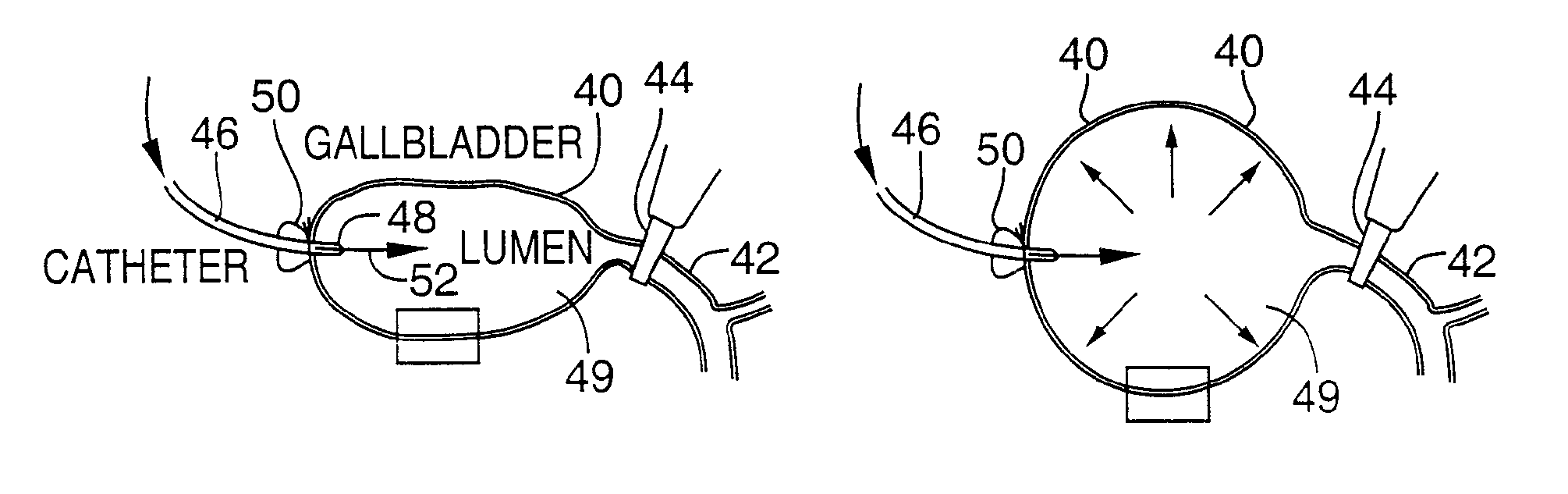
[0082]FIG. 4 illustrates an apparatus used for demonstrating the effect of retrograde biliary infusion on intrabiliary pressure, and the site specific delivery of agents to the cells. Twenty to forty gram anesthesized CD-1 male mice (Charles River) were used as experimental subjects. Following a midline laparotomy, the gallbladder 40 was manually drained through the cystic duct 42. A cholecystotomy catheter 60 (silastic tubing, 0.012″ ID / 0.025 OD) was introduced through the wall of the gallbladder 40, and secured within the gallbladder lumen with the catheter tip 48 advanced so that it was immediately proximal to the junction with the cystic duct 42. An absorbent cellulose (X0-Med, Jacksonville, μl) packing 62 was packed around the entrance site of the catheter into the gallbladder to prevent bile from leaking into the peritoneum.

[0083]A twenty-three gauge needle 63 was used to make an opening in the duodenum 64 and to perform a sphincterotomy on the...

example 2

Radiopaque Tracer Studies

[0092]A silastic catheter was placed in the gallbladder as described above. Straight (1 mm×3 mm) or curved (1 mm×5 mm) Kleinert-Kutz microvascular clips (MVC; Pilling-Weck, Research Triangle, North Carolina) were then placed rostral to the junction of the superior pancreatic duct with the common bile duct to turn the hepatobiliary system into a closed pressure system. Infusions were administered as in Example 1, and the microvascular clip occlusion caused the infusion to move retrograde into the hepatic duct and then into smaller hepatic ducts and ductules. At the end of the administration period (infusion plus dwell time) the clip was removed and the cholecystotomy catheter was withdrawn.

[0093]To evaluate the impact of hepatic venous drainage on the distribution of radioopaque dye and adenovirus following retrograde biliary infusion, the suprahepatic inferior vena cava was temporarily occluded for 5 to 10 minutes with a curved microvascular clip at a level ...

example 3

Latex Microsphere as Model for Vector Delivery

[0098]In order to both corroborate the digital fluoroscopic studies and histologically evaluate the distribution of infusate, 100 nm and 200 nm diameter fluorescent latex microspheres were administered by retrograde biliary infusion. Spheres of this diameter were selected since they are close in diameter to adenoviral (80 nm) and liposomal (200-500 nm) vectors. Yellow green (490 nm peak excitation wavelength) carboxylate-modified fluorescent latex mircrospheres (Molecular Probes, Eugene, Oreg.) were diluted in 1× PBS and extensively sonicated prior to use. Sphere concentration was maintained constant at 1×10¹¹ spheres per animal, while the volume and rate of infusion were varied between animals. Following the completion of infusion, fresh frozen sections were prepared from the liver and lung and evaluated under fluorescent microscopy. To visualize histologic detail more completely, some slides were stained with Evans Blue (0.05% for 20 s...


PUM


Abstract

Methods and devices are disclosed for selective delivery of therapeutic substances to specific histologic or microanatomic areas of organs. Introduction of the therapeutic substance into a hollow organ space (such as an hepatobiliary duct or the gallbladder lumen) at a controlled pressure, volume or rate allows the substance to reach a predetermined cellular layer (such as the ephithelium or sub-epithelial space). The volume or flow rate of the substance can be controlled so that the intralumenal pressure reaches a predetermined threshold level beyond which subsequent subepithelial delivery of the substance occurs. Alternatively, a lower pressure is selected that does not exceed the threshold level, so that delivery occurs substantially only to the epithelial layer. Such site specific delivery of therapeutic agents permits localized delivery of substances (for example to the interstitial tissue of an organ) in concentrations that may otherwise produce systemic toxicity. Occlusion of venous or lymphatic drainage from the organ can also help prevent systemic administration of therapeutic substances, and increase selective delivery to superficial epithelial cellular layers. Delivery of genetic vectors can also be better targeted to cells where gene expression is desired. The access device comprises a cannula with a wall piercing tracar within the lumen. Two axially spaced inflatable balloons engage the wall securing the cannula and sealing the puncture site. A catheter equipped with an occlusion balloon is guided through the cannula to the location where the therapeutic substance is to be delivered.

Description

[0001]This is a continuation of U.S. patent application Ser. No. 12 / 009,646, filed Jan. 17, 2008, now abandoned, which is a divisional of U.S. patent application Ser. No. 09 / 700,999, filed Dec. 4, 2000, (now abandoned) which is a §371 U.S. national stage of PCT / US99 / 11277, filed May 21, 1999, which was published in English under PCT Article 21(2), which in turn claims the benefit of U.S. Provisional Application 60 / 086,565, filed May 21, 1998 and U.S. Provisional Application No. 60 / 087,099, filed May 28, 1998, all of which are incorporated herein in their entirety.FIELD OF THE INVENTION[0002]This invention concerns selective delivery of therapeutic agents, such as drugs or genetic vectors, to specific organs, tissue compartments or cell types.BACKGROUND OF THE INVENTION[0003]Local delivery of therapeutic agents to target organs or tissues is a very desirable technique for delivering drugs with minimal side effects. U.S. Pat. No. 5,087,244 is an example of such targeted drug delivery,...

Claims


Application Information

Patent Timeline
Patent Type & Authority Patents(United States)
IPC IPC(8): A61M31/00A61B17/34A61F2/958A61M1/00A61M25/00A61M25/02A61M25/04
CPCA61B17/3417A61M25/02A61M25/1011A61B2017/3425A61M25/04A61M2025/0233
Inventor WIENER, STEPHEN M.HOYT, JR., ROBERT F.DELEONARDIS, JOHN R.CLEVENGER, RANDALL R.LUTZ, ROBERT J.CHRISTINI, DOUGLAS V.SAFER, BRIAN
Owner UNITED STATES OF AMERICA
Why Eureka
Try Eureka