Method for quantifying organ motion, apparatus therefor, method for estimating organ position, apparatus therefor, method for irradiating radiation, apparatus therefor, and apparatus for detecting abnormal organ

a technology for organs and motions, applied in the field of methods for quantifying organ motion, can solve the problems of insufficient clinical practice, tumors may be irradiated to an excessively large extent, and the analysis of two-dimensional images is difficult, and achieve the effect of accurately quantifying the motion of an organ

Inactive Publication Date: 2013-06-04
NAT INST FOR QUANTUM & RADIOLOGICAL SCI & TECH +1
View PDF9 Cites 8 Cited by
  • Summary
  • Abstract
  • Description
  • Claims
  • Application Information

AI Technical Summary

Benefits of technology

[0014]The present invention has been made for solving the above problems, an object of which is to accurately quantify the motion of an organ.

Problems solved by technology

Therefore, a more accurate irradiation is required to ascertain the motion of the organ at individual positions (the speed and direction of movement), the analysis of which is, however, difficult by referring to two-dimensional images.
In particular, where a lung at which a tumor and normal tissues are greatly different in density is subjected to particle beam therapy, the variation of the organ not only in a direction perpendicular to the irradiation beam (herein after simply referred to as beam) but also in an axial direction of the beam greatly influences a range of the beam, thus, resulting in a fear that the tumor may be irradiated to an excessively small extent or normal tissues may be irradiated to an excessively great extent.
However, such a technology has not been established with current clinical practices.
The metal marker is, however, implanted into the body of a patient, thus giving a greater burden to the patient.
The marker is also restricted in the position and number, thereby making it difficult to ascertain the motion of an organ in detail.
However, the technology described in Non-patent Document 1 has a problem that the outer configuration of an organ does not necessarily coincide with the deformation inside the organ.
Furthermore, the technology described in Non-patent Document 2 does not track an anatomical feature inside an organ, thereby posing a problem regarding the accuracy of volume-of-interest deformation.

Method used


Image

  • Method for quantifying organ motion, apparatus therefor, method for estimating organ position, apparatus therefor, method for irradiating radiation, apparatus therefor, and apparatus for detecting abnormal organ
  • Method for quantifying organ motion, apparatus therefor, method for estimating organ position, apparatus therefor, method for irradiating radiation, apparatus therefor, and apparatus for detecting abnormal organ
  • Method for quantifying organ motion, apparatus therefor, method for estimating organ position, apparatus therefor, method for irradiating radiation, apparatus therefor, and apparatus for detecting abnormal organ

Examples


first embodiment

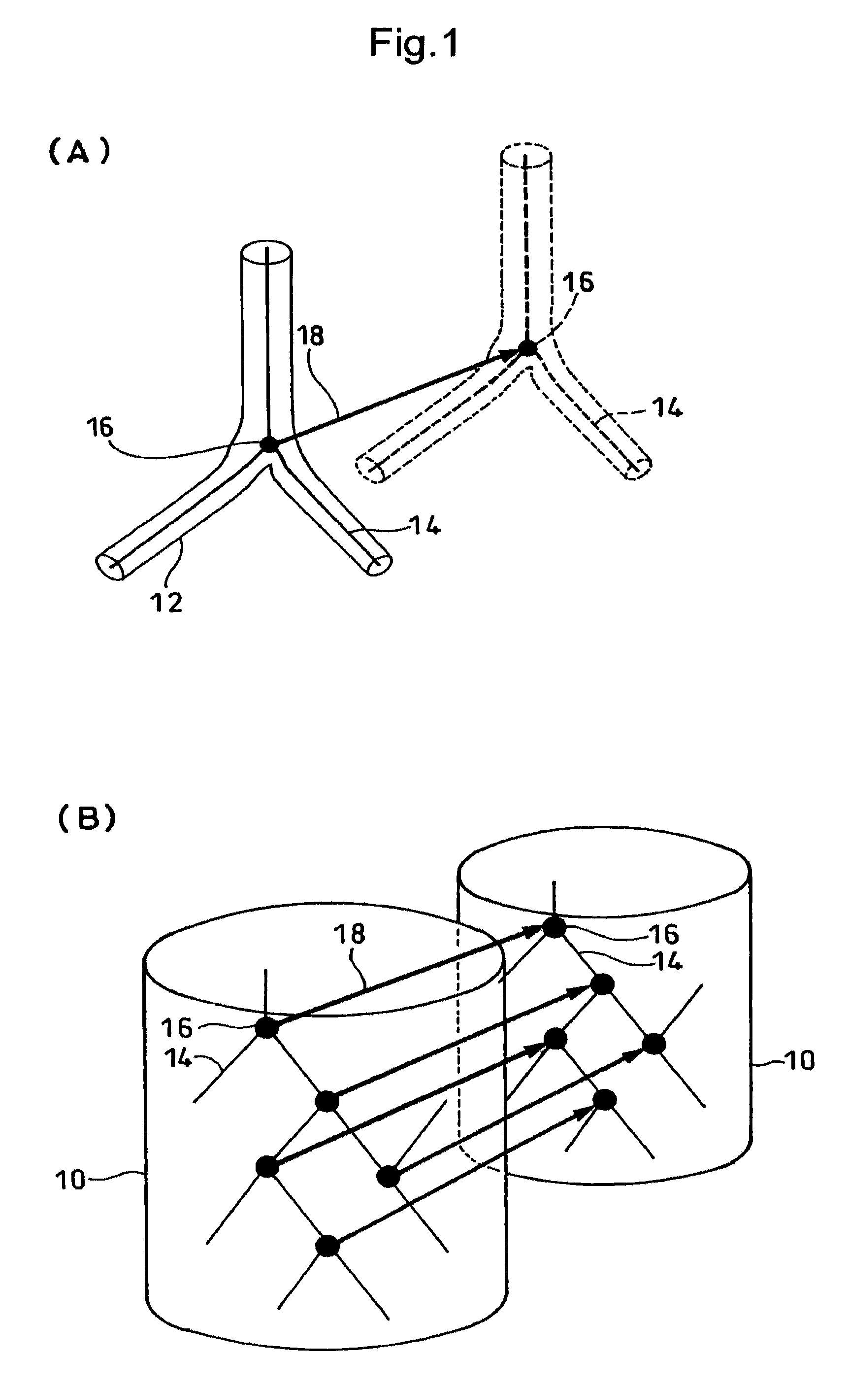
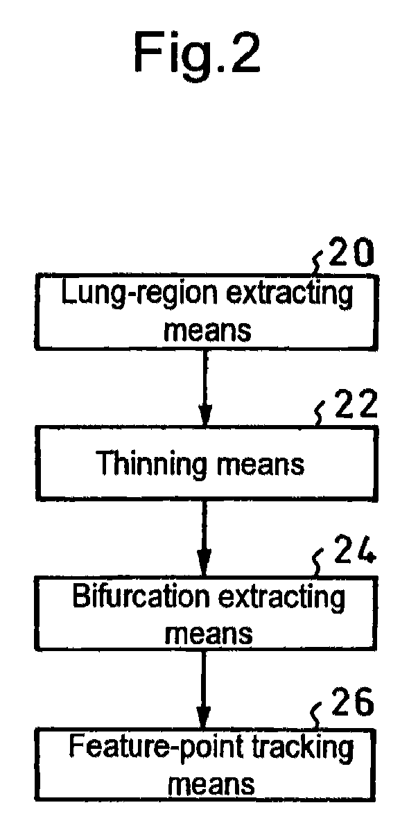
[0055]An apparatus for executing the present invention is that which is constituted as illustrated in FIG. 2 by using, for example, a computer, including lung-region extracting means 20, thinning means 22, bifurcation extracting means 24 and feature-point tracking means 26.

[0056]In the thinning means 22, thresholding is conducted by referring to a certain CT value as a threshold value, and the thus obtained figure having one voxel value is cut. However, if parts such as flesh and bones around a lung are left as a result of the thresholding, these parts are also subjected to the thinning process, and they are substantially greater in volume (voxel number) than the bronchial tubes and blood vessels in the lung, thus resulting in greatly extending the calculation time.

[0057]Therefore, in order to shorten the calculation time and calculate only a necessary lung region, the lung region is first extracted by using the lung-region extracting means 20.

[0058]Specifically, the lung region can...

second embodiment

[0103]Next, an explanation will be made by referring to FIG. 10 for the present invention in which the present invention is used to estimate an organ position so as to irradiate radiation thereto.

[0104]In the present embodiment as well, as illustrated on the left side in FIG. 10, X-ray 37 is irradiated from an X-ray tube 36 to a patient 30 on a bed 34 inside a CT gantry 32. Then, as with the first embodiment, a four-dimensional CT (4D-CT) 40 is used to process an image obtained by an X-ray detector 38, thereby obtaining an image of organ motion (four-dimensional CT image). At this time, for example, movement-information obtaining means 52 is used to simultaneously calculate a respiratory waveform by referring to a respiration sensor 50 fixed on the body surface of the patient 30, thereby obtaining a temporal correlation between them.

[0105]Then, the technology of the first embodiment is used to quantify motions from the four-dimensional CT image at a processing section 60, thereby ob...

third embodiment

[0109]Next, with reference to FIG. 11 (flowchart showing the procedures) and FIG. 12 (schematic diagram), an explanation will be made for the present invention in which the present invention is utilized to detect an abnormal organ.

[0110]In the present embodiment, as the procedures are shown in FIG. 11, displacement vectors for each voxel obtained by the technique of the first embodiment are classified into regions, depending on the vector component concerned, as illustrated in FIG. 12. In other words, as with contour lines obtained depending on the height on each point on a map, regions are classified, depending on the size of components on a vector map, thereby making it possible to extract a region different in displacement. Thereby an abnormal region can be detected.


PUM


Abstract

A plurality of CT images of an organ (10) which has undergone a subtle variation or deformation are used as input data, blood vessels and trachea 12 distributed inside the organ are extracted and subjected to thinning, the thus thinned images 14 are used to extract the coordinates of bifurcations 16 and connections, the thus extracted coordinates are used as feature points to track the motion of individual points between a plurality of CT images in a three dimensional space, thereby measuring the movement of the organ (10). Thus, it is possible to realize a local motion tracking at an arbitrary point over an entire region inside an organ, which would be impossible by using a metal marker.

Description

TECHNICAL FIELD[0001]The present invention relates to a method for quantifying organ motion for quantifying the motion at each voxel position with reference to a three-dimensional digital image at each time point obtained at certain intervals by using high-speed three-dimensional computed tomography (CT) (so-called four-dimensional CT) for the purpose of ascertaining the matching (registration) of each portion inside an organ with respect to an organ such as a lung which undergoes variation or deformation in association with respiration or the like, the deformation and position of volume of interest of tumors or the like in correlation with a phase of respiratory waveform in radiological diagnosis, treatment planning and medical treatment, an apparatus therefor, a method for estimating an organ position by using the above-described method, an apparatus therefor, a method for irradiating radiation, an apparatus therefor and an apparatus for detecting an abnormal organ.BACKGROUND ART[...

Claims


Application Information

Patent Timeline
Patent Type & Authority Patents(United States)
IPC IPC(8): G06K9/62
CPCA61B6/466A61B6/541A61N5/1049G06T7/0028G06T7/0034A61N5/1037A61N5/1064G06T2207/10076G06T2207/10081G06T2207/20164G06T2207/30004G06T2207/30061G06T7/33G06T7/35
Inventor TASHIRO, MUTSUMIMINOHARA, SHINICHI
Owner NAT INST FOR QUANTUM & RADIOLOGICAL SCI & TECH
Why Eureka
Try Eureka