
Diagnosis and treatment of preeclampsia

a preeclampsia and biomarker technology, applied in the direction of instruments, extracellular fluid disorder, peptide/protein ingredients, etc., can solve the problems of general endothelial cell damage in the placenta, the most common cause of death for both children and mothers, and the increase of maternal mortality and morbidity

Inactive Publication Date: 2013-10-29
GUARD THERAPEUTICS INT AB
View PDF4 Cites 5 Cited by
  • Summary
  • Abstract
  • Description
  • Claims
  • Application Information

AI Technical Summary

Benefits of technology

The method enables early detection of preeclampsia, reducing unnecessary hospitalizations and the risk of premature birth, and provides a candidate target for treatment by reducing free fetal haemoglobin levels, potentially alleviating disease progression.

Problems solved by technology

It is today the most common cause of death for both children and mothers during pregnancy.
Decreased placental perfusion, in combination with oxidative stress, causes general endothelial cell damage within the placenta.
Preeclampsia is a leading cause of maternal mortality and morbidity.
It is also associated with a high perinatal mortality and morbidity, due primarily to iatrogenic prematurity.
As mentioned above, it is believed that inadequate placentation, resulting in reduced placental perfusion, is an early step in the development of PE.
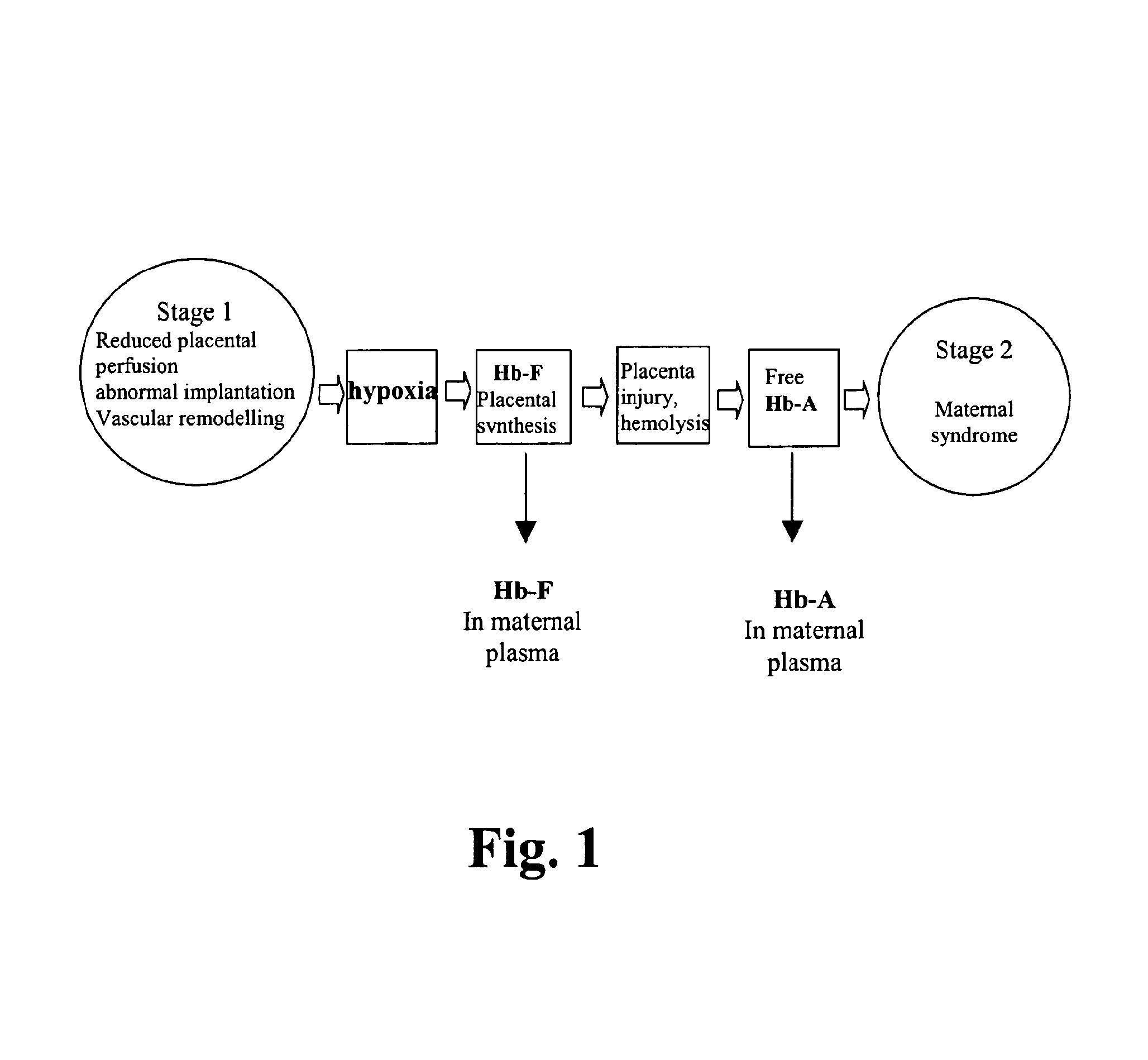
Poor placental perfusion appears to result in a cascade of pathological changes: decreased oxygen delivery, oxidative stress, formation of reactive oxygen species, endothelial damage, increased vascular permeability, and inflammation (see FIG. 1).
However, these monitoring methods are ineffective for diagnosis of the syndrome at an early stage.
Some methods of diagnosis have been described in the prior art but none of these have yet been successfully used in the clinic on a large scale.
Currently, there are no known cures for preeclampsia.
Clearly, the lack of therapy for preeclampsia coupled with the age-old lack of ability to diagnose it until the the disease has progressed to a more severe form, prompts the need for novel approaches for diagnosing and treating preeclampsia, which is a significant public health problem.

Method used


Image

  • Diagnosis and treatment of preeclampsia
  • Diagnosis and treatment of preeclampsia
  • Diagnosis and treatment of preeclampsia

Examples


example 1

Detection of Hb RNA and Protein in Placenta

[0158]Quantitative RT-PCR, In situ hybridization and immunohistochemistry was performed to analyze Hbα, Hbβ and Hbγ mRNA and protein expression in placental samples in PE vs. control subjects.

Sample Collection

[0159]Placental tissue was collected at the Department of Obstetrics and Gynaecology, Lund University Hospital. The sampling, performed with written consent, was approved by the Ethical Committee Review Board for studies in human subjects. Placental tissue from 10 preeclamptic, 15 normal pregnancies, 5 patients with bilateral notch and 5 patients with bilateral notch as well as preeclampsia were included in the study. Placental bed samples (see below) from 5 of the patients with PE and 5 of the controls were also collected. Preeclampsia was defined as blood pressure >140 / 90 mm Hg and proteinuria >0.3 g / L. Patients with essential hypertension or other systemic diseases were excluded. Placenta samples were collected at birth, immediately...

example 2

Detection of Fetal Hb in Maternal Blood

[0178]Quantitative RT-PCR was performed to analyze Hbγ mRNA in blood samples in PE subjects.

Real-Time PCR

[0179]RNA was extracted using QIAamp Viral RNA mini kit (Qiagen) according to manufacturer's instructions. Briefly, 3.6 ml of AVL Buffer was mixed with 36 μl of carrier-RNA (Qiagen) by inverting the tube 10 times. 1 ml of the plasma sample was spun at 1150 g for 10 minutes. 900 μl of the plasma and 3.6 μl of 99% ethanol was added to the AVL buffer solution. Approximately 650 μl of the solution was added to a QlAamp column and spun at 6000 g for 1 minute. This was repeated until the total plasma volume had been added to the column. Column was washed once with AW1 buffer, then spun at 6000 g for 1 minute, followed by a wash with AW2 buffer, then spun at 20,000 g for 3 minutes. RNA was eluated with 50 μl RNAse free water.

[0180]Fetal Hb RNA was quantified with real-time PCR.

Results

[0181]FIG. 7 shows a scatter plot of fetal Hbγ mRNA levels in mat...

example 3

Protein Expression Profiling of the Preeclamptic Placenta Using 2D-Gel Electrophoresis

[0182]In order to screen for differentially expressed proteins in the PE placenta compared to control placentas, we collected placenta samples at delivery from women with PE (n=30) and healthy controls (n=30). Using proteomics technology (2-dimensional gel electrophoresis) we compared haemoglobin delta (Hbδ) expression levels in the different placenta samples.

Patients and Sample Collection

[0183]60 women admitted at the Department of Obstetrics and Gynaecology, Lund University Hospital were included, and assigned to two groups; PE (n=30) and control (n=30) (Table 2). PE was defined as blood pressure of >140 / 90 mmHg and proteinuria of >0.3 g / l or rise in blood pressure above 20 mmHg from the first trimester of pregnancy. A 10×10×10 mm cube of placenta tissue was collected immediately after removal of the placenta. Samples were immediately frozen on dry ice and stored at −80° C. Patients with other sy...


PUM

PropertyMeasurementUnit
diastolic blood pressureaaaaaaaaaa
concentrationsaaaaaaaaaa
pHaaaaaaaaaa

Abstract

The present invention relates to biomarkers for preeclampsia as well as treatment of this disease. In particular, the invention relates to methods for diagnosis or aiding in the diagnosis of preeclampsia of a pregnant female mammal to detect elevated levels of free haemoglobin, particularly free fetal haemoglobin. This facilitates and makes possible early diagnosis and clinical intervention when a preeclamptic condition is found. In addition, the invention relates to a method to treat female mammals with preeclampsia with the purpose to reverse the pathological conditions associated with this disease.

Description

FIELD OF THE INVENTION[0001]The present invention relates to biomarkers for preeclampsia as well as treatment of this disease. In particular, the invention relates to methods for diagnosis or aiding in the diagnosis of preeclampsia of a pregnant female mammal to detect elevated levels of particular substances. This facilitates and makes possible early diagnosis and clinical intervention when a preeclamptic condition is found. In addition, the invention relates to a method to treat female mammals with preeclampsia with the purpose to reverse the pathological conditions associated with this disease.BACKGROUND OF THE INVENTION[0002]Preeclampsia (PE), gestational proteinuric hypertension, complicates 3-7% of all pregnancies, and is a multisystem maternal disorder. Yearly 8,500,000 cases are reported worldwide, of which 5,000 are in Sweden. It is today the most common cause of death for both children and mothers during pregnancy.[0003]PE has been named the disease of theories [Roberts, 2...

Claims


Application Information

Patent Timeline
Patent Type & Authority Patents(United States)
IPC IPC(8): G01N33/00A61K38/42
CPCG01N33/689G01N33/721A61K38/1709A61K38/1774G01N33/00A61K38/40A61K38/42A61K38/44C12Y114/99003A61K38/38G01N2800/368A61P43/00G01N33/68G01N33/72A61P7/00A61P9/12A61P13/02A61P15/00G01N33/569
Inventor HANSSON, STEFANAKERSTROM, BO
Owner GUARD THERAPEUTICS INT AB
Learn More