Device for high-throughput stimulation, immunostaining, and visualization of single cells

a single cell, high-throughput technology, applied in the field of microfluidic devices, can solve the problems of low throughput, laborious cell assays, low throughput, and need for a large volume of reagents, and achieve the effect of improving the sealing of the fluid channel

Active Publication Date: 2014-09-02
THE JOHN HOPKINS UNIV SCHOOL OF MEDICINE
View PDF58 Cites 2 Cited by
  • Summary
  • Abstract
  • Description
  • Claims
  • Application Information

AI Technical Summary

Problems solved by technology

Cell stimulation and staining methods frequently involve the use of antibodies or other reagents that are expensive or available in limited quantities.
Immunofluorescence staining is typically performed on cells adhered to glass slides, resulting in the need for a large volume of reagents.
Such complex methods also make such cell assays labor intensive and low throughput.
However, cells can only be sorted and observed as a group using both FACS and ELISA.
Although fluids can be loaded and mixed using the device, it is not large enough to accommodate eukaryotic cells and seeding of cells with an even distribution would not be possible using the device.
Moreover, precise control of fluid flow through channels is somewhat limited.
This simplifies the fabrication of the device, but substantially limits the number of channels that can be accessed through single ports, and limits the density of the valves in the device, both of which decrease the throughput of the device.

Method used


Image

  • Device for high-throughput stimulation, immunostaining, and visualization of single cells
  • Device for high-throughput stimulation, immunostaining, and visualization of single cells

Examples


example 1

Fabrication of a Device for High-Throughput Stimulation, Immunostaining and Visualization of a Single Cell

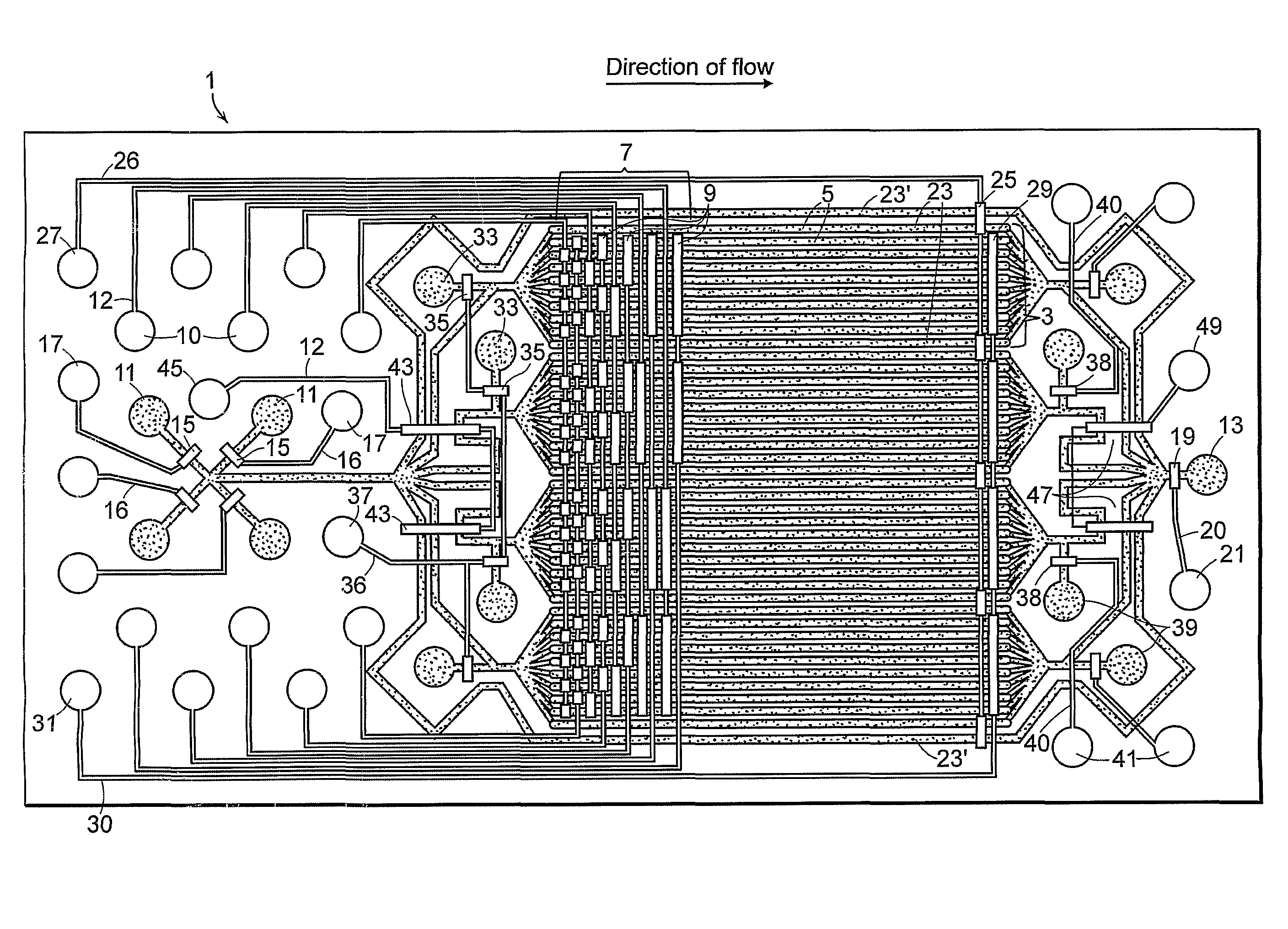
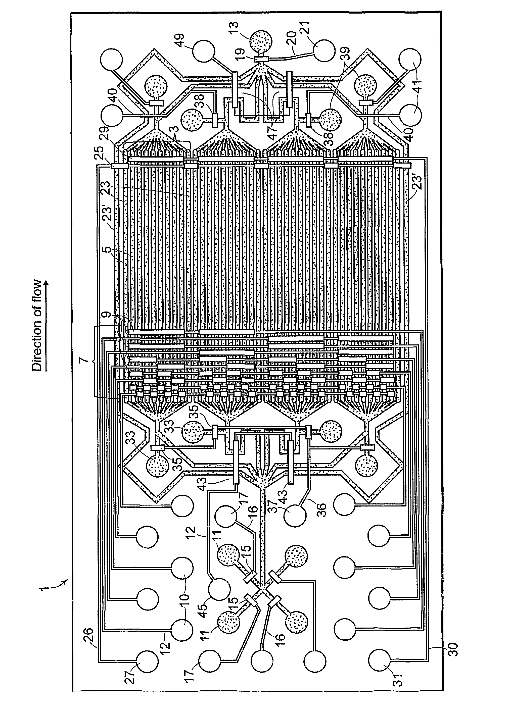
[0043]The device was made using standard soft lithography techniques. The method of fabrication of the device is not a limitation of the invention.

[0044]Elastomeric devices have been developed for use in biological studies (for review see Sia and Whitesides 2003. Electrophoresis 24:3563-3576). Such devices are can be biocompatible and can be prepared relatively easily and inexpensively by methods well known to those skilled in the art (See e.g., reviews Whitesides et al., 2001. Ann. Rev. Biomed. Eng. 3:335-373, incorporated herein by reference). Elastomeric devices made by the process of soft lithography are produced by casting of polydimethylsulfoxide (PDMS) or other silicone rubber onto micromachined molds or coated silicone wafers patterned using contact lithography; however, other materials and methods can be used.

[0045]Briefly, for fabrication of the device of FIG. 1, the f...

example 2

Cell Culture, Stimulation, and Immunofluorescence Staining

[0049]The device was fabricated as described in Example 1. The device was filled with sterile 0.1% gelatin with all of the valves open through an inlet port 11 to coat the parallel fluid channels to promote cell adhesion. Valves were primed with filtered, distilled water. Alternatively, filling may be done with the ports closed as trapped air can be pushed out of the gas-permeable PDMS with hydrostatic pressure. The device was flushed with to remove the coating agent. Well-separated NIH3T3 mouse fibroblasts at a concentration of about 9 million cells per ml were introduced into the parallel fluid channels through an inlet port and the device was transferred to a standard 5% CO2, 37° C. cell culture incubator for 3-4 hours to allow the cells to adhere to the glass bottom of the parallel fluid channels. Cells were observed at various time points after seeding and were found to be viable and have normal morphology for at least 1...

example 3

Detailed 32-Point Timecourse of the Response of One or More Proteins to Persistently Applied Stimulus or Drug

[0052]The method is most readily accomplished using a device having four reaction units with eight parallel fluid channels per reaction unit such as the device shown in FIG. 1. A device can be readily fabricated to allow for variation in the number of reaction units and parallel fluid channels. The device was flushed with DPBS before each step to remove the chemical agent form the preceding step.

[0053]The device was coated the device with an agent that promotes cell adherence (e.g. gelatin, poly L-lysine, fibronectin, collagen, etc.). Cells were introduced into the device and allowed to adhere. Unattached cells were removed by flushing the parallel fluid channels with media. Valves were primed with filtered distilled water and flow is shut off all channels. Media containing an active agent was introduced into the device via any of the main inlets. The multiplexer was actuated...


PUM

PropertyMeasurementUnit
lengthaaaaaaaaaa
lengthaaaaaaaaaa
widthaaaaaaaaaa

Abstract

Cell stimulation, staining, and visualization are common techniques in both clinical and research settings. The invention is directed to microfluidic devices for in situ cell stimulation, staining, and / or visualization, and related methods for applying one or more stimuli to the cells, and fixing and staining of cells in situ. The device allows for high-throughput screening of living cells using a minimal quantity of reagents where the fate of individual cells can be followed over time.

Description

RELATED APPLICATIONS[0001]This application claims the benefit of U.S. Provisional Application No. 60 / 725,415 filed Oct. 11, 2005, having the same title as the instant application. The entire contents of the aforementioned application are hereby incorporated herein by reference.FIELD OF THE INVENTION[0002]The present invention relates to microfluidics devices and methods for their use, including cell stimulation, staining, and / or visualization. More particularly, the invention relates to devices and methods for applying one or more stimuli to living cells, in parallel, and fixing, staining and / or visualizing cells in situ.BACKGROUND OF THE INVENTION[0003]Cell stimulation, staining, and visualization are common techniques in both clinical and research settings. Cell stimulation and staining methods frequently involve the use of antibodies or other reagents that are expensive or available in limited quantities. Immunofluorescence staining is typically performed on cells adhered to glas...

Claims


Application Information

Patent Timeline
Patent Type & Authority Patents(United States)
IPC IPC(8): B01L1/00B01L3/00B01L7/00G01N15/14
CPCB01L3/5025B01L7/00G01N15/14B01L2300/0816B01L2200/06B01L2300/0864B01L2400/0487B01L3/502738B01L2300/123B01L2400/0655G01N15/1463G01N15/1484G01N2015/1006G01N2035/00158G05D7/0694
Inventor LEVCHENKO, ANDRECHEONG, RAYMONDWANG, CHIAOCHUN J.
Owner THE JOHN HOPKINS UNIV SCHOOL OF MEDICINE
Why Eureka
Try Eureka