
Look-locker IR-SSFP for cardiac MR imaging with simultaneous generation of cardiac T1 maps, cine images and IR-prepared images

Inactive Publication Date: 2015-04-14
DEUTES HERZZENT BERLIN
View PDF13 Cites 9 Cited by
  • Summary
  • Abstract
  • Description
  • Claims
  • Application Information

AI Technical Summary

Benefits of technology

The present invention provides a method that allows for the simultaneous generation of T1 maps, cine, and IR-prepared images from one single data set. This approach saves acquisition time and facilitates registration of different modalities. The method also includes increasing the NSA to improve signal-to-noise ratio.

Problems solved by technology

However, such techniques use non-segmented acquisitions with durations of approximately 150 to 200 ms for each raw image.
While it could be shown that this acquisition duration is adequate to acquire images at end-diastole for heart rates up to 100 beats per minute (bpm) with high reproducibility, this approach is not suitable for use in small animals where heart rates in the range of 200 to 600 bpm are to be expected.
Consequently, alternative approaches are warranted for these situations, which usually lead to a significant rise in over-all acquisition time.
This might not be desirable for imaging protocols where other types of MR images are to be collected within the same session, causing long scan times that might potentially exceed the limits where the animals can be studied safely under stable conditions.

Method used


Image

  • Look-locker IR-SSFP for cardiac MR imaging with simultaneous generation of cardiac T1 maps, cine images and IR-prepared images
  • Look-locker IR-SSFP for cardiac MR imaging with simultaneous generation of cardiac T1 maps, cine images and IR-prepared images
  • Look-locker IR-SSFP for cardiac MR imaging with simultaneous generation of cardiac T1 maps, cine images and IR-prepared images

Examples


example 1

Methods:

[0055]The method of the invention combines an adapted segmented, retrospectively gated, IR-prepared Look-Locker type pulse sequence with a multimodal reconstruction framework.

Pulse Sequence Details:

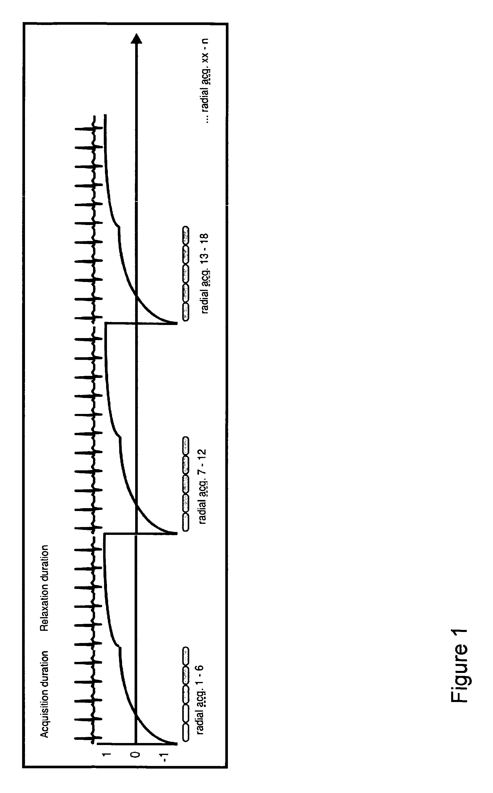
[0056]FIG. 1 illustrates the pulse sequence scheme. After an ECG-triggered non-selective (adiabatic) inversion pulse, n radial profiles of the first phase of the cardiac cycle are read out by a non-balanced steady-state free precession (SSFP) sequence, immediately followed by the read-out of the same n radial profiles of the second phase of the cardiac cycle and so on for a predefined period of time (“acquisition duration”). Upon completion of a predefined waiting period (“relaxation duration”), the next set of n radial profiles of the first phase of the cardiac cycle is read out, immediately followed by the same next set of n profiles of the second phase of the cardiac cycle and so on. The number of cardiac cycles (cc) covered by each acquisition step is given by cc=acquisition d...

example 2

Materials and Methods

[0067]Animal studies were approved by the local animal care committee and performed according to Good Laboratory Practice guidelines.

[0068]All experiments were carried out on a whole-body 3 Tesla MR system (Achieva, Philips Medical Systems, Best, The Netherlands) equipped with a QuasarDual gradient system (80 mT / m, 200 mT / m / ms slew rate) and a 70 mm solenoid coil for rat hearts.

Pulse Sequence Scheme

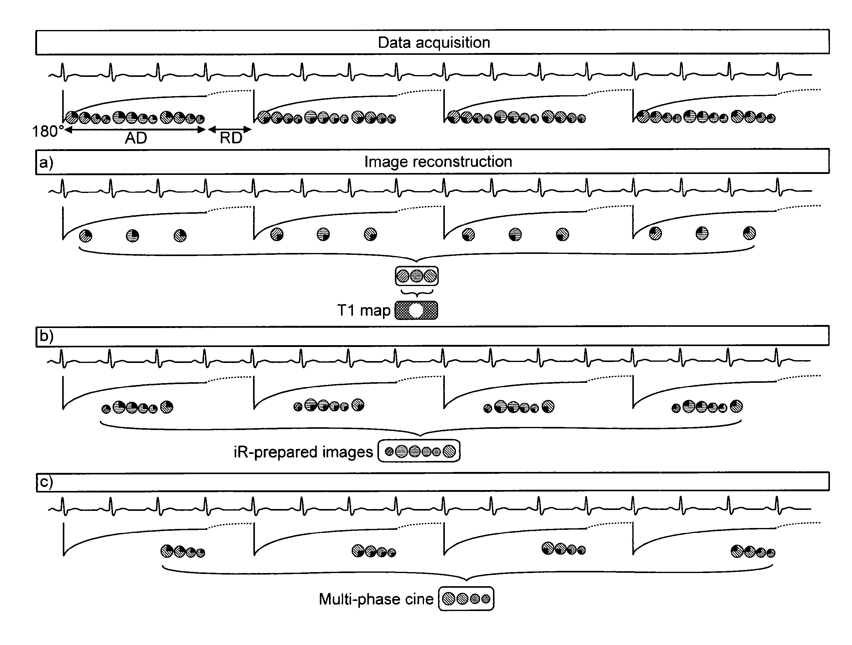
[0069]FIG. 4 shows the pulse sequence scheme for fully sampled data sets (top) and reconstruction of multi-modal images (a-c). FIG. 5 illustrates the principles of “temporal undersampling”, which was implemented to accelerate the acquisition.

[0070]Reconstruction of the acquired image data was performed using a customized reconstruction framework (ReconFrame, Gyrotools, Zurich, Switzerland) implemented with Matlab (The MathWorks, Natick Mass., USA). For T1 maps, 3-parameter curve fitting was performed for each pixel as used for standard Look-Locker pulse sequences usin...


PUM


Abstract

The present invention is directed to a method for use in conducting cardiac MR imaging which allows for reconstruction of T1 maps, cine images and IR-prepared images from one raw data set, wherein the method comprises the following steps: a) acquisition of raw data by use of an ECG-triggered, segmented, inversion recovery (IR) -prepared Look-Locker type pulse sequence for data acquisition, wherein the pulse sequence encompasses more than one shot, wherein each shot comprises: i) an ECG-triggered inversion pulse; ii) SSFP cine data acquisition of radial segmented profiles over more than one RR-interval for a predefined acquisition duration AD; and iii) a relaxation duration RD, during which no data is acquired; b) retrospective gating of raw data by sorting acquired raw data for each RR-interval into a pre-determined number of heart phases by definition of specific time windows within the RR-intervals and sampling of raw data acquired during the time windows respectively; c) image reconstruction, wherein the retrospectively gated raw data is used for reconstruction of T1 maps, cine images and / or IR-prepared images.

Description

RELATED APPLICATIONS[0001]This is a U.S. national stage of application No. PCT / EP10 / 067191, filed on Nov. 10, 2010. Priority is claimed on the following application: U.S. Application Ser. No. 61 / 259,680 filed on Nov. 10, 2009, the contents of which is herein incorporated here by reference.BACKGROUND OF THE INVENTION[0002]T1 mapping provides quantitative information on tissue alterations and gadolinium kinetics. In the heart, the ability to identify myocardial edema and the potential to identify diffuse interstitial fibrosis are of particular interest.[0003]Mapping of T1 relaxation times typically requires the acquisition of multiple images with different inversion times in order to allow for an accurate fitting of the underlying T1 curve. Dedicated cardiac T1 mapping schemes typically acquire a sufficient number of images within one breath-hold. Dedicated ECG (electrocardiogram)-triggered Look-Locker Inversion Recovery (IR) techniques have already been described for cardiac magnetic...

Claims


Application Information

Patent Timeline
IPC IPC(8): A61B5/055G01R33/48G01R33/50G01R33/56G01R33/561G01R33/563G01R33/567
CPCG01R33/50A61B5/055G01R33/4818G01R33/4824G01R33/5602G01R33/5614G01R33/56308G01R33/56325G01R33/5619G01R33/5673
Inventor MESSROGHLI, DANIEL
Owner DEUTES HERZZENT BERLIN
Learn More