Acquisition of high-temporal free-breathing MR images

a free-breathing, high-temporal technology, applied in the field of methods, can solve the problems of limited clinical value of cardiac mr imaging methods, particularly difficult imaging, moving objects, etc., and achieve the effects of short total acquisition time, increased cardiac stress, and increased heart ra

Inactive Publication Date: 2005-05-03
GENERAL ELECTRIC CO
View PDF8 Cites 17 Cited by
  • Summary
  • Abstract
  • Description
  • Claims
  • Application Information

AI Technical Summary

Benefits of technology

[0013]The invention includes segmenting k-space acquisition to permit cardiovascular examinations which were previously not feasible due to the requirements of high temporal resolution and a short total acquisition time. The invention provides an examination using MR-based stress functions, which is an MR version of the widely-used ECG-based stress test. The patient is first subjected to successively increasing levels of cardiac stress leading to increased heart rates. Stress can be induced either by physical exercise or by the administration of a pharmaceutical, such as dobutamine. Once the heart rate has stabilized for a given stress level, the R—R interval is measured and recorded using standard ECG equipment or peripheral pulse gating equipment. Once recorded, the ECG reading is disregarded by the MR scanner, and an MR scan is commenced in which MR data is acquired using the recorded R—R interval as a time period for acquisition irrespective of the start or end of the actual R—R interval. Data acquisition for the current segment continues as in a segmented CINE acquisition until the end of the expected R—R interval time is reached. The k-space views are updated for the next segment and the process continues until all data have been acquired. Image reconstruction and display commences when all data necessary to reconstruct the CINE data set have been acquired. By segmenting data and acquiring different segments and successive acquisitions, higher resolution images can be achieved.
[0014]This method uses a virtual cardiac trigger by estimating an occurrence of the end of a cardiac cycle by the measurement of the cardiac cycle time immediately prior to the start of scan. In this manner, it is not necessary to actually wait for an ECG or peripheral gate trigger to complete a fast segmented k-space CINE scan with high temporal resolution. If an adequate ECG-trigger exists during scanning or if peripheral pulse gating is used, the MR scanner may update the cardiac interval time periodically during the continuous acquisition. This updating of the cardiac cycle or interval time can be automated or be placed under manual, operator control.

Problems solved by technology

Moving objects are particularly difficult to image, especially if an imaging plane is set in space with the object moving in and out of the imaging plane.
Such imaging is especially difficult when a second periodic motion is added thereto.
For example, imaging of objects in a subject which is breathing causes a periodic motion of internal structures, which is also further complicated by the beating motion of the heart if the structure is near the heart.
Until recently, cardiac MR imaging methods have been of limited clinical value for several reasons.
First, such methods had a relatively long acquisition time relative to the cardiac cycle which resulted in cardiac motion blurring.
Second, the long duration of the imaging scan requires patients to hold their breath for unreasonably long periods to avoid motion artifacts due to respiratory motion.
While monitoring can be performed using the aforementioned breath-held technique, repeated breath-holding can be very exhausting for this class of patients.
ECG-gating poses yet another obstacle.
Such gating suffers from a number of problems such as operator dependence, inter-patient variability, detached ECG leads, and corruption of ECG signals due to noise from the imaging gradients.
However, peripheral gating suffers from the limitation of introducing a delay relative to the ECG-gating, so it is not possible to identify the exact moment of the cardiac R-wave.
Such resolutions are unacceptable for heart rates of the order of 150-180 beats per minute (bpm) to effectively visualize the systolic phase.
However, this still poses a problem in that it requires a large number of heartbeats to complete an acquisition, during which breathing artifacts can be significant.
Therefore, this is also not a desirable solution to the problem.

Method used


Image

  • Acquisition of high-temporal free-breathing MR images
  • Acquisition of high-temporal free-breathing MR images
  • Acquisition of high-temporal free-breathing MR images

Examples


Embodiment Construction

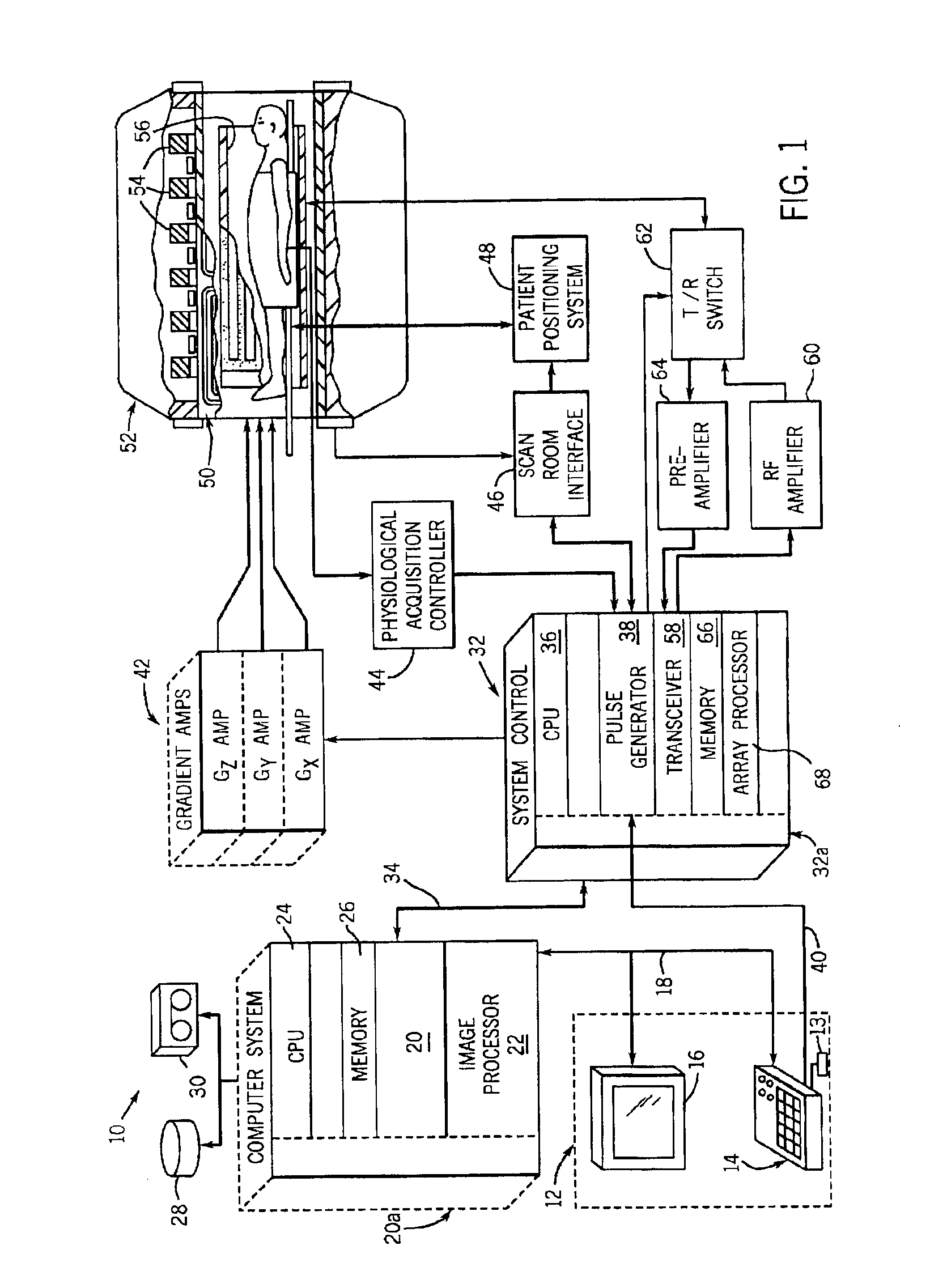
[0025]Referring to FIG. 1, the major components of a preferred MRI system 10 incorporating the present invention are shown. The operation of the system is controlled from an operator console 12 which includes a keyboard or other input device 13, a control panel 14, and a display 16. The console 12 communicates through a link 18 with a separate computer system 20 that enables an operator to control the production and display of images on the screen 16. The computer system 20 includes a number of modules which communicate with each other through a backplane 20a. These include an image processor module 22, a CPU module 24 and a memory module 26, known in the art as a frame buffer for storing image data arrays. The computer system 20 is linked to a disk storage 28 and a tape drive 30 for storage of image data and programs, and it communicates with a separate system control 32 through a high speed serial link 34. The input device 13 can include a mouse, joystick, keyboard, track ball, to...


PUM


Abstract

A system and method are disclosed to acquire high temporal resolution free-breathing cardiac MR images. The technique includes monitoring heart rate of a patient just prior to image acquisition to acquire a time period of an R—R interval, and using this time period from the heart rate monitoring to prospectively estimate future R—R intervals. The acquisition of MR data can then commence at any point in an R—R interval and extend for the time period recorded. The data acquisition can be segmented and acquired in successive R—R intervals, then combined to create high temporal resolution images.

Description

BACKGROUND OF INVENTION[0001]The present invention relates generally to an improved method for acquiring magnetic resonance images (MRI), and more particularly, to a method and apparatus to acquire high temporal resolution MR images that is particularly useful in cardiovascular MR examinations.[0002]When a substance such as human tissue is subjected to a uniform magnetic field (polarizing field B0), the individual magnetic moments of the spins in the tissue attempt to align with this polarizing field, but precess about it in random order at their characteristic Larmor frequency. If the substance, or tissue, is subjected to a magnetic field (excitation field B1) which is in the x-y plane and which is near the Larmor frequency, the net aligned moment, or longitudinal magnetization, MZ, may be rotated, or tipped, into the x-y plane to produce a net transverse magnetic moment Mt. A signal is emitted by the excited spins after the excitation signal B1 is terminated and this signal may be...

Claims


Application Information

Patent Timeline
Patent Type & Authority Patents(United States)
IPC IPC(8): G01R33/567G01R33/54G01R33/48A61B5/055
CPCG01R33/567
Inventor SARANATHAN, MANOJKUMARFOO, THOMAS K. F.DERBYSHIRE, J. ANDREW
Owner GENERAL ELECTRIC CO
Why Eureka
Try Eureka