Apparatus and methods for treating intracorporeal fluid accumulation

a technology of intracorporeal fluid and apparatus, applied in the direction of wound drain, diagnostics, diagnostic recording/measuring, etc., can solve the problems of increasing morbidity and mortality, and affecting the treatment effect of patients, so as to reduce the risk of infection, and reduce the effect of infection

Active Publication Date: 2015-05-26
SEQUANA MEDICAL NV
View PDF36 Cites 19 Cited by
  • Summary
  • Abstract
  • Description
  • Claims
  • Application Information

AI Technical Summary

Benefits of technology

[0015]The present invention overcomes the drawbacks of previously-known fluid management systems for treating ascites by providing a fluid management system that automatically and autonomously removes ascites accumulations with little patient involvement. The fluid management system of the present invention preferably comprises an implantable device including a pump, a controller, a battery and a transceiver; a charging and communication system configured to periodically charge the battery of, and communicate with, the implantable device; and monitoring and control software, suitable for use with a conventional personal computer, for configuring and controlling operation of the implantable device and charging and communication system. Preferably, the monitoring and control software is available only to the treating physician, such that the patient generally interacts with the implantable device only via the charging and communication system for purposes of recharging the implantable device. In accordance with one aspect of the present invention, the implantable device is configured to pump fluid in small increments, at relatively high flow rates, during predetermined times of the day to achieve a target volume, and further is configured to periodically alter the pump position to reduce the risk of clogging of the implantable device during non-pumping intervals. The pump also may be programmed to perform a rapid sequence of backward and forward movements if a blockage is detected, thereby clearing the blockage. Additionally, the fluid management system may include one or more sensors configured to detect indicia of the onset of infection, e.g., an increase in temperature, respiratory rate, or the viscosity of ascitic fluid, and one or more alarms configured to indicate to the physician a prediction or detection of infection based on the output(s) of those sensors.
[0017]In one preferred embodiment, the implantable device includes an electrically-driven mechanical gear pump configured for subcutaneous implantation. The pump has an inlet port coupled to an inflow catheter and an outlet port coupled to a bladder catheter. In accordance with one aspect of the present invention, the pump employs a pair of floating gears that function as a positive displacement pump, wherein a driving gear is coupled to a splined shaft of an electric motor to minimize power consumption arising due to manufacturing variations or shaft eccentricity. The inflow catheter comprises a tube having a first end configured to be coupled to the pump inlet and a second end configured to be positioned in a selected cavity, e.g., peritoneum, pleura or pericardial sac. The second end of the inflow catheter includes a plurality of through-wall apertures that permit fluid accumulating to pass into the catheter. The bladder or outflow catheter comprises a tube having a first end configured to be coupled to the pump and a second end configured to be inserted through the wall of, and fixed within, a patient's bladder. Alternatively, for treating pleural or pericardial effusions, the second end may be configured for placement in the peritoneal cavity, rather than the bladder. The fluid circuit further includes sensors arranged to monitor ambient pressure, pressure at the pump inlet, pressure at the pump outlet, pressure in the bladder (or peritoneal cavity, if this is used as a sink), and optionally the temperature of the ascitic fluid and the respiratory rate of the patient. The inflow and outflow catheters include connectors configured to reduce the risk of improper implantation.
[0018]The implantable device further comprises a controller, packaged together with the pump, electric motor, battery, charging coil, and radio transceiver within a low volume sealed housing. The controller is coupled to the pump motor, battery, transceiver and a plurality of sensors to continually monitor pressure, temperature, humidity, charge status, pump status, patient movement and other environmental and system related parameters. The controller preferably comprises a processor, nonvolatile memory for storing firmware, implant identification information, and system and environmental data, and volatile memory that serves as a buffer for computations and instructions during execution and firmware updating. The pump motor is configured for extended use and low power consumption, and preferably includes Hall effect sensors for position sensing and to determine the direction of rotation (and correspondingly, flow and fluid viscosity). The battery preferably is a long-lasting lithium-ion or lithium polymer battery that is coupled to an inductive charging circuit, thereby enabling the battery to be recharged using the external charging and communication system. A radio frequency transceiver preferably is employed in the device for transmitting system information to, and receiving information from, the external charging and communication system, including system performance data, commands, and firmware upgrades. All of the foregoing components preferable are disposed within the housing, which further includes a filler having a low permeability for water, thereby reducing infiltration of moisture into the housing.
[0021]It is contemplated that the system of the present invention may avoid difficulties typically associated with the previously-known apparatus and methods for addressing ascites. It is expected, for instance, that the system and methods of the present invention will enable small quantities of peritoneal fluid to be moved to the bladder without the inconvenience and complications generally associated with use of pharmaceuticals or paracenteses. In particular, because the apparatus and methods of the present invention avoid repeated, periodic removal of large quantities of fluid, as occurs with paracenteses, the tendency to generate additional ascites to offset the removed fluid will be reduced. These effects in turn are expected to obviate the need to infuse plasma expanders, such as human albumin, into the patient following paracentesis, thereby resulting in significant cost savings to the patient and health care system. The prediction or detection of infection, particularly at an early stage of infection, further may improve patient outcomes and reduce the need for more expensive treatments. Finally, the apparatus and methods of the present invention are expected to provide improved quality of life for chronic ascites patients, allowing such patients to pursue less sedentary lifestyles than would otherwise be possible, and encouraging better compliance with medically-directed dietary and exercise regimes.

Problems solved by technology

Chronic ascites, pleural effusion and pericardial effusion are conditions in which chronic fluid collections persist and result in increased morbidity and mortality.
If left untreated, the fluid accumulation may interfere with proper lung function, significantly increasing morbidity and mortality.
Although a relatively simple procedure, placement of an intercostal drain is associated with a relatively high rate of major complications, including hemorrhage and infection.
However, this procedure requires a lengthy hospital stay and is reported to be associated with the onset of adult respiratory distress syndrome, a potentially life-threatening complication.
In pericardial effusion, fluid accumulates in the pericardial sac and may lead to increased intrapercardial pressure and reduced cardiac output.
Although this procedure is usually well tolerated by patients, the pericardial window may close, requiring re-operation.
Untreated ascites can result in respiratory compromise, compression of the inferior vena cava (a vital blood vessel) and spontaneous bacterial peritonitis (a life-threatening condition).
The drugs often employed to treat ascites are usually long-term and often result in complications.
The difficulty with this treatment, however, is that fluid is removed from the entire body, including the circulating volume of blood.
This in turn may result in excessive loss of fluid required to perfuse the vital organs of the human body.
Thus, even with frequent application, the medicines frequently provide unsatisfactory results.
The regular accumulation and drainage by paracentesis of large quantities of fluid in the peritoneal cavity adversely impacts patient quality of life and often can interfere with the patient's ability to combat the underlying disease, such as cirrhosis.
Moreover, repeated paracenteses place the patient at increased risk for a life-threatening infection of the peritoneal cavity.
Other surgical / invasive procedures typically involve treatment of the cause of the ascites (for example, the Transjugular Intrahepatic Portosystemic Shunt) but these measures also frequently result in serious and life-threatening complications or become ineffective over time.
Consequently, such procedures are performed infrequently.
In particular, the presence of large quantities of fluid within the peritoneal cavity frequently disturbs the patient's fluid equilibrium, such that the patient's body attempts to compensate for fluid loss due to paracentesis by increasing ascites production.
Consequently, regularly scheduled paracenteses followed by infusion of human albumin impose significant economic burdens on the patient and the health care system.
Such previously known devices suffer from a variety of drawbacks, including fibrous encapsulation, frequent clogging and infection.
Such devices provided little improvement over periodic paracenteses, and resulted in increased rates of infection, re-operation or other complications if left in place for any length of time.
Moreover, a key drawback of such previously-known systems is the requirement that the patient must repeatedly locate and manually actuate the pumping mechanism on a daily basis.
Such activity may be difficult for patients, especially the elderly and obese, and further complicated by an ascites-distended abdomen.
Consequently, the difficulty of manipulating such previously-known systems promotes patient non-compliance, in turn leading to clogging and infection.

Method used


Image

  • Apparatus and methods for treating intracorporeal fluid accumulation
  • Apparatus and methods for treating intracorporeal fluid accumulation
  • Apparatus and methods for treating intracorporeal fluid accumulation

Examples


first embodiment

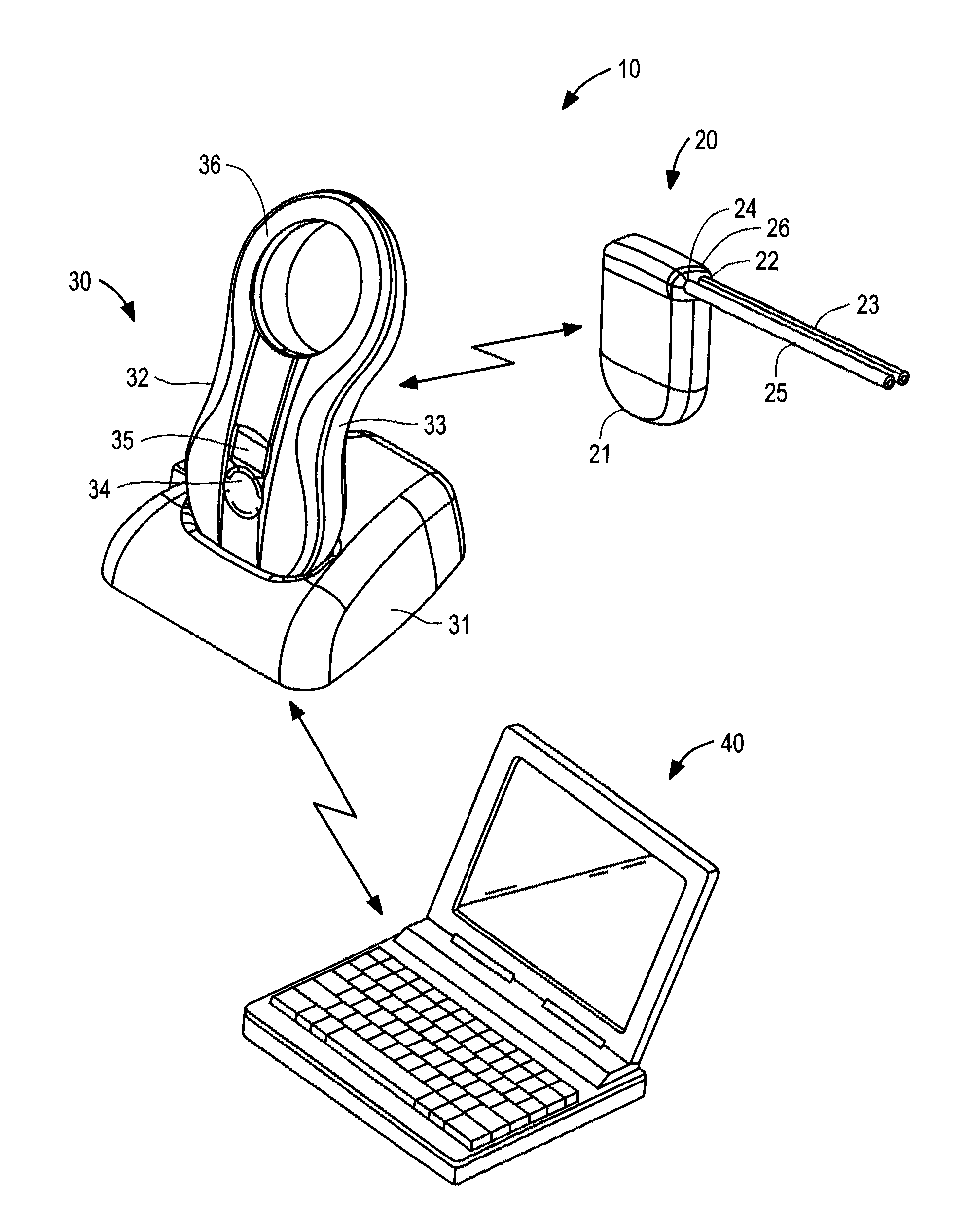
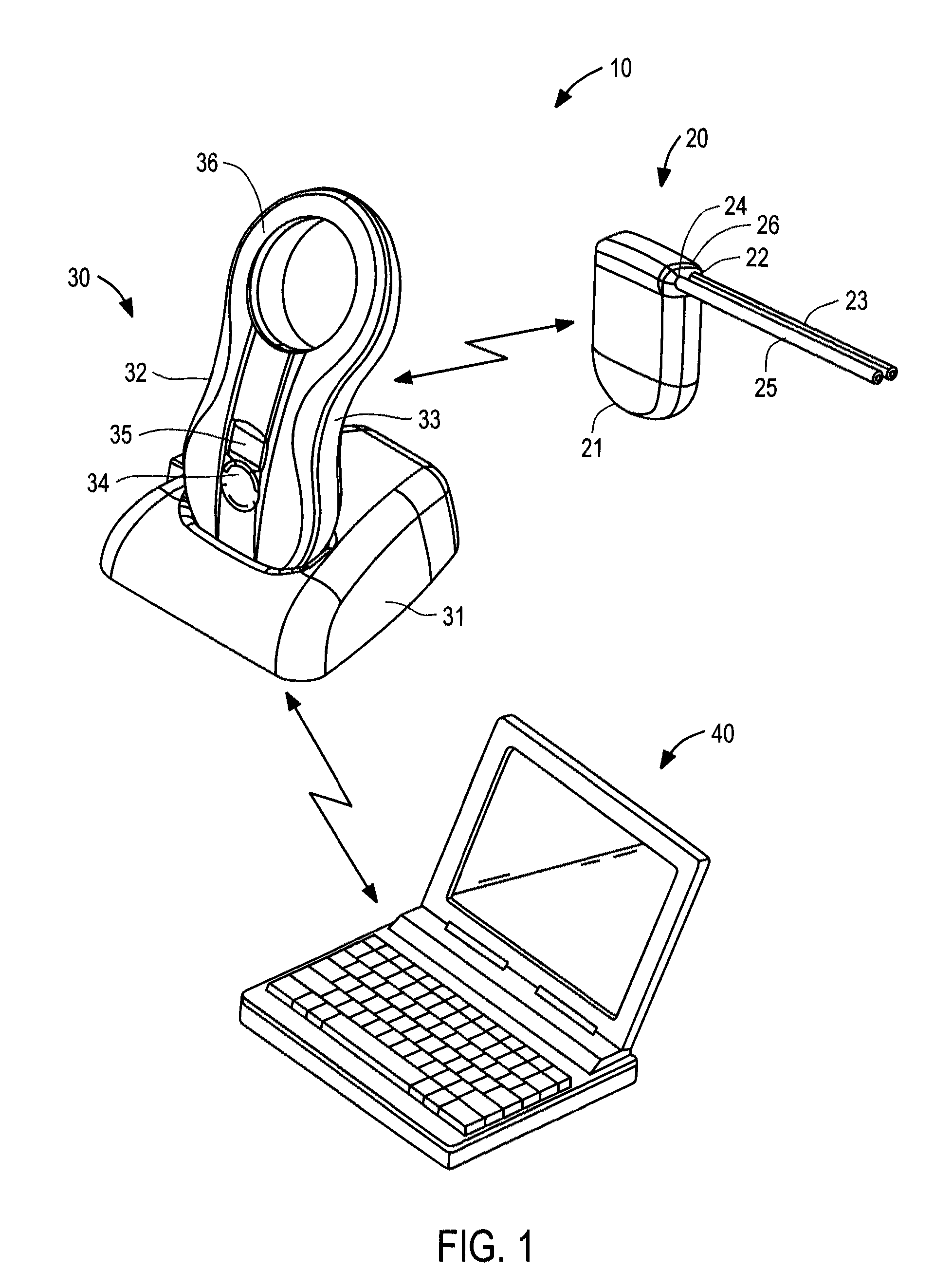
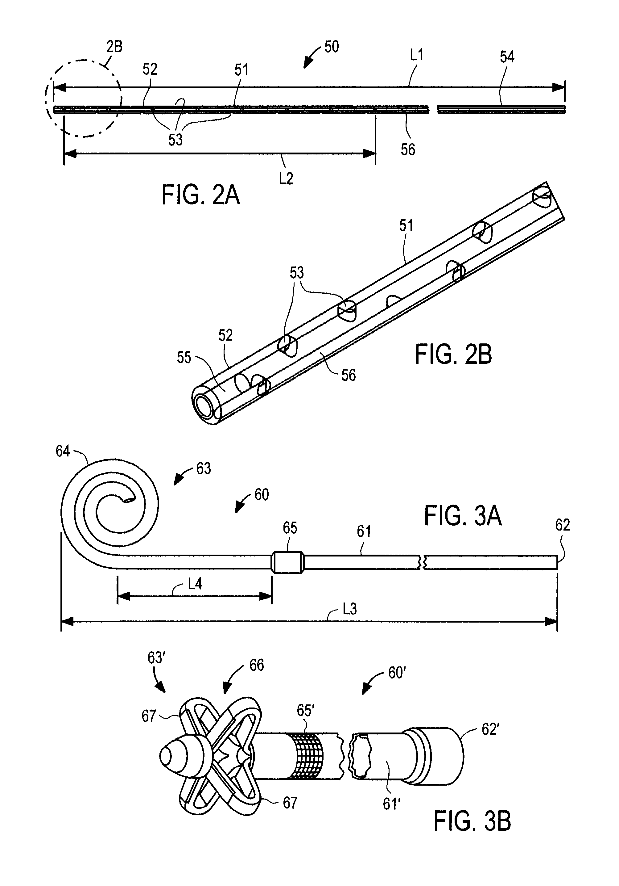
[0048]With respect to FIG. 3A, outflow catheter 60 of the present invention is described, corresponding to bladder catheter 25 of FIG. 1. Outflow catheter 60 preferably comprises tube 61 of medical-grade silicone having inlet end 62 and outlet end 63 including spiral structure 64, and polyester ingrowth cuff 65. Outflow catheter 60 includes a single internal lumen that extends from inlet end 62 to a single outlet at the tip of spiral structure 64, commonly referred to as a “pigtail” design. Inlet end 62 may include a connector for securing the inlet end of the outflow catheter to implantable device 20, or may have a length that can be trimmed to fit a particular patient.

[0049]When configured for use as the outflow catheter in an ascites treatment system, outflow catheter may have length L3 of about 45 cm, with cuff 65 placed length L4 of about 5 to 6 cm from spiral structure 64. Outflow catheter 60 may be loaded onto a stylet with spiral structure 64 straightened, and implanted usin...

second embodiment

[0053]With respect to FIG. 3B, an outflow catheter of the present invention is described, in which similar components are identified with like-primed numbers. Outflow catheter 60′ preferably comprises tube 61′ of medical-grade silicone having inlet end 62′, outlet end 63′ and polyester ingrowth cuff 65′. In accordance with this embodiment, outlet end 63′ includes malecot structure 66, illustratively comprising four resilient wings 67 that expand laterally away from the axis of the catheter to reduce the risk that outlet end 63′ of the catheter will be inadvertently pulled loose after placement. Inlet end 62′ may include a connector for securing the inlet end of the outflow catheter to implantable device 20, or may have a length that can be trimmed to fit a particular patient.

[0054]Malecot structure 66 preferably is constructed so that wings 67 deform to a substantially flattened configuration when a stylet is inserted through the lumen of the catheter. In this manner, outflow cathet...


PUM


Abstract

A fluid management system for the treatment of ascites, pleural effusion or pericardial effusion is provided including an implantable device including a pump, control circuitry, battery and transceiver; a charging and communication system configured to periodically charge the battery and communicate with the implantable device to retrieve performance data; and monitoring and control software, suitable for use with conventional personal computers, for configuring and controlling operation of the implantable device and charging and communication system. The implantable device includes a number of features that provide automated movement of fluid to the bladder with reduced risk of clogging, with no patient involvement other than occasional recharging of the battery of the implantable device. The monitoring and control software is available only to the treating physician, such that the physician interacts with the implantable device via the charging and communication system.

Description

I. CROSS-REFERENCE TO RELATED APPLICATIONS[0001]This application claims the benefit of U.S. Provisional Patent Application No. 61 / 443,668, filed Feb. 16, 2011 and entitled “Apparatus and Methods for Treating Intracorporeal Fluid Accumulation,” the entire contents of which are incorporated herein by reference.II. FIELD OF THE INVENTION[0002]This application relates to apparatus and methods for treating intracorporeal fluid accumulations, such ascites, pleural effusion and pericardial effusion.III. BACKGROUND OF THE INVENTION[0003]There are a variety of conditions which result in pathologic chronic collection of bodily fluids within the peritoneum, pleura or pericardial sac. Chronic ascites, pleural effusion and pericardial effusion are conditions in which chronic fluid collections persist and result in increased morbidity and mortality.[0004]These foregoing conditions currently are treated typically by one of three methods: 1) external drainage, which poses a risk of infection and lo...

Claims


Application Information

Patent Timeline
Patent Type & Authority Patents(United States)
IPC IPC(8): A61M1/00A61M5/142A61M27/00G16H40/63
CPCA61M27/002A61M5/14276A61M5/14236A61M2205/3507A61M2205/3569G06F19/3468A61M2205/8243A61M1/0023A61M2205/04A61M2205/33A61M2205/18A61M2205/8237A61M2205/8206A61M2210/1017A61M2210/101A61M2210/122A61M2210/1085A61M1/84A61M1/28G16H40/63A61M2205/10A61M2205/3368A61M2205/3379A61M2205/502A61M2205/52A61M2230/005A61M2230/42G16H20/17
Inventor DEGEN, THOMAS WERNERTHOMMEN, DANIEL THOMASJOHNSON, NOEL L.
Owner SEQUANA MEDICAL NV
Why Eureka
Try Eureka