Biosensor for detection and visualisation of single-stranded DNA

a biosensor and dna technology, applied in the field of single stranded dna detection and visualisation, can solve the problems of limitations of assays relying on intrinsic tryptophan fluorescence changes, and achieve the effect of rapid reaction time of methods

Inactive Publication Date: 2015-09-01
RGT UNIV OF CALIFORNIA +1
View PDF2 Cites 0 Cited by
  • Summary
  • Abstract
  • Description
  • Claims
  • Application Information

AI Technical Summary

Benefits of technology

[0021]Attachment of the detectable label to the SSB may sometimes reduce the affinity of the protein for single stranded DNA. However, this does not prevent an alteration of the detectable characteristics on binding single stranded DNA which can be quantitatively measured.
[0037]The invention also provides a SSB wherein the label is attached to a region of the protein surface. Regions of the protein such as a subunit-subunit interaction surface are not exposed and are thus unsuitable for labelling purposes. Surface located residues are more easily accessible for labelling purposes and are less likely to disrupt the overall shape of the protein when labelled.
[0044]Fluorophores will rarely be attached to an amino acid directly, but will instead be attached via a linker. The choice of linker can also have an effect on the way the labelled SSB functions, as the size, shape and flexibility of the linker can change the ability of a linker to come into proximity with other groups.
[0047]After attachment of the label, labelled protein will usually be purified to separate it from free label and from any mis-labelled protein. The mis-labelled protein may be unlabelled protein with which label did not react or protein where label has attached in the wrong position (either in place of or in addition to the desired label). During purification of the labelled protein, treatment with a thiol reagent may be included, such as β-mercaptoethanol, dithiothreitol or sodium 2-mercaptoethanesulfonate as this can improve the fluorescence response of the protein.
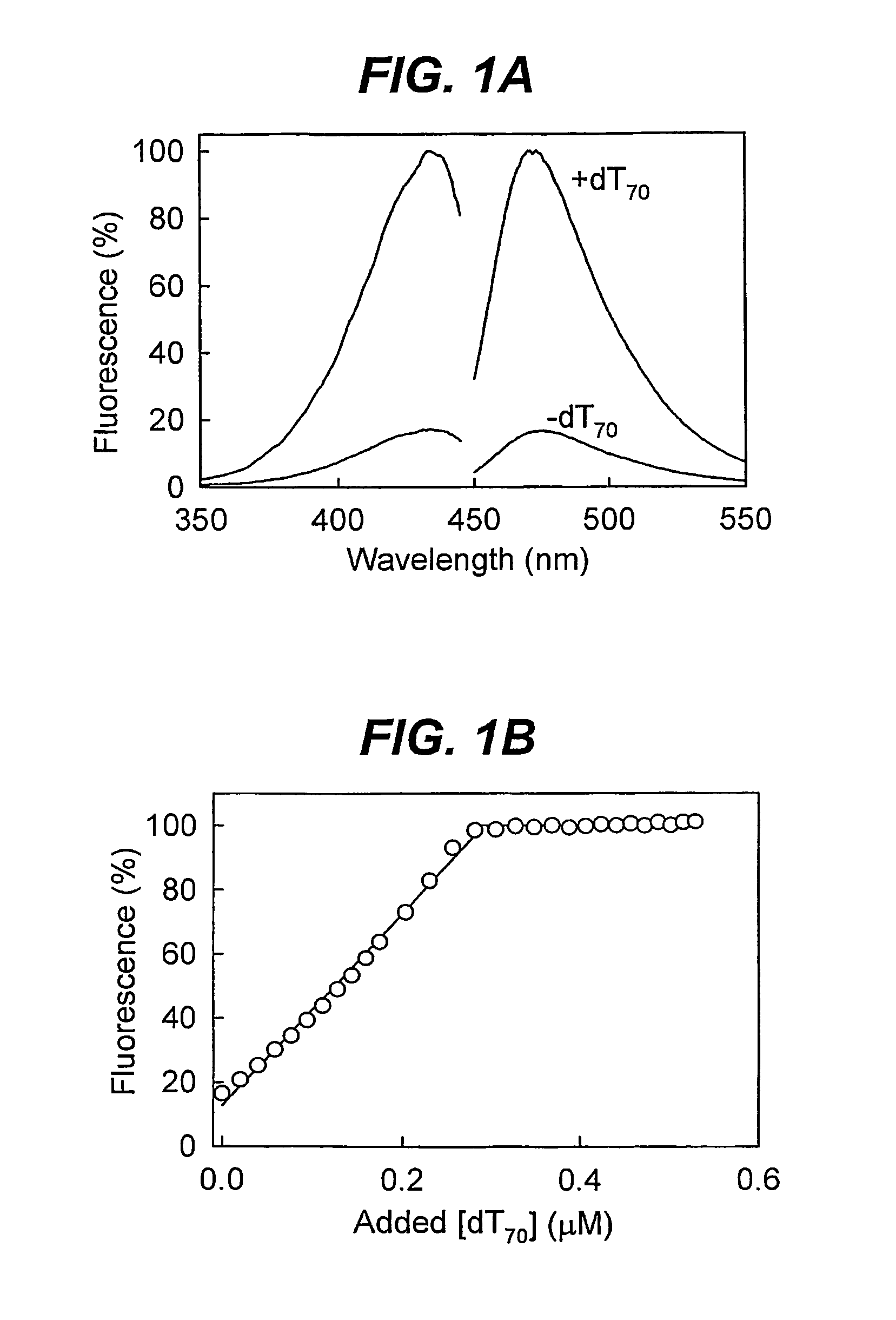
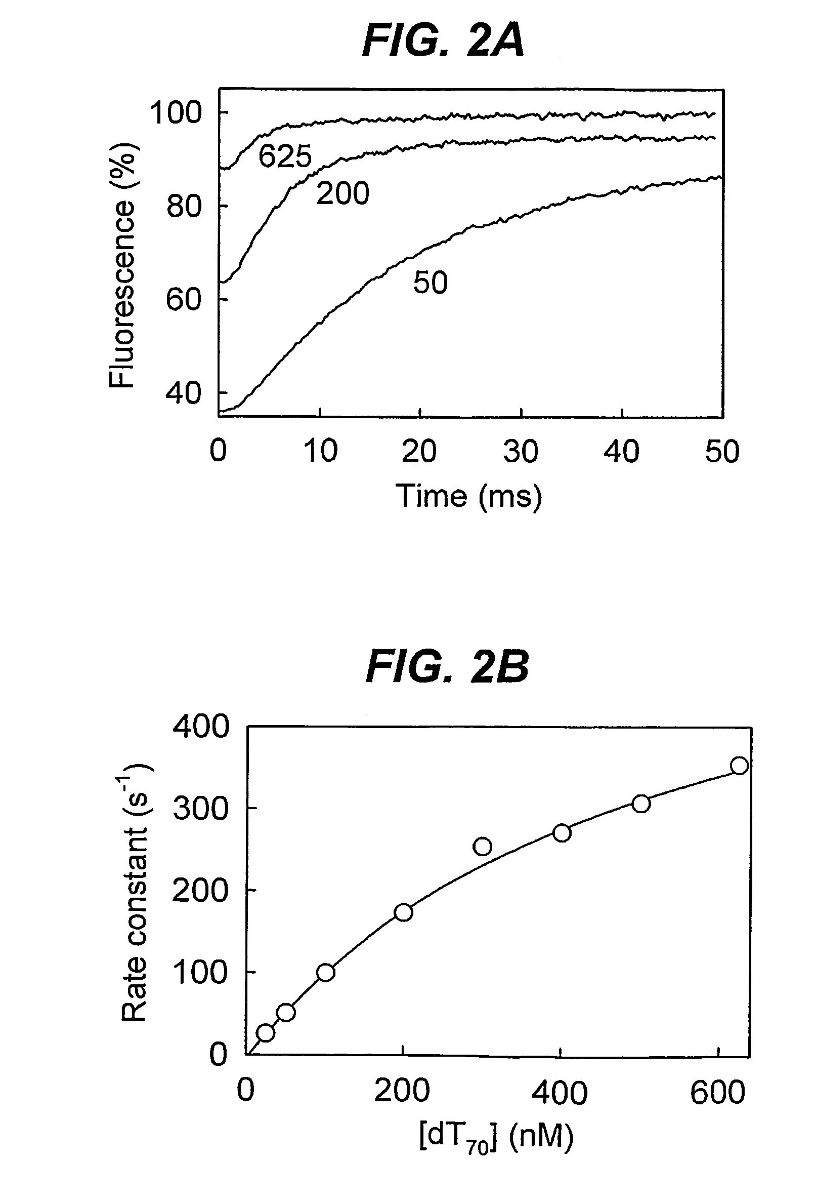
[0059]By employing this method, using a modified SSB, it is possible to follow the kinetics of biological systems due to the extremely rapid reaction time of the method.

Problems solved by technology

Furthermore, assays relying on the intrinsic tryptophan fluorescence changes have limitations due to photobleaching and interference from other species that absorb in the same UV range as tryptophan.

Method used


Image

  • Biosensor for detection and visualisation  of single-stranded DNA
  • Biosensor for detection and visualisation  of single-stranded DNA
  • Biosensor for detection and visualisation  of single-stranded DNA

Examples


Embodiment Construction

Preparation of mutant E. coli SSB proteins

[0075]The wild type E. coli ssb gene was cloned into a pET15b vector using PCR with primers containing flanking NdeI and BamHI restrictions sites. This vector was used as a template for site directed mutagenesis to produce genes for G26C, S92C or W88C E. coli SSB. The pET15b constructs were sequenced to confirm the presence of the desired mutations and the integrity of the remainder of the ssb gene. The mutated genes were then transferred to pET22b vectors using the NdeI and BamHI restriction sites and these were used to transform BL21 pLysS for overexpression using the T7 promoter system. Cells were grown in 21 of LB at 37° C. to mid-log phase and expression was induced with 1 mM IPTG. After three hours, the cells were harvested, resuspended in 50 ml of 50 mM Tris.HCl pH 7.5, 200 mM NaCl, 1 mM EDTA, 1 mM dithiothreitol and 10% sucrose, and stored at −80° C.

[0076]The mutant E. coli SSB proteins were purified at 4° C. using a modification of ...


PUM

PropertyMeasurementUnit
excitation wavelengthsaaaaaaaaaa
pHaaaaaaaaaa

Abstract

A modified protein of the single strand DNA-binding domain, SSB family comprising a detectable label is disclosed. The label has detectable characteristics which alter on binding single stranded DNA. The protein is thus useful in an assay for single stranded DNA.

Description

[0001]This application is a U.S. national stage of International Application No. PCT / GB2008 / 001995 filed Jun. 11, 2008.[0002]All documents cited herein are incorporated by reference in their entirety.TECHNICAL FIELD[0003]This invention relates to assays for single stranded DNA, particularly the detection and visualisation of single stranded DNA in biological solutions. More particularly, the present invention relates to a modified protein of the single strand DNA-binding domain, SSB family, and to the use of such a protein in a biological assay.BACKGROUND ART[0004]The formation and maintenance of single-stranded DNA (ssDNA) is an important part of many processes involving DNA. For instance, strand separation of double-stranded DNA (dsDNA) is catalyzed by helicases and this exposure of the bases on the DNA allows further processing, such as repair or replication. Assays of helicase activity are an important part of probing the related biological processes.[0005]Although there are met...

Claims


Application Information

Patent Timeline
Patent Type & Authority Patents(United States)
IPC IPC(8): C07K1/00A61K39/108C07K14/245G01N33/53G01N33/533
CPCC07K14/245G01N33/533G01N33/5308G01N2500/00
Inventor DILLINGHAM, MARKKOWALCZYKOWSKI, STEPHEN C.WEBB, MARTIN
Owner RGT UNIV OF CALIFORNIA
Why Eureka
Try Eureka