Molecular beacon based assay for the detection of biomarkers for breast cancer metastasis

a breast cancer and biomarker technology, applied in biochemistry apparatus and processes, organic chemistry, sugar derivatives, etc., can solve the problems of inability to detect low number of tumor cells, and relatively painless serum sampling technique, so as to achieve the effect of robustness of assay, and reduce the total analysis tim

Inactive Publication Date: 2016-03-29
FURCHAK JENNIFER +2
View PDF3 Cites 28 Cited by
  • Summary
  • Abstract
  • Description
  • Claims
  • Application Information

AI Technical Summary

Benefits of technology

[0033]In an embodiment, products comprising the molecular beacons are contemplated. The product may be in the form of a kit for determining the presence of and / or the levels of expression of PIP mRNA, HER2 mRNA and / or hMAM mRNA, biomarkers for breast cancer metastasis, the kit comprising a molecular beacon. The assay for determining the presence of and / or the levels of biomarker mRNA expression is sensitive and reproducible with a limit of detection in the pM range. In the presence of non-target mRNA, the assay shows similar sensitivity, indicating that the PIP molecular beacon is specific to its target mRNA. The robustness of the assay is demonstrated by the similar limit of detection and sensitivity in undiluted serum samples.
[0034]Total analysis time for the MB assay is significantly shorter than protocols based on RT-PCR. A single sample in buffer can be analyzed in 15 minutes compared to at least 5 hours in RT-PCR. The decrease in total analysis time is even more significant if the sample is present in a complex matrix. The assay can be preformed directly in undiluted serum, eliminating the need for days of sample preparation.
[0035]With a limit of detection in the picomolar range, the assay does not compare with current protocols based on RT-PCR in terms of detection limit. RT-PCR relies on amplification to increase concentration of mRNA in serum to a detectable level and therefore is able to detect very low concentrations. However, the absence of an amplification step the instant method decreases the possibility of false positive and negative results.
[0036]Strategies for improving the detection of biomarker mRNA include preconcentrating the mRNA or tumor cells with a fast and efficient procedure before detection with a molecular beacon.
[0037]The potential impact of this work in detection and understanding of breast cancer metastasis lies in improvements in simplicity, accuracy, and speed over current methods. This dramatically reduces the total time for sample analysis from days to hours. The ability to sensitively and specifically detect biomarkers directly in serum samples, as well as in biopsy material, in minimal time and with a straightforward procedure could allow for improved patient treatment and prognoses. Ultimately, simplification of biomarker detection will allow higher sample throughput, resulting in better understanding of disease progression.

Problems solved by technology

A combination of better surgical techniques for removal of tumors, improved treatment options, and earlier detection of cancer has lead to a decline in cancer mortality over the last two decades.
Of these, serum sampling is a relatively painless technique that allows for frequent sampling.
This method cannot detect low numbers of tumor cells.
This method is time consuming and requires trained scientists for analysis.
However, it is costly and time consuming due to the use of multiple antibodies and washing steps in analysis.
Even in light of several advantages, RT-PCR has a few significant disadvantages in terms of analysis time, efficiency, and accuracy.
The process is too time consuming for rapid analysis in hospital or clinical settings.
Separation and visualization by slab gel electrophoresis and ethidium bromide staining has limited ability for quantitation, which is crucial to developing patient-specific treatment regimens.
To compound the issues further, there are difficulties in multiplex analysis.
Analysis of three or more biomarkers proves difficult due to unequal amplification of the sequences due to sample conditions, extracellular serum factors, and the formation of primer-dimers resulting in false-positive and false-negative results.
Q-RT-PCR, therefore, improves quantification in RT-PCR but brings its own disadvantages to the analysis.

Method used


Image

  • Molecular beacon based assay for the detection of biomarkers for breast cancer metastasis
  • Molecular beacon based assay for the detection of biomarkers for breast cancer metastasis
  • Molecular beacon based assay for the detection of biomarkers for breast cancer metastasis

Examples


example 1

Preparation of Human PIP cDNA and Human PIP mRNA

[0068]All nucleic acids were purified by polyacrylamide gel electrophoresis (PAGE) before use. Nucleic acids were denatured at 70° C. for 2 minutes, mixed 2:1 with loading dye (8 M urea; 20 mM EDTA; 5 mM Tris-HCl, pH 7.5; 0.5% w / v xylene cyanol; and 0.5% w / v bromophenol blue), and separated on 20 percent polyacrylamide gels (24 g urea; 25 mL 40% polyacrylamide; 10 mL 5×TBE) at 17 W per gel for approximately 3 hours. DNA was visualized with UV shadowing (100-280 nm). RNA was visualized by ethidium bromide staining of a thin, vertical strip of gel. DNA and unstained RNA were extracted by cutting bands from the gels, crushing, and tumbling over night in water. This process was performed twice and the eluate from both extractions was combined. Extracts were purified by chloroform extraction (24:1 chloroform:isoamyl alcohol solution saturated with TE buffer) and ethanol precipitation. Purified products were reconstituted in 50 μL of sterile...

example 2

Preparation of a PIP Molecular Beacon

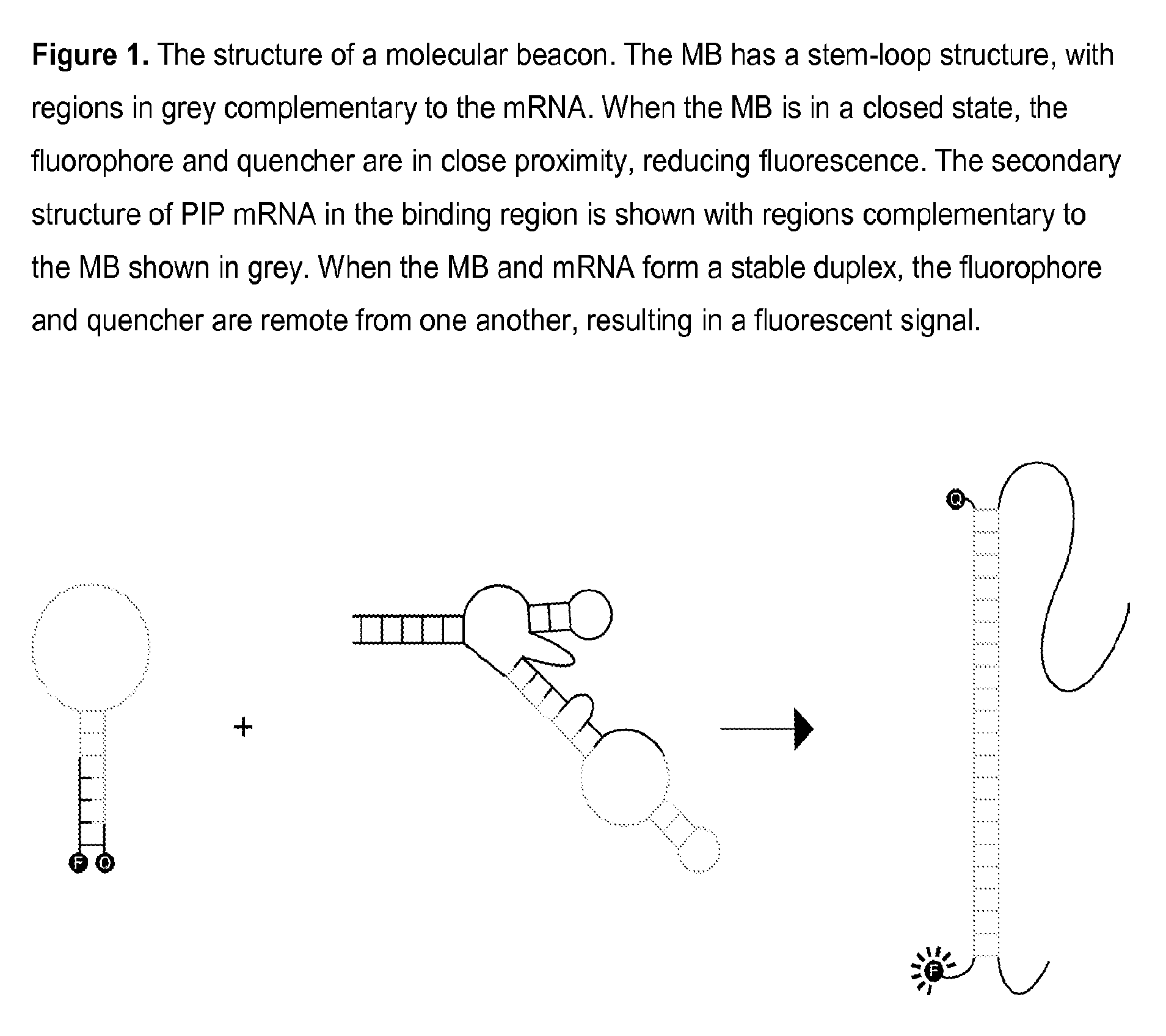
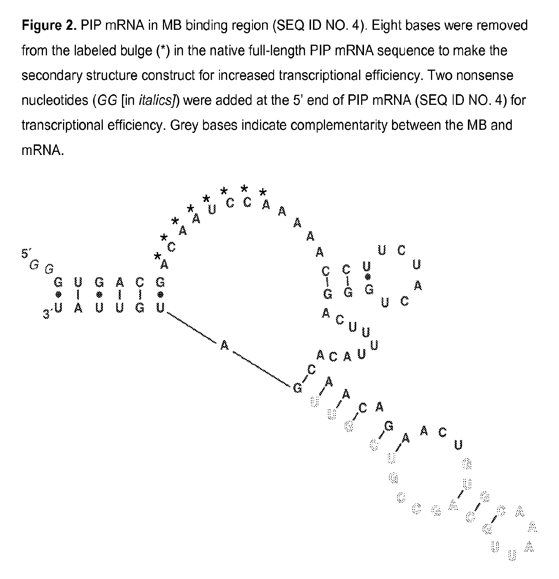
[0074]A PIP MB was designed based on the mRNA sequence for PIP mRNA (NCBI Accession NM_002652) which was determined using GenBank entries and a literature reference. Expected mRNA secondary structures were modeled using mfold energy minimization and a region of secondary structure that was conserved in the majority of possible structures was found. The MB sequence is complementary to the region of conserved secondary structure and incorporates nucleotides at the 5′ and 3′ ends which are self-complementary, thereby forming a closed, stem-loop structure. A PIP MB DNA, 5′-TGTGCAACGACGGCTGCAATTTGCACA-3′ (SEQ ID NO. 6), was chemically synthesized and modified to incorporate a fluorophore and a quencher at the 5′ and 3′ ends, respectively. A PIP MB of the invention consists essentially of tetrachloro-6-carboxyfluorescein-TGTGCAACGACGGCTGCAATTTGCACA (SEQ ID NO. 6)-BLACK HOLE QUENCHER® 1.

example 3

Analysis of the Binding of the PIP Molecular Beacon to PIP mRNA

[0075]Samples were prepared to give final concentrations of 50 mM HEPES, pH 7.5; 100 mM MgCl2; 200 mM KCl; 25 mM DTT; 20 nM MB. All solutions were heated to 95° C. for 2 minutes and allowed to anneal at room temperature prior to analysis. Fluorescence emission was monitored with excitation at 521+ / −5 nm and emission at 535+ / −5 nm at 25° C. using a Cary Eclipse fluorescence spectrophotometer (Agilent Technologies, Inc., Santa Clara, Calif.).

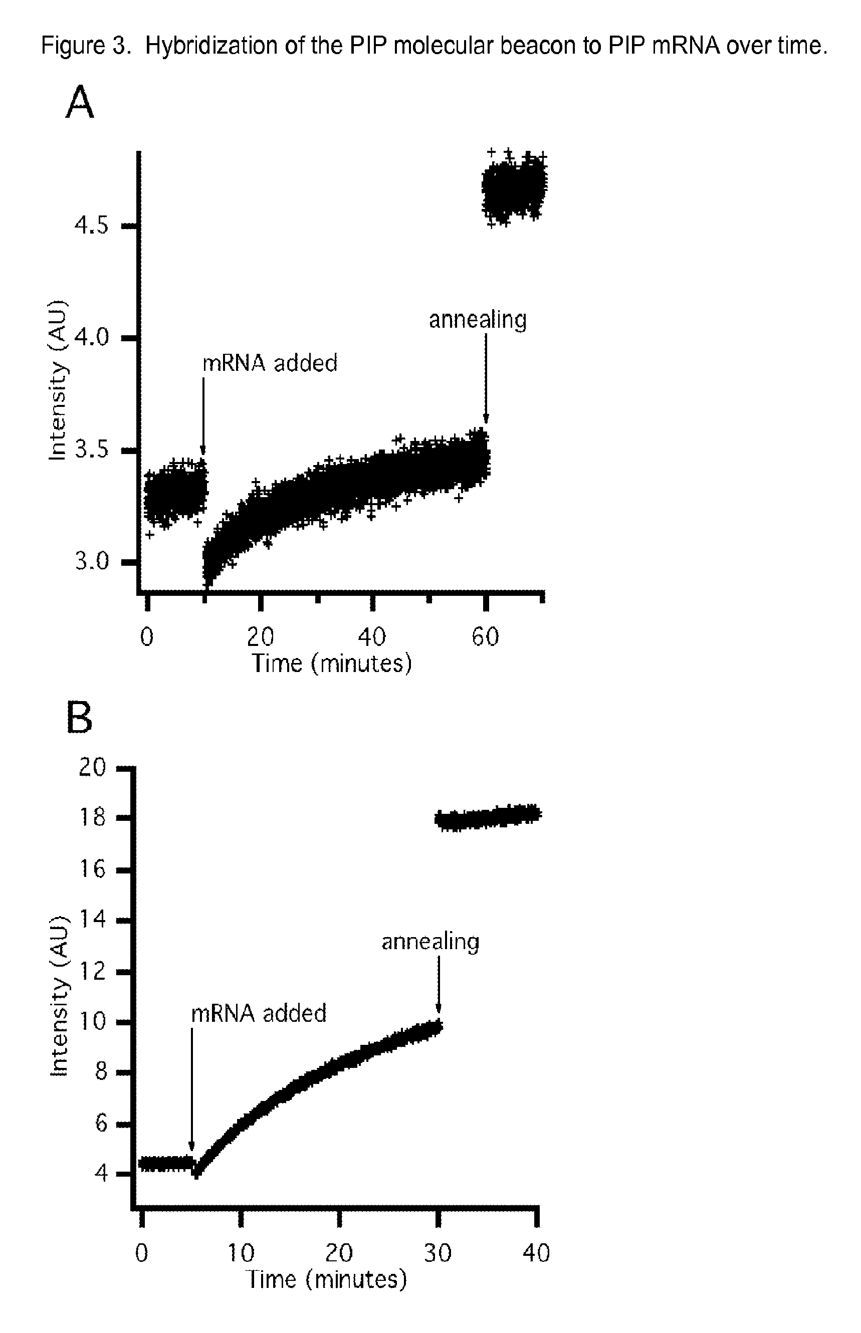
[0076]For temporal analysis of MB-mRNA hybridization, each MB sample was spiked with mRNA after establishing a fluorescence baseline. Fluorescence emission was then monitored for 30-60 minutes before the samples were heated to 95° C. for 2 minutes and annealed at room temperature. Emission was monitored for 10 minutes after annealing.

[0077]To evaluate the in vitro MB binding to mRNA, the baseline fluorescence intensity was established, target mRNA was then added, the nucleic acids were...


PUM

PropertyMeasurementUnit
quenching wavelength rangeaaaaaaaaaa
quenching wavelengthaaaaaaaaaa
quenching wavelength rangeaaaaaaaaaa

Abstract

The invention encompasses molecular beacon (MB) probes for monitoring the presence of human breast cancer biomarkers and for the analysis of breast cancer metastasis. The molecular beacon is an oligonucleotide probe which sensitively and specifically identifies biomarker mRNA in samples, in the presence of serum, in minimal time using fluorescence detection. The molecular beacons may be comprised in kits for the detection / quantitation of cancer biomarkers in clinical samples. The invention provides improvements in simplicity, accuracy, and speed over current methods, which could allow for improved patient treatment and prognoses.

Description

BACKGROUND OF THE INVENTION[0001]Breast cancer is a prevalent form of cancer among women in the United States. The detection, removal, and treatment of breast carcinoma are important focuses of the medical and scientific community. A combination of better surgical techniques for removal of tumors, improved treatment options, and earlier detection of cancer has lead to a decline in cancer mortality over the last two decades. Mortality is increasingly linked to early, undetected metastatic cancer.[0002]Traditionally, analysis of disease progression and metastasis is achieved through physical tumor staging parameters, such as tumor size and presence of nodal or distal metastases, using the tumor-node-metastases staging system. This method is qualitative, changes with advances in diagnosis, and requires a large pool of data (i.e. more patients) to provide accurate results. An analytical detection method would be advantageous for detection of metastatic breast cancer and accurate quantit...

Claims


Application Information

Patent Timeline
Patent Type & Authority Patents(United States)
IPC IPC(8): C12Q1/68C07H21/02C07H21/04
CPCC12Q1/6818C12Q1/6886C12Q2600/158C12Q2525/301C12Q2525/307C12Q2565/1015
Inventor FURCHAK, JENNIFERGUETSCHOW, ERIKBLACK, WILLIAM, ALEXANDER
Owner FURCHAK JENNIFER
Why Eureka
Try Eureka