Systems, processes and computer-accessible medium for providing hybrid flourescence and optical coherence tomography imaging

a technology of optical coherence tomography and hybrid flourescence, applied in the field of medical imaging, can solve the problems of not being able to provide quantitative or three-dimensional information, not being able to facilitate intravascular imaging, and prior techniques and systems may not be appropriate for three-dimensional or quantitative imaging of hollow organs, etc., to achieve accurate co-registration, improve quantification, and accurate fluorescence images

Active Publication Date: 2016-05-10
THE GENERAL HOSPITAL CORP
View PDF275 Cites 36 Cited by
  • Summary
  • Abstract
  • Description
  • Claims
  • Application Information

AI Technical Summary

Benefits of technology

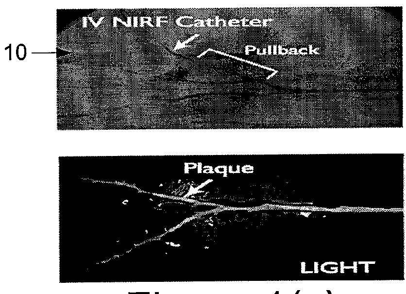
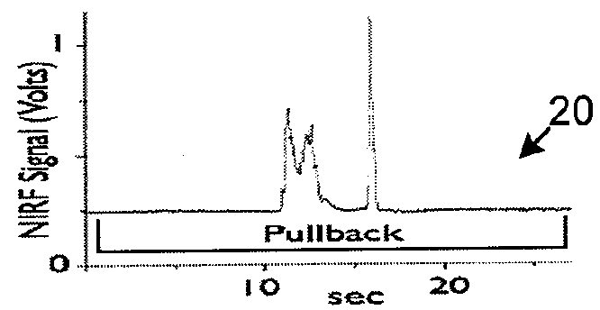
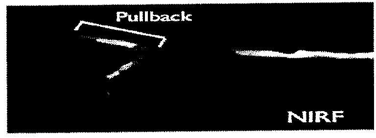
[0015]In addition, according to one exemplary embodiment of the present invention, the apparatus and process can be provided via hybrid imaging, e.g., three-dimensional, quantitative fluorescence images of hollow organs by employing concomitant / integrated imaging of the hollow structure architecture. In another exemplary embodiment of the present invention, a common optical path system may be used for some or most of the acquisition to facilitate an accurate co-registration, although different components are necessary for architectural and fluorescence imaging. Further, it is possible to appropriately illuminate, synchronize and process the signals in order to obtain accurate fluorescence images. In addition, architectural hollow organ information may be incorporated into the fluorescence imaging model to likely improve the quantification.
[0016]One exemplary embodiment of the system according to the present invention may operate on, e.g., (a) approximately 360-degree fluorescence imaging, (2) concomitant structural imaging with integrated OCT (and / or possibly another modality including but not limited to ultrasound, MRI, NIR or other spectroscopy, nuclear imaging); and / or (3) depth-resolved and depth and optical property corrected fluorescence imaging using OCT registration. This exemplary system (e.g., catheter) may be used on humans with a clinical availability of OCT, the long track record of NIR fluorochromes in humans (e.g. indocyanine green), planned clinical trials of a protease-activatable and other NIRF imaging agents, and a rapid OCT approach (e.g., optical frequency domain imaging (OFDI) techniques), that may facilitate comprehensive coronary arterial imaging during a saline injection, as opposed to prior prolonged balloon occlusions.

Problems solved by technology

However, the prior techniques and systems may not be appropriate for three-dimensional or quantitative imaging of hollow organs, for example, in intra-vascular or gastro-intestinal applications.
Such systems rely predominantly on surface information from fluorescence reflectance imaging, but likely lack the ability to provide quantitative or three-dimensional information.
Lasers In Surgery and Medicine 38:305-313 (2006)), such spectroscopic arrangement / technique does not (a) form images of fluorochrome distribution, (b) utilize the OCT information to effectuate fluorescence imaging, as well as quantitative or three-dimensional images, (c) may not be suitable for intravascular imaging given its large size (e.g., 2.0 mm diameter) and (d) does not address the ability to resolve multiple fluorochromes via multispectral / deconvolution methods described herein.
However the geometrical size and other aspects of such system did not facilitate its use for intravascular imaging.
None of the systems described above however provide the imaging robustness of or teach on hybrid anatomical molecular imaging for offering a highly accurate diagnostic, monitoring or treatment system and importantly for utilizing anatomical information to correct for photon propagation related events in tissue and offer quantitative information that is independent of (a) catheter placement in the hollow organ, b) variation of optical properties that may affect the fluorescence signal and contrast achieved, and c) variation of the depth of the activity that can similarly affect the fluorescence signal and contrast achieved.

Method used


Image

  • Systems, processes and computer-accessible medium for providing hybrid flourescence and optical coherence tomography imaging
  • Systems, processes and computer-accessible medium for providing hybrid flourescence and optical coherence tomography imaging
  • Systems, processes and computer-accessible medium for providing hybrid flourescence and optical coherence tomography imaging

Examples


Embodiment Construction

)

[0028]Catheter

[0029]According to one exemplary embodiment, it is possible to obtain three-dimensional fluorescence and architectural images of a hollow organ. This can be achieved with an exemplary embodiment of the system of the present invention as shown in FIGS. 2(a) and 2(b) that can be used as a catheter 200, 200′ which may utilize a rotating fiber 210 inside an appropriate catheter sheath. Such exemplary system / catheter 200 can be combined with the ability of performing congruent optical coherence imaging as shown in FIGS. 2(a) and 2(b), providing two alternative exemplary implementations. The fiber 210 of the exemplary system 200 / 200′ can be a common fiber that may combine single mode (e.g., for OCT) and multi-mode operations (e.g., for fluorescence detection).

[0030]As shown in FIGS. 2(a) and 2(b), this exemplary system / catheter 200 / 200′ may include a light collection system 220, 220′ for the multimode detection (e.g., fluorescence collection), an interferometer system 230, ...


PUM

PropertyMeasurementUnit
diameteraaaaaaaaaa
spectral widthaaaaaaaaaa

Abstract

Apparatus and method can be provided for obtaining information regarding at least one portion of a biological structure. For example, using a first arrangement, it is possible to generate a first electro-magnetic radiation to be forwarded to the portion and receive, from the portion, a second fluorescent radiation associated with the first electro-magnetic radiation Further, using a second arrangement, it is possible to obtain information associated with the second fluorescent radiation, and generate at least one image of the portion as a function of the second fluorescent radiation. According to an exemplary embodiment of the present invention, the first arrangement can be inserted into at least one lumen, and the image of the portion may be associated with the at least one lumen.

Description

FIELD OF THE INVENTION[0001]The present invention relates generally to medical imaging and, more particularly, to apparatus and process for generating fluorescence images representative of functional or molecular activity which may indicate, e.g., a disease in hollow organs using a catheter approach. According to one exemplary embodiment of the present invention, it is possible to combine fluorescence imaging technique(s) with optical coherence tomography technique(s) utilizing certain optical components, and other anatomical imaging techniques can be utilized as well.BACKGROUND INFORMATION[0002]Fluorescence images can be generated in-vivo for imaging of physiological or molecular function in live biological tissues. One exemplary technology that has revolutionized the ways tissue and disease processes can be visualized relates to the use of externally administered fluorescence probes with sensitivity and specificity to certain molecular, cellular or physiological targets. For examp...

Claims


Application Information

Patent Timeline
Patent Type & Authority Patents(United States)
IPC IPC(8): A61B5/00G01N21/47G01N21/64
CPCA61B5/6852A61B5/0066A61B5/0073G01N21/4795G01N21/6456
Inventor JAFFER, FAROUC A.NTZIACHRISTOS, VASILIS
Owner THE GENERAL HOSPITAL CORP
Why Eureka
Try Eureka