
Methods of promoting wound healing and attenuating contact hypersensitivity with gingiva-derived mesenchymal stem cells

a stem cell and mesenchymal stem cell technology, applied in the field of stem cell therapy, can solve the problems of difficult identification of allergens to improve contact avoidance, abnormal wound healing in diabetic patients, scarring and fibrotic diseases, etc., and achieve the effect of rapid ex vivo expansion and ease of isolation

Active Publication Date: 2017-06-20
UNIV OF SOUTHERN CALIFORNIA
View PDF5 Cites 1 Cited by
  • Summary
  • Abstract
  • Description
  • Claims
  • Application Information

AI Technical Summary

Benefits of technology

The patent text describes a unique population of stem cells derived from human gingival tissue that can be easily isolated and expanded in the lab. These cells have stem cell-like properties and can help treat skin wounds by promoting faster healing, re-epithelialization, and improved blood vessel growth. When these cells are used in treatment, they can reduce inflammation and production of certain proteins that can cause slow healing. The human gingiva-derived mesenchymal stem cells (GMSCs) can be an effective treatment for cutaneous wounds and can help improve the overall quality of skin tissue.

Problems solved by technology

Upon tissue insult, the immediate inflammatory response is characterized by infiltration and activation of leukocytes, whereas a delayed or excessive inflammatory response may lead to abnormal wound healing in diabetic patients, scarring and fibrotic diseases.
Currently, topical application of corticosteroid is the first-line palliative measure for ACD with short-term outcome, while allergen identification to improve contact avoidance is still challenging.

Method used


Image

  • Methods of promoting wound healing and attenuating contact hypersensitivity with gingiva-derived mesenchymal stem cells
  • Methods of promoting wound healing and attenuating contact hypersensitivity with gingiva-derived mesenchymal stem cells
  • Methods of promoting wound healing and attenuating contact hypersensitivity with gingiva-derived mesenchymal stem cells

Examples


example i.b

GMSCs Induce Anti-Inflammatory Immune Profile in Macrophages in Co-Culture

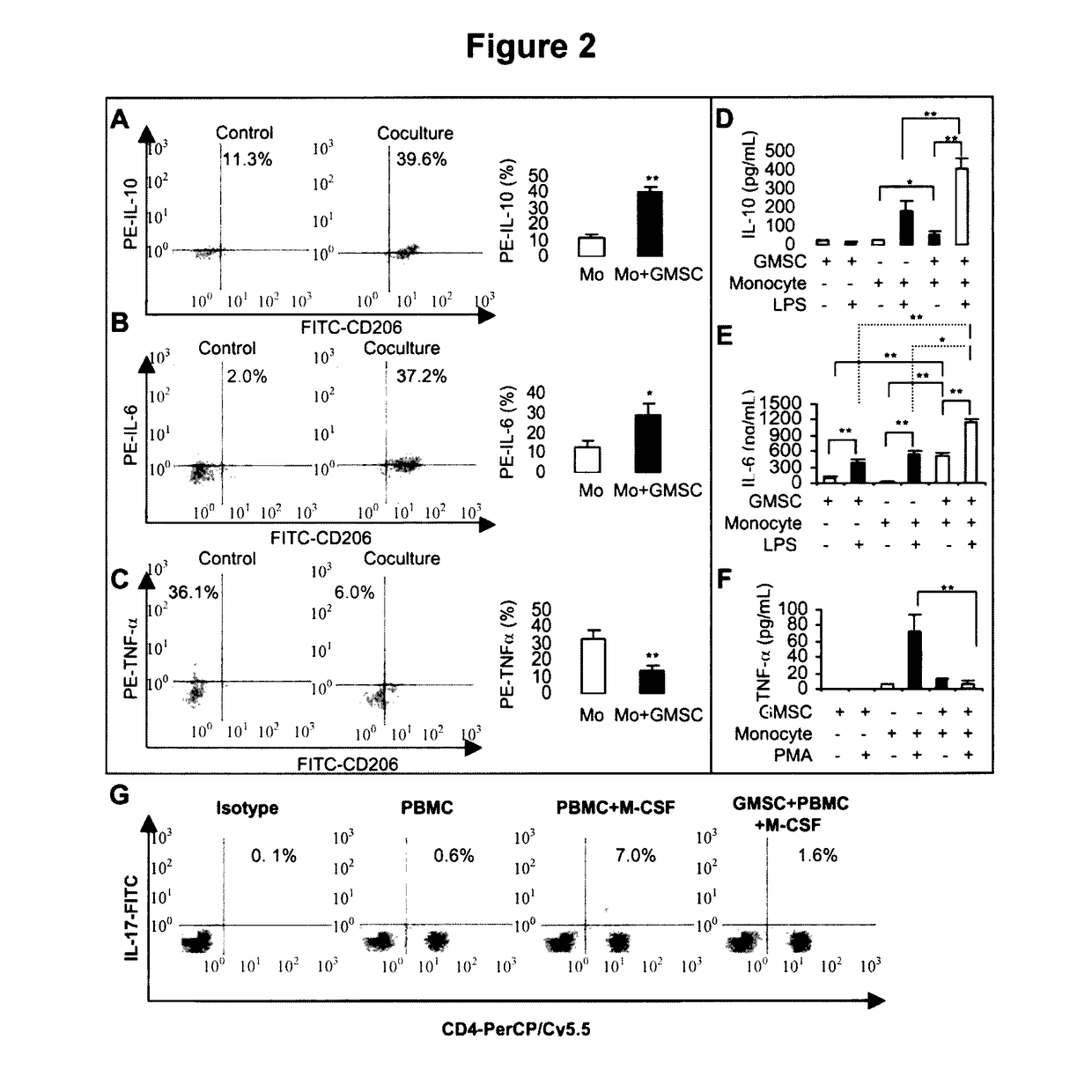
[0059]Next, we determined the cytokine expression profile by macrophages co-cultured with GMSCs. Flow cytometric analysis showed that, in comparison with macrophages cultured alone, co-culture with GMSCs significantly increased the percentage of macrophages expressing IL-10 (FIG. 2A) and IL-6 (FIG. 2B), while decreased TNF-α-positive macrophages (FIG. 2C) following stimulation using different protocols as previously described [20]. The differential cytokine profile of secretory IL-10, IL-6, and TNF-α in the supernatants of macrophages, GMSCs, and their co-cultures, was further confirmed by ELISA, respectively. Minimal release of cytokines was observed in macrophages or GMSCs alone in the absence of stimuli (FIG. 2D, 2F). Addition of stimulating agents triggered a burst of IL-10 and TNF-α secretion by macrophages, but had minimal effect on GMSCs cultured alone (FIG. 2D, 2F). The increased secretion of IL-10 tri...

example 1

Interplay Between GMSCs and Macrophages Regulated the Local Inflammatory Response During Skin Wound Healing

[0064]We next investigated the in vivo effects of GMSCs on inflammatory cell response and production of local inflammatory cytokines in skin wounds. Analysis of skin wounds on day 3, 5, and 7 following GMSC injection indicated that infiltration of inflammatory cells was significantly decreased in GMSC-treated wounds as compared to controls (FIG. 6C). GMSC treatment decreased neutrophil infiltration as represented by a time-dependent decrease in MPO activity in wounded skin at several time points post-wounding (FIG. 6Da). In addition, ELISA analysis showed that GMSC treatment significantly decreased the local levels of both proinflammatory cytokines, TNF-α and IL-6, and increased the anti-inflammatory cytokine IL-10 (FIG. 6Db-d). Without being limited by theory, it is believed that GMSC treatment promotes skin wound healing, at least in part, by suppressing inflammatory cell inf...

example i

Materials and Methods Used in Example I

[0066]Animals.

[0067]C57BL / 6J mice (male, 8-10 week-old) were obtained from Jackson Laboratories (Bar Harbor, Me., http: / / www.jax.org) and group-housed at the Animal Facility of University of Southern California (USC). All animal care and experiments were performed under the institutional protocols approved by the Institutional Animal Care and Use Committee (IACUC) at USC.

[0068]Cytokines and Reagents.

[0069]Recombinant human IL-4, CCL-2 (MCP-1), IL-6 and M-CSF were purchased from PeproTech (Rocky Hill, N.J., http: / / www.peprotech.com). Lipopolysaccharide (LPS) from Escherichia coli 055:B5, phorbol 12-myristate 13-acetate (PMA), Brefeldin A were obtained from Sigma-Aldrich (St. Louis, Mo., http: / / www.sigmaaldrich.com). Antibodies include anti-CD14 allophycocyanin (APC), anti-CD11a fluorescein isothiocyanate (FITC), anti-CD90 peridinin chlorophyll protein (PerCp)-Cy5.5, anti-IL-6-PE, anti-IL-10-PE, anti-TNFα-PE and anti-IL-17-FITC (eBiosciences, San...


PUM


Abstract

Disclosed are methods of polarizing macrophages to exhibit M2 phenotype, including introducing an effective amount of gingiva-derived mesenchymal stem cells to an environment comprising a population of macrophages such that the macrophages are in fluid communication with the gingiva-derived mesenchymal stem cells. Also disclosed are methods of promoting cutaneous wound healing including administering to a patient an effective amount of human gingiva-derived mesenchymal stem cells, thereby resulting in at least one of accelerated wound closure, rapid re-epithelialization, improved angiogenesis and improved tissue remodeling relative to untreated controls. Also disclosed are methods for attenuating contact hypersensitivity in a patient, the methods including administering to a patient an effective amount of human gingiva-derived mesenchymal stem cells at a time at least timeframe selected from the group consisting of before sensitization, after sensitization and before challenge and after challenge, thereby attenuating contact hypersensitivity.

Description

CROSS-REFERENCE TO RELATED APPLICATIONS[0001]This application claims the benefit of U.S. Provisional Application No. 61 / 364,339, filed Jul. 14, 2010, the entire contents of which are incorporated herein by reference in its entirety.STATEMENT REGARDING FEDERALLY SPONSORED RESEARCH OR DEVELOPMENT[0002]This invention was made with government support under Contract Nos. CA82422, R01 DE 019932 awarded by the National Institutes of Health. The government has certain rights in the invention.FIELD OF THE INVENTION[0003]The invention pertains to the field of stem cell therapy. One aspect of the invention pertains to the use of human gingiva-derived mesenchymal stem cells to enhance cutaneous wound healing via their immunomodulatory effects on the innate immune cells, specifically driving the polarization of M2 macrophages.BACKGROUND OF THE INVENTION[0004]Cutaneous wound healing represents a highly coordinated process to achieve tissue homeostasis, which involves complex interactions of diffe...

Claims


Application Information

Patent Timeline
Patent Type & Authority Patents(United States)
IPC IPC(8): A01N63/00A61K35/28
CPCA61K35/28
Inventor LE, ANH D.ZHANG, QUNZHOU
Owner UNIV OF SOUTHERN CALIFORNIA
Learn More