System and method for electrical impedance spectroscopy

a technology system and method, applied in the field of system and method can solve the problems of limited availability of ambulance, brain injury, intracranial hemorrhage, etc., and achieve the effects of improving the accuracy of electrical impedance spectroscopy, sensitively assessing the impedance of deep brain tissue, and reducing the risk of brain injury

Active Publication Date: 2017-11-21
THE GENERAL HOSPITAL CORP
View PDF1 Cites 4 Cited by
  • Summary
  • Abstract
  • Description
  • Claims
  • Application Information

AI Technical Summary

Benefits of technology

[0022]The present invention generally provides systems and methods for portably providing rapid, affordable point-of-care detection, assessment, and monitoring of brain injuries. These systems and methods include an improvement of electrical impedance spectroscopy (EIS) that employs the Stochastic Gabor Function (SGF) as an EIS stimulus current shape, either as a single pulse or dual pulses. For example, an optimized EIS stimulus paradigm based on dual energy pulses using the SGF can more sensitively assess deep brain tissue impedance than other single pulse EIS paradigms. The substantially increased range of depth penetration facilitated by subtraction of dual energy SGF pulses can also result in greater sensitivity and specificity for distinguishing hemorrhagic from non-hemorrhagic brain injury, and greater accuracy for the anatomic localization and size assessment of intracranial lesions. The present systems and methods may be provided as portable and noninvasive devices that may be employed in ambulance, battlefield, and intensive care unit settings.

Problems solved by technology

Intracranial hemorrhage, stroke, and traumatic brain injury are major public health problems.
Computed Tomography (CT) and Magnetic Resonance Imaging (MRI) are currently the first line modalities for the evaluation of acute brain injury, including hemorrhage and stroke, but are limited in their ambulance, battlefield, and intensive care unit (ICU) availability.
Furthermore, with increasing use of improvised explosive devices (IEDs), blast-related concussive injury has become common.
Moreover, even the most advanced current generation MRI pulse sequences, such as diffusion tensor imaging (DTI), magnetization transfer imaging (MT), and MR spectroscopy (MRS), are incapable of detecting the subtle histological changes (for example, micro-edema and cytoarchitectural axonal disruption) associated with acute mild to moderate traumatic brain injury on an individual patient basis.
Even when portable CT scanners are available, they remain a limited resource with undependable technical support and few contingency options for equipment breakdown, cannot be used for continuous monitoring, and cannot be deployed by corpsmen in the field for emergent triage of individuals with—otherwise unapparent—subdural and epidural hematomas.
Monitoring by serial clinical neurological exam is difficult or infeasible in intubated, sedated patients, is nonspecific, difficult to quantify, and has relatively poor reproducibility.
As expected of any clinical scale, NIHSS has significant limitations.
Transportation of critically ill patients is a huge challenge; moreover, radiation exposure is a growing concern.
Both of these modalities require large and expensive machines, even with limited-use “portable” CT.
Moreover, CT scanning exposes patients to ionizing radiation.
MRI scanning generates strong electromagnetic fields, which make it unsuitable for many patients, and MRI is incompatible with the majority of metallic and electronic hardware.
Thus, it is prohibitively expensive and infeasible to continually monitor a patient in an intensive care unit using CT or MRI.
For example, invasive intracranial pressure (ICP) monitors, such as “bolts” or ventricular catheters, detect only the most catastrophic changes after ischemic stroke and do not provide information as to whether brain lesions have undergone hemorrhagic transformation.
Invasive tools are also unappealing because they require invasive craniotomy and, in addition to the obvious surgical risks, carry risks of infection in this era of super-resistant bacteria (such MRSA and VRE).
Even more importantly, they do not provide information regarding spatial localization.
Hence, in most ICUs, ICP monitoring is currently limited to only the highest risk patients.
However, such a method is relatively inaccurate, requires a high level of operator expertise, measures only one vessel location at a time, is logistically difficult to implement, and thus impractical.
However, these methods are limited in both their depth of skull penetration (typically on the order of millimeters) and their extent of lateral coverage (a few square centimeters, depending on the probe array size).
As such, these methods may potentially introduce greater user-dependent variability than other methods.
However, such a method lacks sufficient sensitivity, specificity, speed, and ease of use required for out-of-hospital diagnosis of stroke or TBI.
Moreover, unfortunately, spatial localization and histological characterization of these brain injuries with existing EIS devices is limited by the disproportionate surface—as opposed to deep—current flows inherent to existing prototypes.
For example, with single frequency EIS systems, one can detect shallow hematomas that create low-impedance anomalies near the skull surface, but detection of deep lesions remains challenging.

Method used


Image

  • System and method for electrical impedance spectroscopy
  • System and method for electrical impedance spectroscopy
  • System and method for electrical impedance spectroscopy

Examples


Embodiment Construction

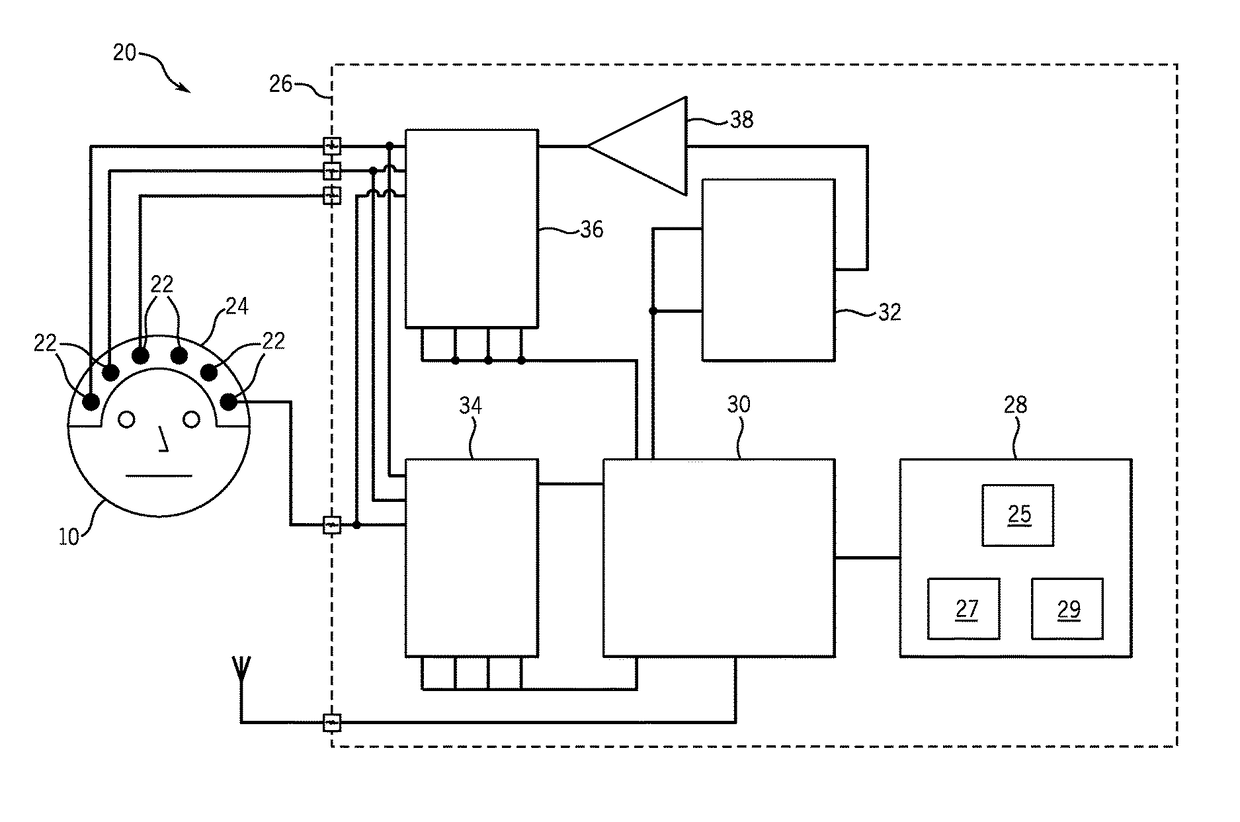
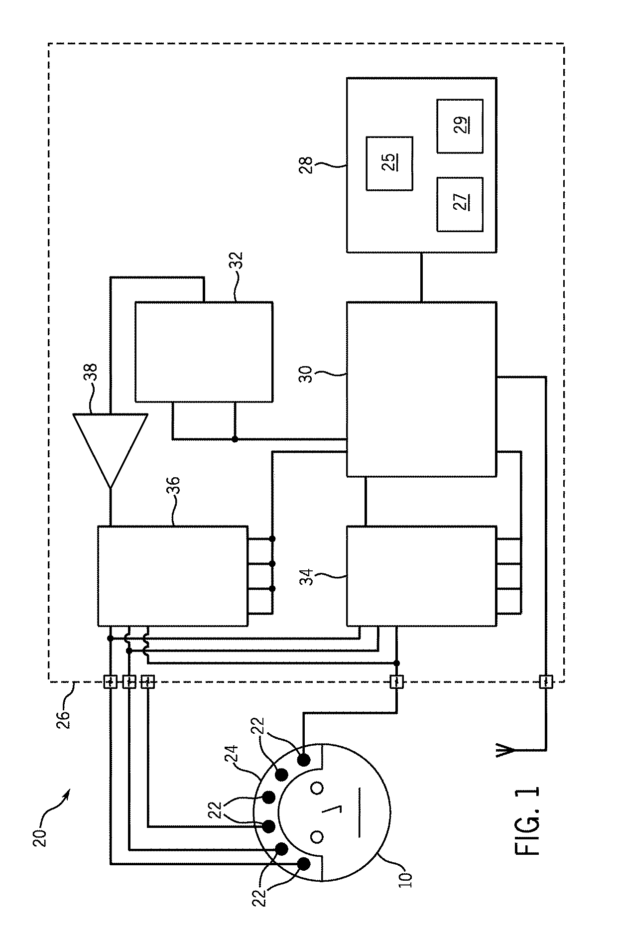
[0043]The present invention provides systems and methods that include an improvement of electrical impedance spectroscopy (EIS) that employs the Stochastic Gabor Function (SGF) as an EIS stimulus current shape. More specifically, the SGF is an excitation waveform that, in the present case, creates excitation pulses during EIS of the brain. The SGF is a uniformly distributed noise, modulated by a Gaussian envelop, with a wide frequency spectrum representation regardless of the stimuli energy, and is least compact in the sample frequency phase plane. The SGF facilitate both shallow and deeper tissue penetration, a capability that not is achieved with conventional stimulus paradigms. In addition, as further described below, an advantage of SGF pulse delivery is the potential for “dual energy” subtraction imaging that can more sensitively assess deep brain tissue impedance than current single pulse paradigms.

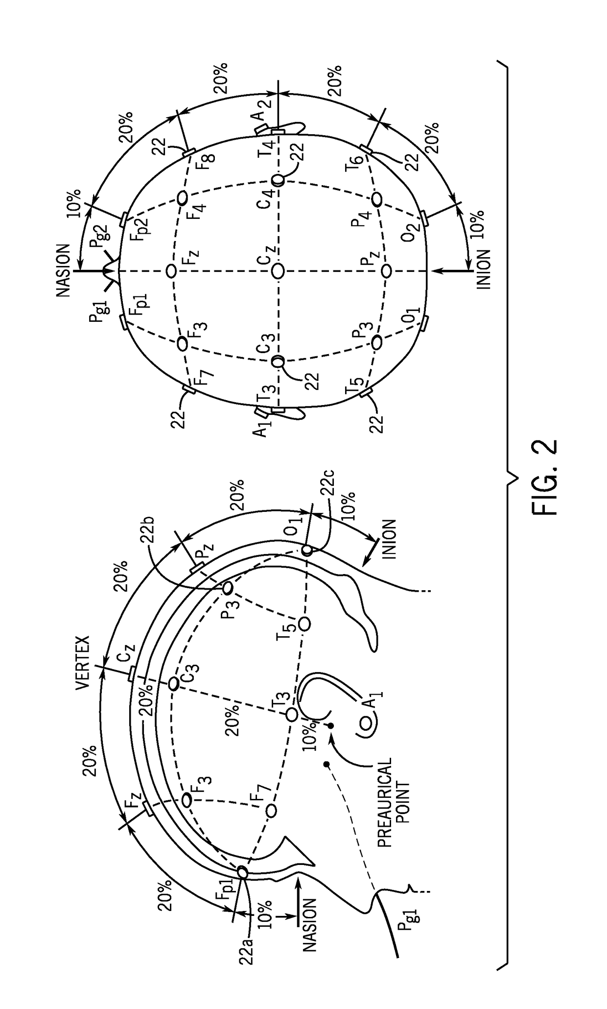
[0044]EIS is maximally sensitive to impedance changes at the electrode-skin int...


PUM


Abstract

Systems and methods for determining brain health of a subject include or employ an electrical stimulator configured to apply a current to at least one pair of electrodes, and the electrodes are positioned on a skull of the subject to apply the current and to receive brain activity of the subject. The electrical stimulator is configured to apply a current having a waveform according to a Stochastic Gabor Function (SGF). A signal processor is configured to record the brain activity of the subject in the form of spectral electrical impedance data, and a computer system having non-transient computer readable media is programmed and configured to process the spectral electrical impedance data and indicate an impedance change within the brain of the subject.

Description

CROSS-REFERENCE TO RELATED APPLICATIONS[0001]This application is a 371 application of PCT / US2013 / 053959 filed Aug. 7, 2013 which claims the benefit of, U.S. Provisional Patent Application Ser. No. 61 / 680,597, filed on Aug. 7, 2012, 61 / 680,947, filed on Aug. 8, 2012, 61 / 693,299, filed on Aug. 26, 2012, and 61 / 782,031, filed on Mar. 14, 2013, all of which are incorporated herein by reference in their entirety and for all purposes.STATEMENT REGARDING FEDERALLY SPONSORED RESEARCH[0002]This invention was made with government support under 5U54EB007954-04, sub-contract 005213, awarded by the National Institute of Biomedical Imaging and Bioengineering of the National Institutes of Health. The government has certain rights in the invention.BACKGROUND OF THE INVENTION[0003]The present disclosure generally relates to systems and methods employing electrical impedance spectroscopy (EIS) for detecting, characterizing, and monitoring brain injuries and, more particularly, to systems and methods ...

Claims


Application Information

Patent Timeline
Patent Type & Authority Patents(United States)
IPC IPC(8): A61B5/04A61B5/00A61B5/053A61B5/0478
CPCA61B5/0478A61B5/04012A61B5/053A61B5/7217A61B5/6814A61B2562/04A61B2562/0214A61B5/291A61B5/316
Inventor BONMASSAR, GIORGIOROSENTHAL, ERIC S.LEV, MICHAEL H.
Owner THE GENERAL HOSPITAL CORP
Why Eureka
Try Eureka