Cartilage repair, preservation and growth by stimulation of bone-chondral interphase and delivery system and methods therefor

a cartilage and interphase technology, applied in the field of cartilage repair, preservation and growth by stimulation of bone-chondral interphase and delivery system and methods therefor, can solve the problems of increased risk of hypertension, increased risk of coronary artery disease, kidney failure, etc., and achieves the effects of preserving cartilage, reducing table time, and repairing cartilag

Active Publication Date: 2017-11-28
VAD SCI
View PDF9 Cites 1 Cited by
  • Summary
  • Abstract
  • Description
  • Claims
  • Application Information

AI Technical Summary

Benefits of technology

[0069]Novel therapeutics and methods of treatment to repair, preserve and grow cartilage are presented. In addition, systems and methods for delivering a therapeutic to a hard to reach anatomical area, such as, for example, the BCI, are presented. In exemplary embodiments of the present invention, a cannulated delivery device provided with a cutting tip and threads on its distal exterior can be provided which has considerable advantages over conventional devices. These include, for example, (i) ease of manufacture, (ii) use of the exemplary device being faster than conventional approaches, with both less table time and less steps, (iii) lesser exposure of internal tissues to ambient air, and thus less risk of infection, and (iv) lesser technical complexity leading to lesser complications. Using such an exemplary device, various novel therapies for joint and cartilage repair, preservation and generation can be implemented. Various versions of such a device are disclosed. Alternatively, for disc repair, a delivery device directed to percutaneous intradiscal annular repair, or “PIARES” device can be used to introduce therapeutics intradiscally. The device is a two-needle device, with a first cannula / needle, with a finger grip at its distal end, and a longer inner needle, which can then penetrate through the outer needle into the disc, and can then, for example, be used to introduce therapeutics, for example, via a syringe. When provided with a septum at the inner needle's proximal end, the PIARES device becomes a completely closed system, and its use minimizes trauma. Thus, in exemplary embodiments of the present invention, a surgical hand tool can be provided, used for the non-invasive placement and delivery of therapeutics, to a targeted site. This can be done through minimally invasive skin incision, or without any incision, as maybe desired. The delivery and placement of the therapeutic can be controlled and does not need a powered drill or guide wire.
[0070]An exemplary device can have a closed pointed end, a threaded portion, and be provided with thread cutting / forming features, such as flute(s), and can have a shaft perforations to the central lumen at a distal end to deliver therapeutics or other preparations. At the proximal end, means can be provided to attach a syringe in communication with the shaft's central lumen, and there may be a keyed engagement feature for attachment of a hand grip. The delivery device can be made of sufficient length to reach bone on either side of a desired or targeted joint, and to easily penetrate soft tissue and cortical bone to reach a targeted site in cancellous bone adjacent to a cartilage defect.

Problems solved by technology

Although these medications decrease inflammation and pain, their prolonged use (i) is thought to have an adverse impact on cartilage and (ii) comes with complications of increased risk of hypertension, coronary artery disease, renal failure (especially in diabetics) and peptic ulcer disease.
However, it has limited success rates in treating knee OA.
Thus, while some studies show good success rates, others show rather poor ones.
Furthermore, success rates decrease substantially in those patients with moderate to severe knee OA.
This technique has seen limited success rates.
However, if done excessively, microfracture can sometimes even accelerate the rate of cartilage loss.
These procedures have significant complication rates of blood clots and infections, are expensive, require hospital stays, have the associated liability of inserting metal in the body, and come with markedly increased healthcare costs.
Each time internal tissues are exposed in this way the risk of infection increases.
Furthermore, this is technically more challenging and time consuming with increased risk of complications.

Method used


Image

  • Cartilage repair, preservation and growth by stimulation of bone-chondral interphase and delivery system and methods therefor
  • Cartilage repair, preservation and growth by stimulation of bone-chondral interphase and delivery system and methods therefor
  • Cartilage repair, preservation and growth by stimulation of bone-chondral interphase and delivery system and methods therefor

Examples


example a

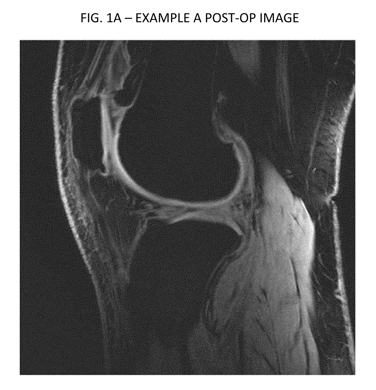
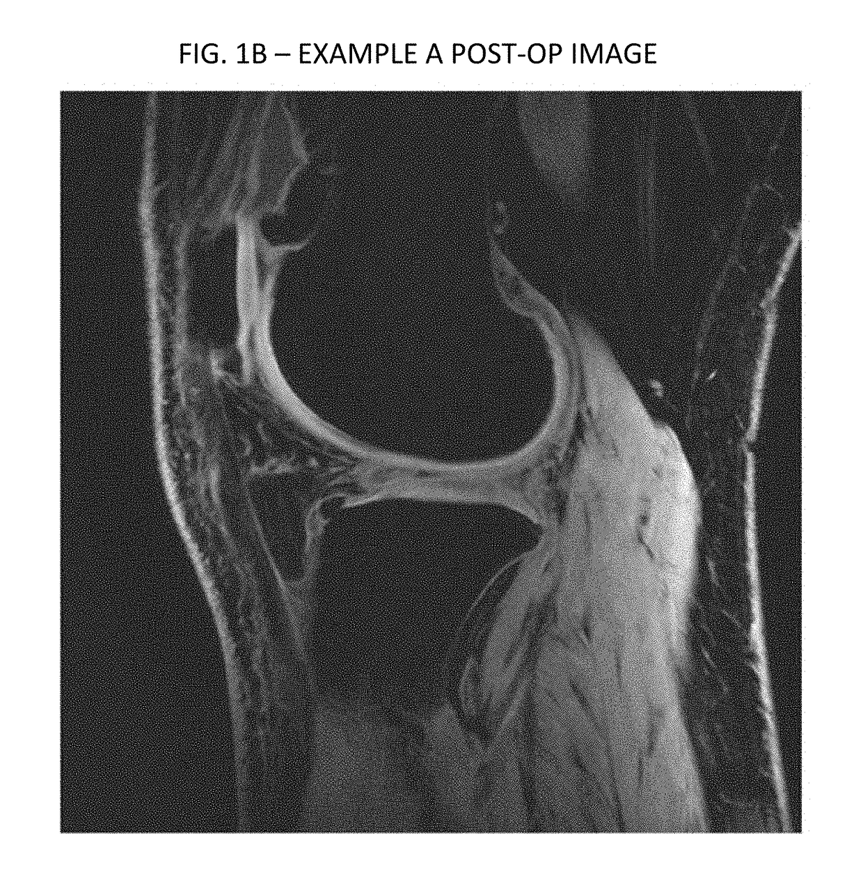
[0109]In an experimental trial of treatment methods according to an exemplary embodiment of the present invention, a 50 year old female with advanced degenerative osteoarthritic disease of the knee was treated. Prior to the treatment, a pre-operative MRI was done. Post treatment a three month follow-up MRI was performed on the patient and read by an independent radiologist. Provided hereto as Appendix A is the independent radiologist's report. As noted in the report, the post operative MRI showed an increase in cartilage matrix from 1.8 mm pre-treatment to 3.7 mm at the follow-up MRI. Thus, the inventive technique shows early promise for cartilage repair and possible regeneration.

example b

[0110]Imaging data from another experimental case are provided in FIGS. 1-4. As can be seen therein, FIGS. 1 and 2 are respectively axial and sagittal images of a patient's knee from a preoperative scan. FIG. 1 depicts an area of decreased blood flow due to an ischemia, as shown by the red arrow, also seen in FIG. 2 pointed to by the red and green arrows. Injections similarly performed according to the above described protocol, to the femoral and tibial compartments by drilling and injecting at the bone-cartilage interface, followed by injections into the knee joint. FIGS. 3 and 4 are corresponding axial and sagittal images form a MRI taken three months following the treatment. As can be seen, significant new cartilage has grown, and the ischemia has been essentially resolved, as shown in FIG. 3.

example c

[0111]FIG. 5 depicts side by side comparisons of sagittal images of a knee of a third patient. The left panel is an image form a preoperative scan, and the right panel a corresponding image from a post operative MRI. As shown in FIG. 5, the post operative MRI showed an increase in cartilage matrix from 1.60 mm to 1.87 mm at the follow-up MRI.

[0112]In exemplary embodiments of the present invention, the therapeutic protocol described above can similarly be used for osteoarthritis and avascular necrosis, as well as for treating meniscal and labral injuries in the joints.

[0113]Exemplary Variations of the Protocol

[0114]In exemplary embodiments of the present invention, variations on the above-described protocols can be used for other anatomical areas. Examples of these are next described.

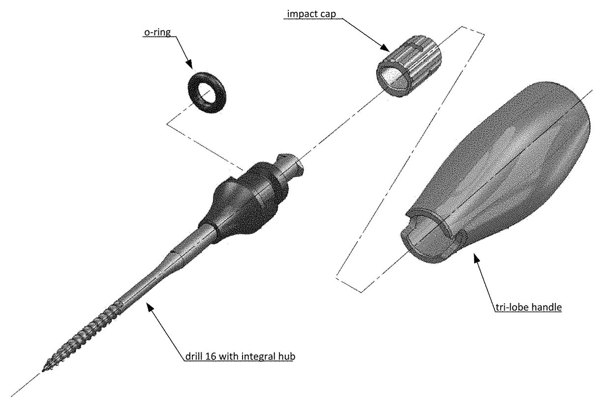
[0115]Joints—for joints, the step of injecting GCSF for stimulating bone marrow may be skipped. The drilling / twisting alone of the delivery device (as described below) will stimulate bone marrow combined...


PUM


Abstract

Therapeutics and methods of treatment to repair, preserve and grow cartilage are presented. In addition, systems and methods for delivering a therapeutic to a hard to reach anatomical area, such as, for example, the BCI, are presented. A cannulated delivery device provided with a cutting tip, cutting flutes and threads on its distal end is disclosed, as well as therapies for joint and cartilage repair, preservation and generation using it. Alternatively, for disc repair, a “PIARES” device for Percutaneous Intradiscal Annular Repair introduces therapeutics intradiscally. The device may have two-needles; a first cannula / needle with a finger grip, and a longer inner needle to penetrate through the outer needle into the disc, and introduce therapeutics via a syringe. When provided with a septum at the inner needle's proximal end, the PIARES device is a completely closed system; using it minimizes trauma.

Description

CROSS-REFERENCE TO RELATED APPLICATIONS[0001]The present application claims priority to U.S. Provisional Patent Applications No. 61 / 686,835, filed on Apr. 11, 2012, and 61 / 800,574, filed on Mar. 15, 2013, each entitled “CARTILAGE REPAIR, PRESERVATION AND GROWTH BY STIMULATION OF BONE-CHONDRAL INTERPHASE AND DELIVERY SYSTEM AND METHODS THEREFOR”, each of which is hereby incorporated herein by reference.TECHNICAL FIELD[0002]The present invention relates to various novel treatments for degenerative joints and discs, and improved devices and therapies for the delivery of therapeutic agents to hard to reach anatomical areas with minimal trauma so as to better implement such novel treatments.BACKGROUND OF THE INVENTION[0003]Conventional Therapies for Degenerative Disc and Other Cartilage Disease[0004]Considering knee degeneration or osteoarthritis (“OA”) as an example, pain in knee OA, defined as loss of articular cartilage in the knee, is thought to be caused by increased pressure on the...

Claims


Application Information

Patent Timeline
Patent Type & Authority Patents(United States)
IPC IPC(8): A61B17/34A61B17/16A61B17/56A61M5/158
CPCA61B17/3472A61M5/158A61M37/00
Inventor VAD, VIJAYBARVE, RAGHAVMULHAUSER, PAULKIRK, III, KARL D.
Owner VAD SCI
Why Eureka
Try Eureka