Preventive and therapeutic drug for cartilaginous hyperplasia and method of screening for the same

a cartilaginous hyperplasia and therapeutic drug technology, applied in the direction of drug composition, post-translational modification detection, instruments, etc., can solve the problems of insufficient effectiveness and no reports of successful in vitro reproduction of the pathological condition of nomid

Active Publication Date: 2018-03-20
KYOTO UNIV
View PDF3 Cites 1 Cited by
  • Summary
  • Abstract
  • Description
  • Claims
  • Application Information

AI Technical Summary

Problems solved by technology

Although such therapy is effective in suppressing systemic inflammation, it is not sufficiently effective against pathological conditions such as epiphyseal hyperplasia of long bones (Non-Patent Document 7).
However, there have been no reports on successful in vitro reproduction of the pathological conditions of NOMID.

Method used


Image

  • Preventive and therapeutic drug for cartilaginous hyperplasia and method of screening for the same
  • Preventive and therapeutic drug for cartilaginous hyperplasia and method of screening for the same
  • Preventive and therapeutic drug for cartilaginous hyperplasia and method of screening for the same

Examples


example 1

[0120]All of the experiments described below were conducted with the approval of the ethics committee of Kyoto University. In addition, sampling from patients was conducted with the preliminary acquisition of informed consent in accordance with the Declaration of Helsinki. iPS cells were produced from two NOMID patients having NLRP3 somatic mosaics (p.Tyr570Cys and p.Gly307Ser). At least three clones of wild-type NLRP3 iPS cells (hereinafter referred to as “wild-type iPS cells”) and mutant-type NLRP3 iPS cells (hereinafter referred to as “mutant-type iPS cells”) were established from each patient and used for the experiments. Every experiment described below was conducted by comparing wild-type cells and mutant-type cells having isogenic backgrounds.

[0121]iPS cells were produced by the following method. Specifically, each of the obtained human dermal fibroblasts (HDFs) was cultured in DMEM (Nacalai Tesque) medium supplemented with 10% FBS (Invitrogen), 0.5% penicillin, and streptomy...

example 2

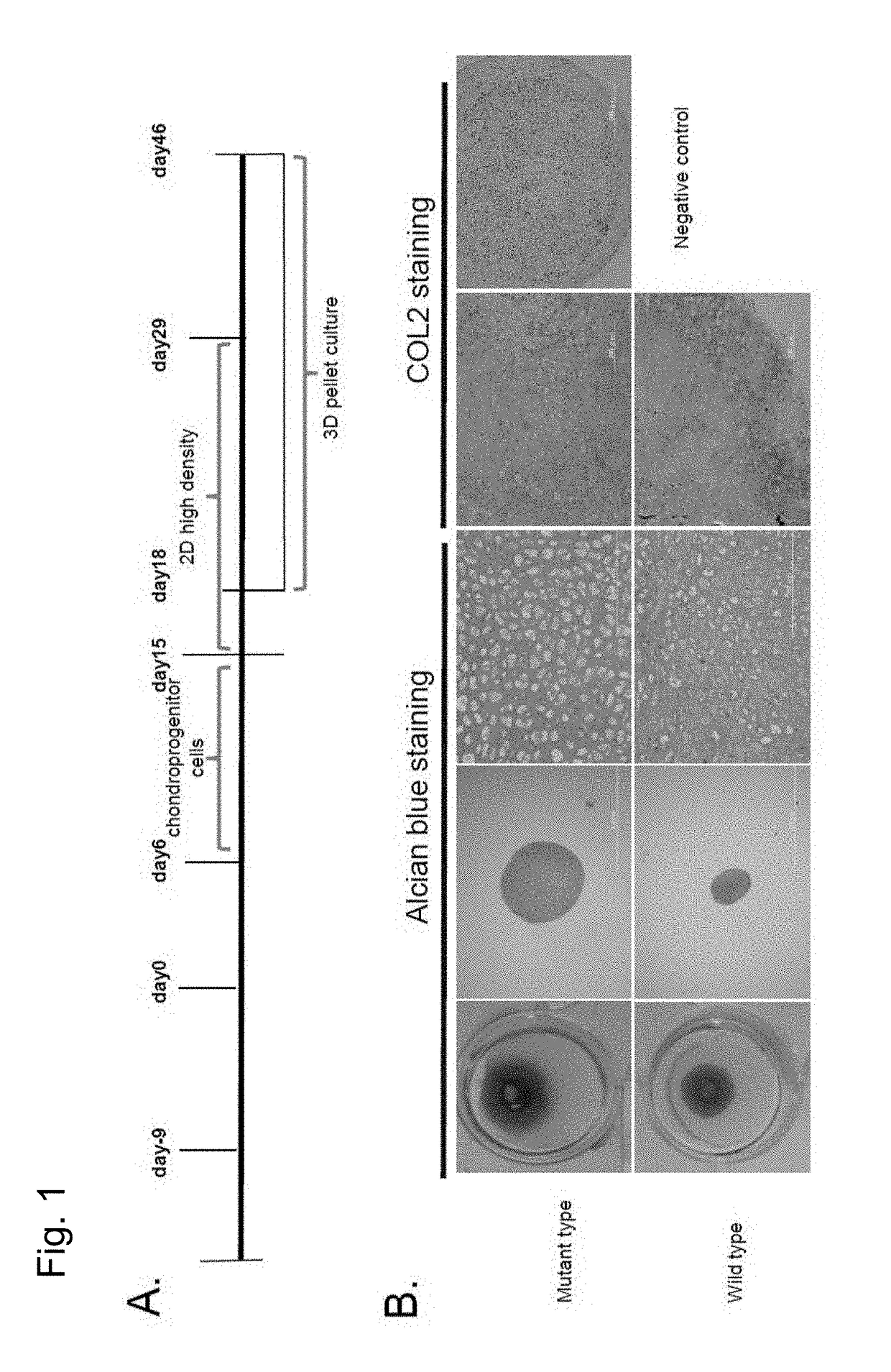
1) Neural Crest Cell Induction (Days 0-8)

[0122]The iPS cells obtained in Example 1 were induced to differentiate into neural crest cells via feeder-free culture. First, the feeder cells were removed using CTK (0.25% Trypsin (Life Technologies), 0.1 mg / ml collagenase IV (Life Technologies), 1 mM CaCl, 20% (v / v) KSR), followed by washing with PBS. Then, the iPS cells were recovered using a scraper, suspended in mTeSR medium (mTeSR Basal Medium (400 ml), 5× Supplement (100 ml), Penicillin / Streptomycin (2.5 ml)) (STEMCELL Technology), and pipetted once. Subsequently, a Matrigel-coated dish (prepared by applying a Matrigel stock solution (×50) (BD Biosciences) diluted with DMEM / F12 medium (Life Technologies) to a dish) was stored overnight at 4° C. Matrigel was removed 36 to 60 minutes before the use of the cells. The cells were dried and seeded (5×10⁴ / 10-cm dish) on mTeSR medium (10 ml) and cultured at 37° C. in 5% CO2. Two days later, the medium was replaced with neural crest induction...

example 3

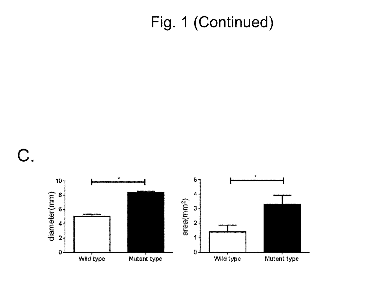
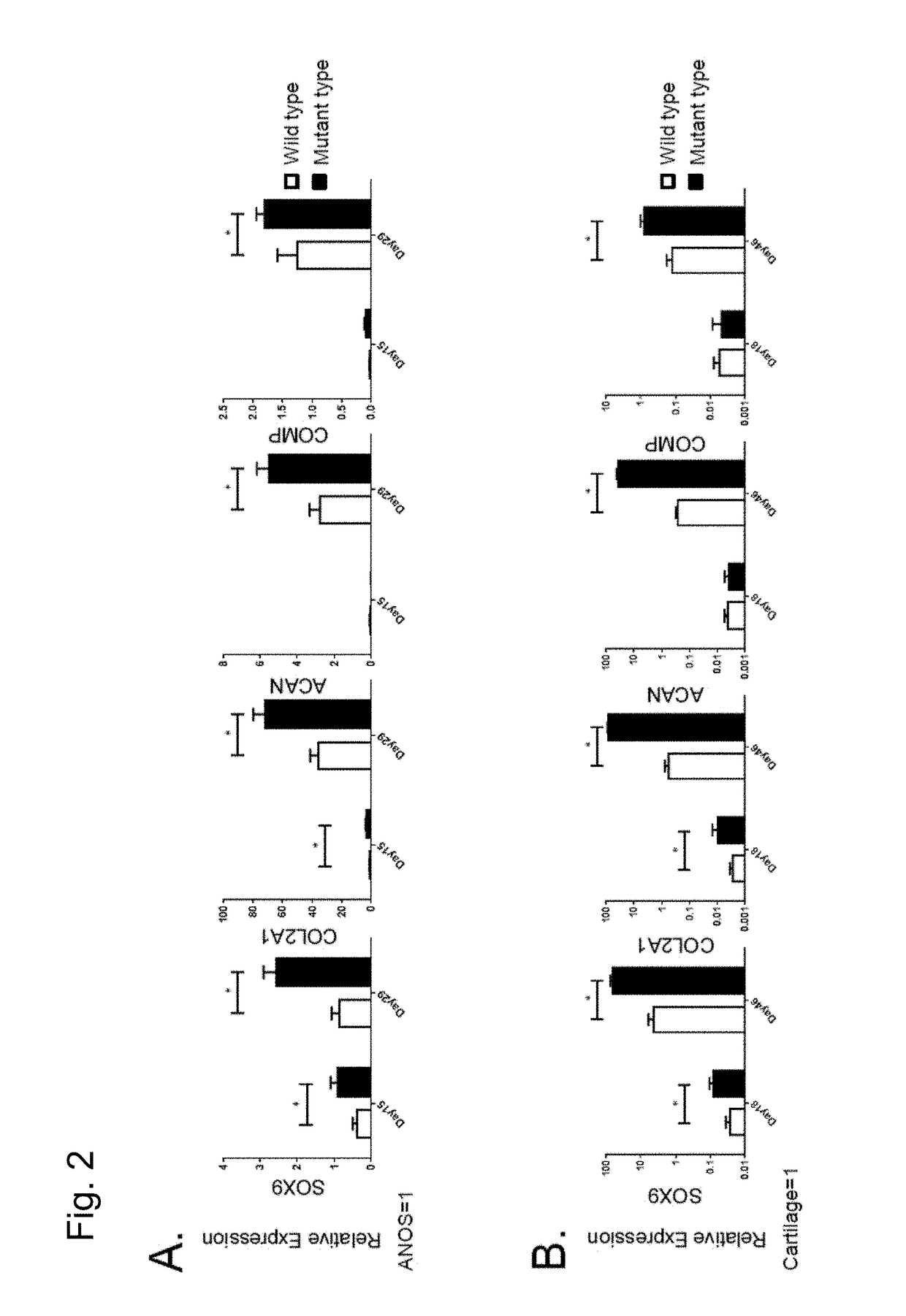
[0129]In order to further examine the details of the state of chondrocytes obtained through differentiation induction by the methods (2D micromass culture and 3D pellet culture) used in Example 2, chondroprogenitor cells before induction of chondrocytes and induced chondrocytes were examined to analyze the expression state of genes specifically expressed in chondrocytes (chondrocyte-specific genes). Gene expression analysis was conducted using the following method.

[0130]mRNA was isolated from each cell using an RNeasy Mini Kit (Qiagen). cDNA was synthesized via reverse transcription using 1 μg of the obtained total RNA as a template and a Superscript III reverse transcriptase (Invitrogen). A standard curve for quantitative real-time PCR was created to conduct analysis. Real-time PCR analysis was conducted using a Power SYBR Green qPCR mastermix (Invitrogen) and a StepOne real-time PCR system (ABI) for quantitative determination. Table 1 lists the primer sequences and assay IDs.

[0131...


PUM

PropertyMeasurementUnit
concentrationaaaaaaaaaa

Abstract

A method of screening for a therapeutic and / or preventive drug for cartilaginous hyperplasia and a therapeutic and / or preventive drug for cartilaginous hyperplasia are provided.The following are provided: a method of screening for a therapeutic and / or preventive drug for cartilaginous hyperplasia, comprising a step of culturing chondroprogenitor cells under conditions in which the cells are brought into contact with a test substance and conditions in which the cells are not brought into contact with the test substance and a step of determining the SOX9 promoter activity, cAMP level, or degree of phosphorylation of CREB in the cells or the extracellular matrix volume in a culture; and a therapeutic and / or preventive drug for cartilaginous hyperplasia, comprising as an active ingredient an adenylate cyclase inhibitor.

Description

CROSS-REFERENCE TO RELATED APPLICATIONS[0001]This application claims priority from Japanese application JP 2014-227500, filed Nov. 7, 2014.[0002]The instant application contains a Sequence Listing which has been submitted in ASCII format via EFS-WEB and is hereby incorporated by reference in its entirety. Said ASCII copy, created on Dec. 22, 2015, is named sequence.txt and is 5 KB.TECHNICAL FIELD[0003]The present invention relates to a method of screening for a preventive and / or therapeutic drug for cartilaginous hyperplasia. The present invention also relates to a therapeutic drug for cartilaginous hyperplasia.BACKGROUND ART[0004]Systemic autoimmune disorder is a disease that is classified as a form of primary immunodeficient syndrome, which is caused by a deficiency in the innate immune system (and especially a deficiency involving pattern-recognition receptors) and is characterized by exhibiting an uncontrollable immune response. Neonatal onset multisystem inflammatory disease (N...

Claims


Application Information

Patent Timeline
Patent Type & Authority Patents(United States)
IPC IPC(8): G01N33/50C07D473/34
CPCG01N33/5041C07D473/34G01N33/5038G01N33/5044G01N2500/10G01N2400/00G01N2400/40G01N2440/14G01N2333/4703A61P19/02A61P19/08A61P43/00
Inventor TOGUCHIDA, JUNYANISHIKOMORI, RYUTAYOKOYAMA, KOJIIKEYA, MAKOTOHEIKE, TOSHIO
Owner KYOTO UNIV
Why Eureka
Try Eureka