
Mouse immune state monitoring reagent and its preparing process

A reagent and state technology, applied in biological testing, material inspection products, instruments, etc., can solve the problems of affecting the judgment of results, low sensitivity, damage, etc., and achieve comprehensive and reliable results

Inactive Publication Date: 2008-03-26
ZHUJIANG HOSPITAL SOUTHERN MEDICAL UNIV
View PDF0 Cites 4 Cited by
  • Summary
  • Abstract
  • Description
  • Claims
  • Application Information

AI Technical Summary

Problems solved by technology

[0009] 6. In the induction of immune tolerance, chimera status or gene therapy may become an indicator of immune detection in the future
51Cr release test is still the most commonly used standard test method for detection of specific T lymphocyte toxicity. The results of this method are accurate and reproducible; but there are also the following shortcomings: ① The use of radioactive 51Cr has the risk of r-ray radiation, which is not conducive to safe operation and waste disposal, and special measuring instruments are required; ②The spontaneous release rate of 51Cr is high, and some cell labeling problems and low sensitivity often affect the result judgment due to the large difference in the labeling efficiency of different target cells; ③The half-life of 51Cr (27.8 days), cannot be used Suitable for animal experiments that require multiple determinations; ④The co-incubation time of cells is short and the test operation steps are many, so it cannot be measured at the level of a single cell
Moreover, the 51Cr release test is an in vitro test. The test itself only measures the activity of CTL and NK cells, which can only reflect the status of cellular immunity to a certain extent, and cannot fully reflect the complex immune environment in the body.
[0014] 2. Using the ELISA method to use the cytokine concentration in the recipient serum as a monitoring index, and the effectiveness of using the RT-PCR method to detect the cytokine mRNA in the graft biopsy specimens have been recognized by some scholars, but cytokines are a Complex network, single cytokine cannot comprehensively reflect immune status
But graft biopsy pathological examination also has its limitation: one. Biopsy tissue detection is a kind of destructive examination method, has certain influence to the normal survival of graft, can not take sample repeatedly, in addition, takes too few samples and will Affect the correctness of pathological slice reading
Second, biopsy pathological examination is a non-quantitative index, which affects the accuracy of clinical diagnosis. Pathological examination is easily affected by the subjective factors and technical level of the reader, resulting in bias and lack of objectivity
3. Pathological biopsy cannot judge the function of infiltrating lymphocytes, which is not conducive to the early diagnosis of rejection. In the early stage of rejection or recipients with graft failure, even clinically stable recipients , pathological biopsy sections can see a large number of T lymphocyte infiltration, or only dotted T cell infiltration

Method used

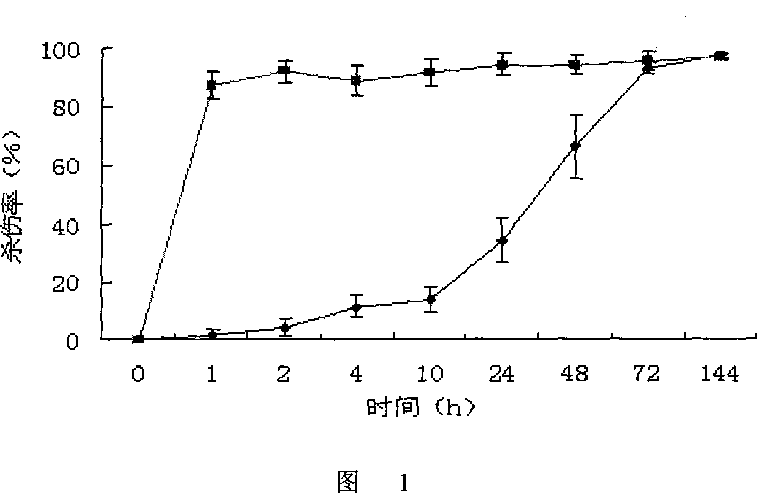
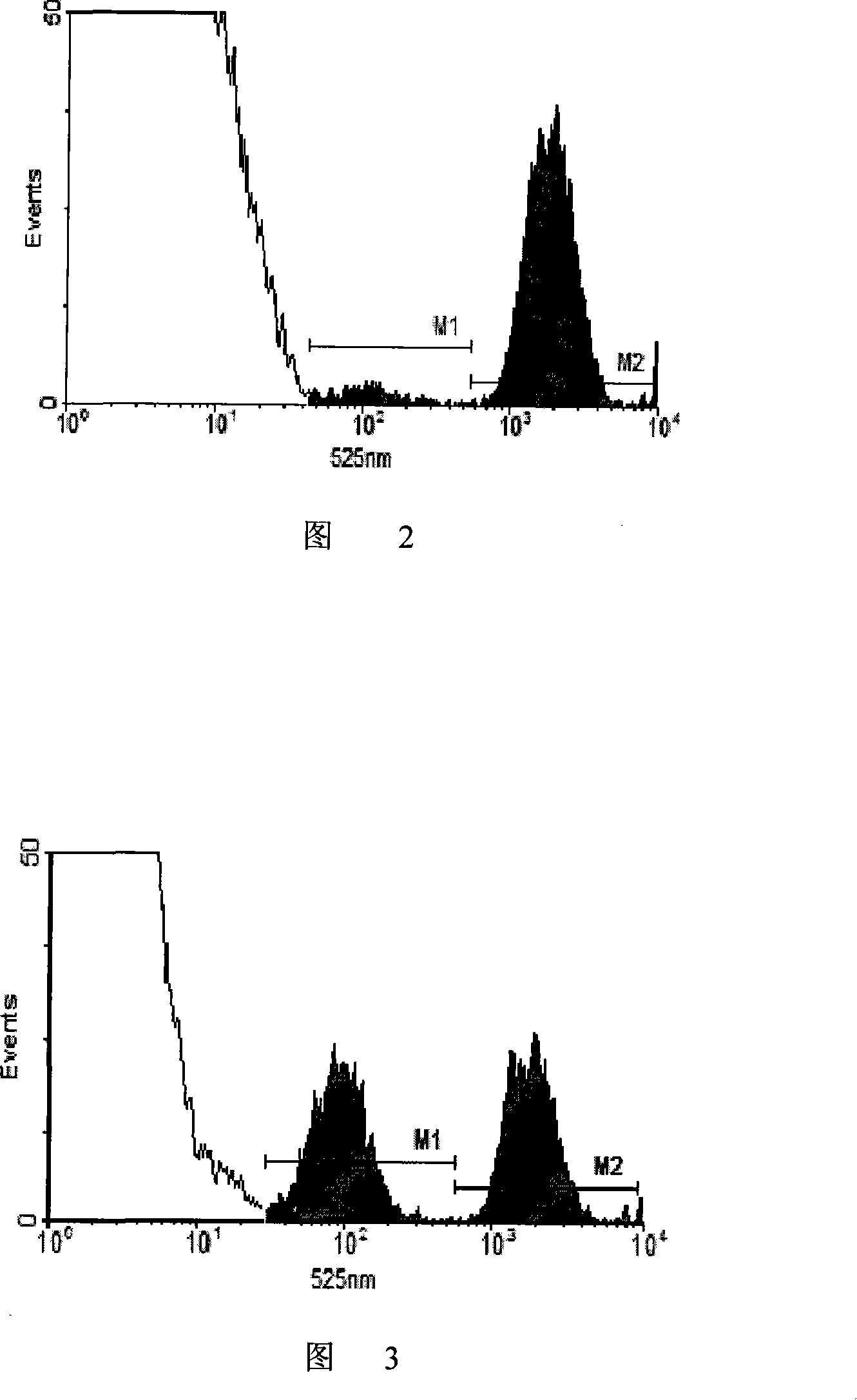
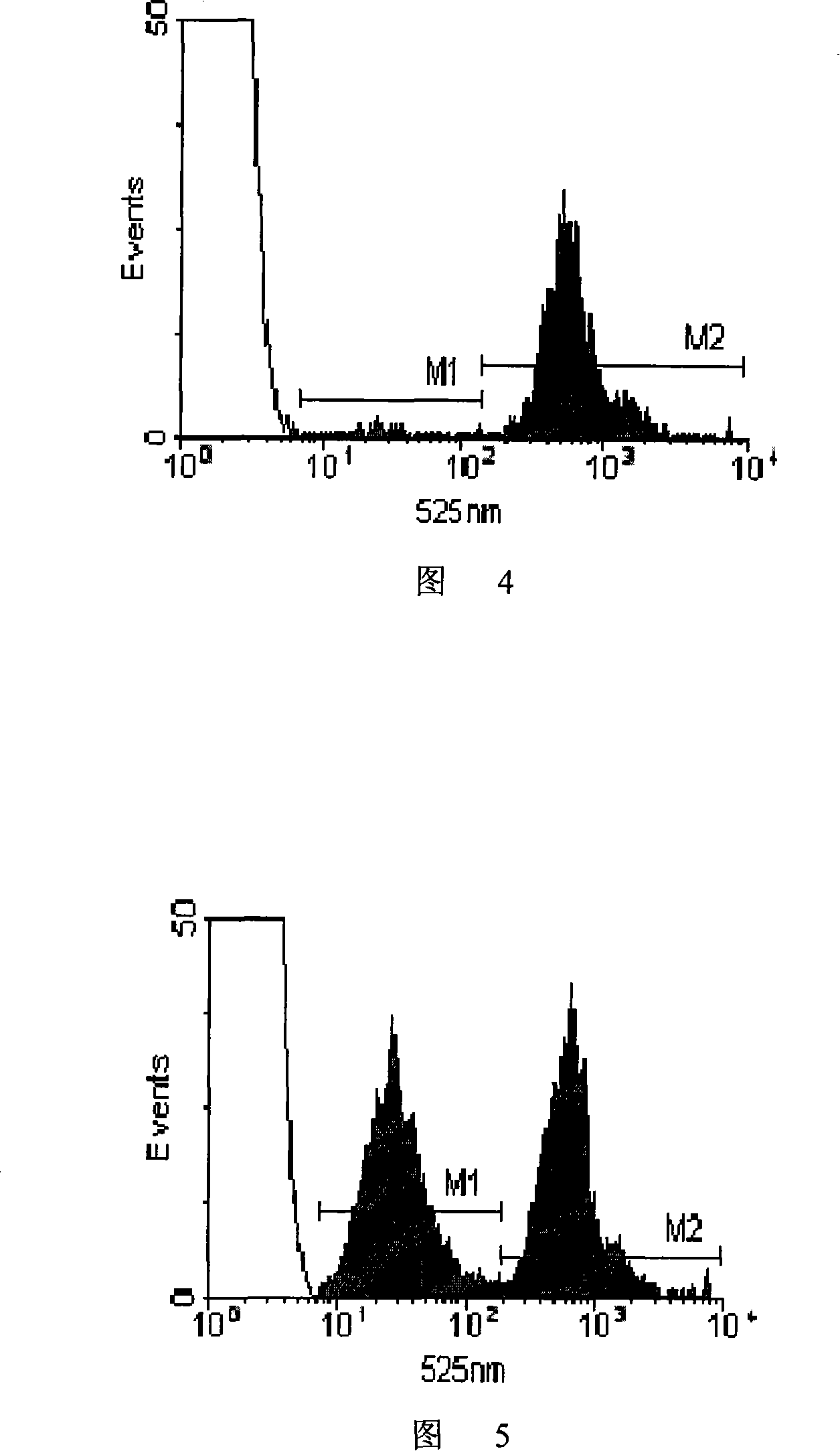

Image

  • Mouse immune state monitoring reagent and its preparing process
  • Mouse immune state monitoring reagent and its preparing process
  • Mouse immune state monitoring reagent and its preparing process

Examples


example 1

[0138] 1) Take splenocytes from C57 mice and BALB / c mice, suspend them in PBS and adjust the cell concentration to 107 cells / mL;

[0139] 2) Dilute the dimethyl sulfoxide solution of CFSE with a concentration of 10 mM with 50 times of PBS to obtain a 200 μM CFSE working solution;

[0140] 3) Add CFSE working solution to the splenocyte suspension of C57 mice for staining so that the final concentration of CFSE is 0.3 μM, add CFSE working solution to the splenocyte suspension of BALB / c mice to make the final concentration of CFSE 6 μM, and then At 37°C, incubate for 15 minutes;

[0141] 4) The stained cells were washed twice with 0.01M 4°C PBS and then resuspended in PBS;

[0142] 5) Mix the two kinds of fluorescently stained cells according to the ratio of cell number 1:1, and adjust the total cell concentration to 5×10⁷ cells / mL.

[0143] Cell viability test: After diluting an appropriate amount of cell suspension, take a drop of the diluted solution and a drop of 0.5% trypa...

example 2

[0147] 1) Take splenocytes from C57 mice and BALB / c mice, suspend them in PBS and adjust the cell concentration to 107 cells / mL;

[0148] 2) Dilute the dimethyl sulfoxide solution of CFSE with a concentration of 10 mM with 50 times of PBS to obtain a 200 μM CFSE working solution;

[0149] 3) Add CFSE working solution to the splenocyte suspension of C57 mice for staining so that the final concentration of CFSE is 0.3 μM, add CFSE working solution to the splenocyte suspension of BALB / c mice to make the final concentration of CFSE 6 μM, and then At 37°C, incubate for 17 minutes;

[0150] 4) The stained cells were washed 3 times with 0.01M 4°C PBS and then resuspended in PBS;

[0151] 5) Mix the two kinds of fluorescently stained cells according to the ratio of cell number 1:1, and adjust the total cell concentration to 8×10⁷ cells / mL.

example 3

[0153] 1) Take splenocytes from C57 mice and BALB / c mice, suspend them in PBS and adjust the cell concentration to 107 cells / mL;

[0154] 2) Dilute the dimethyl sulfoxide solution of CFSE with a concentration of 10 mM with 50 times of PBS to obtain a 200 μM CFSE working solution;

[0155] 3) Add CFSE working solution to the splenocyte suspension of C57 mice for staining so that the final concentration of CFSE is 0.3 μM, add CFSE working solution to the splenocyte suspension of BALB / c mice to make the final concentration of CFSE 6 μM, and then At 37°C, incubate for 20 minutes;

[0156] 4) The stained cells were washed 3 times with 0.01M 4°C PBS and then resuspended in PBS;

[0157] 5) Mix the two kinds of fluorescently stained cells according to the ratio of cell number 1:1, and adjust the total cell concentration to 1×10⁸ cells / mL.


PUM


Abstract

This invention provides a kind of mouse immunity state test reagent, this reagent is PBS suspension of mouse spleen cell in two inbreeding system which is stained by CFSE, the total concentration of the two kind of mouse spleen cell is 0.5-1 multiply 10 to the power 8 per ml, the proportion is 1:1, fluorescence tinction differential concentration is 1:20. Mainline the reagent in the model mouse; it can judge the immunity state of the model mouse by the change of the proportion of the two cell in this reagent when letting blood to test.

Description

technical field [0001] The present invention relates to medical preparations, in particular to medical preparations derived from animal materials. Background technique [0002] Transplantation rejection is the loss of graft function or damage to the recipient's body caused by the immune response induced by transplantation antigens. It is the main reason for clinical transplantation failure and the key problem that transplantation immunology is trying to overcome. In allografts, even if the surgery is successful, the graft will not survive long-term due to rejection. Therefore, transplant rejection has always been the focus of research in the medical field. In the research process of transplant rejection, the detection of the recipient's immune status is an essential link. However, there is still no reliable reagent / method to detect the immune status of the receptor. The existing detection of the immune status of the receptor mainly reflects the immune status indirectly by d...

Claims


Application Information

Patent Timeline
IPC IPC(8): G01N33/49G01N15/00
Inventor 孙尔维蒋泽生高毅潘明新王海澜袁小澎马聪
Owner ZHUJIANG HOSPITAL SOUTHERN MEDICAL UNIV
Learn More