Hydroxyapatite micrometre tube and method of producing the same

A technology of hydroxyapatite and microtubes, applied in chemical instruments and methods, phosphorus compounds, medical science, etc., to achieve the effects of convenient operation, cost reduction, and simple preparation process

Inactive Publication Date: 2008-05-07
SHANGHAI INST OF CERAMIC CHEM & TECH CHINESE ACAD OF SCI
View PDF0 Cites 19 Cited by
  • Summary
  • Abstract
  • Description
  • Claims
  • Application Information

AI Technical Summary

Problems solved by technology

However, the morphology of the obtained hydroxyapatite is generally needle-like and rod-like, and there is no report on the preparation of tubular hydroxyapatite by wet chemical method.

Method used


Image

  • Hydroxyapatite micrometre tube and method of producing the same
  • Hydroxyapatite micrometre tube and method of producing the same
  • Hydroxyapatite micrometre tube and method of producing the same

Examples


Embodiment 1

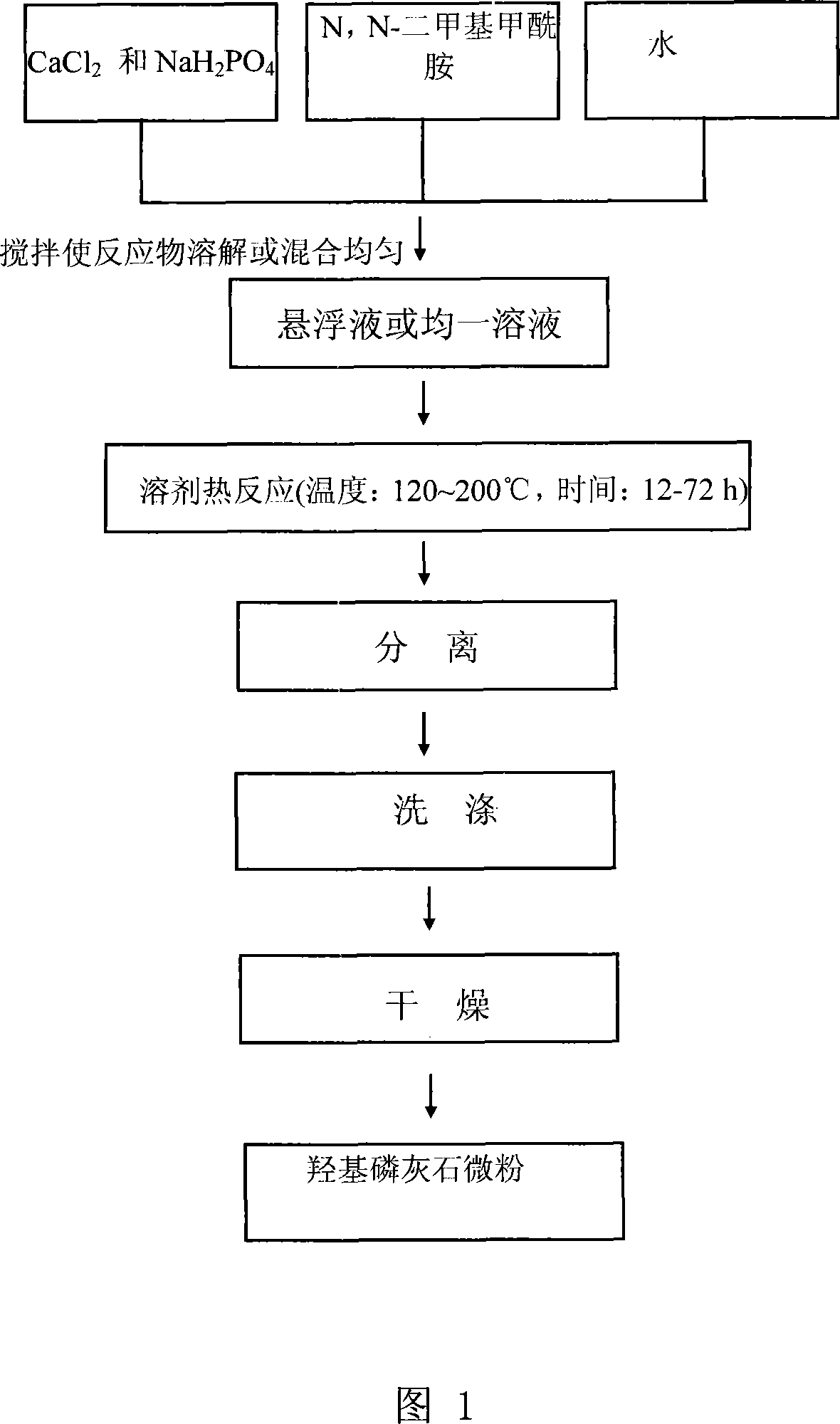
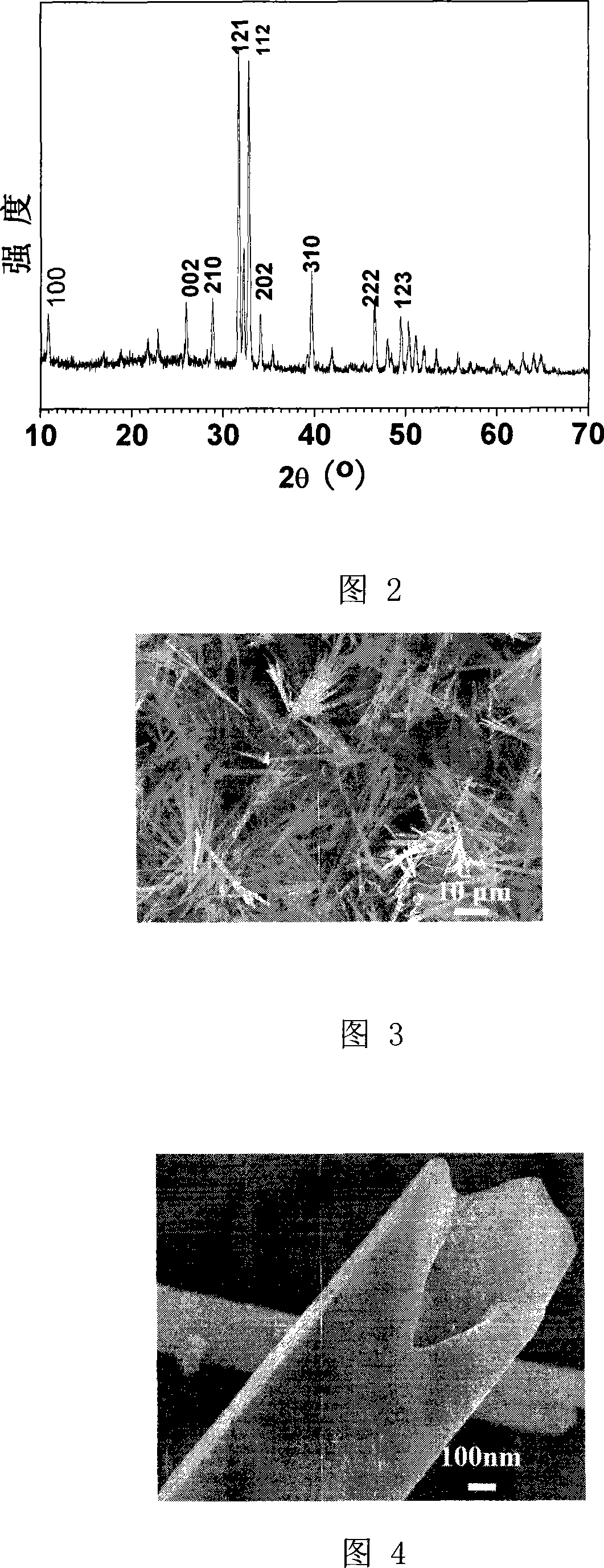
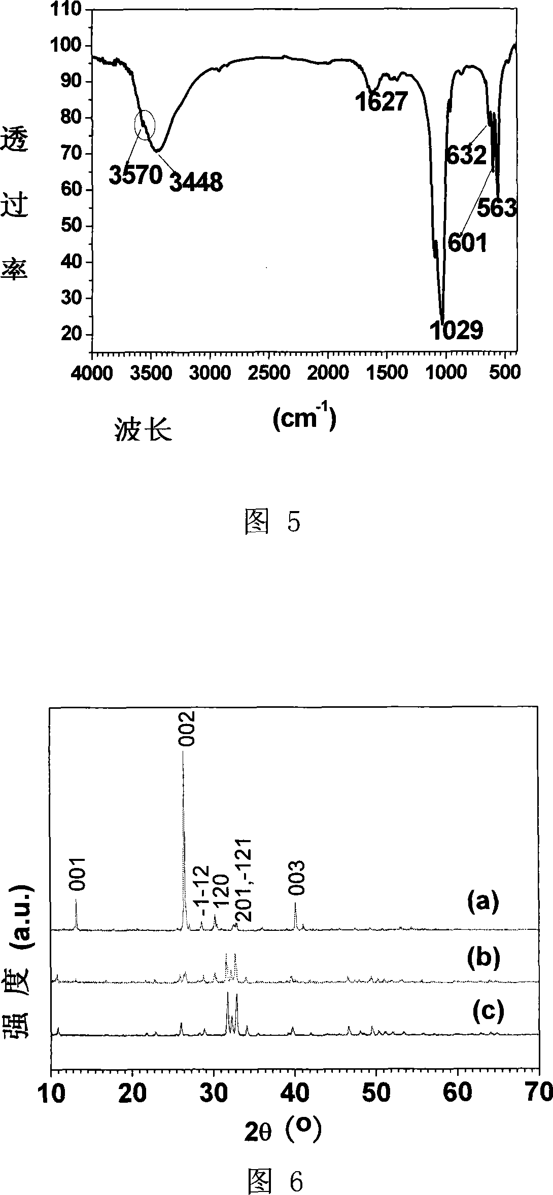
[0029] At room temperature, 0.110g CaCl 2 and 0.094g NaH 2 PO 4 Added to 5 mL of distilled water, followed by 10 mL of N,N-dimethylformamide to obtain a homogeneously dispersed suspension. The suspension was transferred to a 20mL reaction kettle and sealed. The reaction kettle was put into an oven and kept at 160° C. for 24 hours. After the reaction kettle was naturally cooled to room temperature, the product was taken out, and the product was separated by centrifugation. The separated product was washed twice with absolute ethanol and distilled water respectively, and dried under vacuum at 60°C. Fig. 2 is the X-ray diffraction spectrum of the hydroxyapatite microtube prepared in this example, no diffraction peaks of any impurities are found in the spectrum, indicating that the product is a single-phase hydroxyapatite microtube. Figures 3 and 4 are scanning electron micrographs of which the tubular structure can be clearly seen. Figure 5 is its infrared spectrum, 3570cm ...

Embodiment 2

[0031] At room temperature, 0.110g CaCl 2 and 0.094g NaH 2 PO 4 Added to 5 mL of distilled water, followed by 10 mL of N,N-dimethylformamide to obtain a homogeneously dispersed suspension. The suspension was transferred to a 20mL reaction kettle and sealed. The reaction kettle was put into an oven and kept at 160° C. for 12 hours. After the reaction kettle was naturally cooled to room temperature, the product was taken out, and the product was separated by centrifugation. The separated product was washed twice with absolute ethanol and distilled water respectively, and dried in vacuum at 60°C to obtain hydroxyapatite microtubes. Fig. 6c is the X-ray diffraction spectrum of the hydroxyapatite microtube prepared in this example, no diffraction peaks of any impurities are found in the spectrum, indicating that the obtained product is a single-phase hydroxyapatite microtube. The hydroxyapatite prepared in this example has a microtube structure.


PUM

PropertyMeasurementUnit
lengthaaaaaaaaaa
diameteraaaaaaaaaa
lengthaaaaaaaaaa

Abstract

The invention relates to a hydroxyapatite micrometer pipe and a preparation method thereof, and belongs to the biologic material field. The method is characterized in that water soluble calcium salt and acid phosphate are taken as raw material, water is taken as the dissolvent, N is added, N-dimethyl formamide is taken as addition agent, the materials are stirred at a room temperature, heat treatment for 12 to 72 hours under 120 to 200 DEG C is performed, and the product after heat treatment is separated, then cleaned and dried. The inner diameter of the hydroxyapatite micrometer pipe prepared by the method is 450 to 550 nm, the outer diameter is 1000 to 1200 nm, the length is 20 to 50 micrometers, and the hydroxyapatite micrometer pipe can be applied in the biologic material field. The invention has the advantages that the cost of the material is low, the process is simple, the operation is convenient, and the appearance can be controlled and so on.

Description

technical field [0001] The invention relates to a hydroxyapatite microtube and a preparation method thereof, more precisely relates to the preparation of hydroxyapatite micropowder by a weakly basic additive, and belongs to the field of biological materials. Background technique [0002] Hydroxyapatite has good biocompatibility and has a chemical composition similar to that of human bones and teeth. It is often used as bone substitute materials, carriers for drug delivery, gas sensors, ion exchanges, catalysts or catalyst carriers, etc. The existing preparation methods include precipitation method, solvothermal method, sol-gel method and microwave-assisted synthesis method. However, the morphology of the obtained hydroxyapatite is generally needle-like and rod-like, and there is no report on the preparation of tubular hydroxyapatite by wet chemical method. Contents of the invention [0003] The purpose of the present invention is to provide a hydroxyapatite microtube and ...

Claims


Application Information

Patent Timeline
Patent Type & Authority Applications(China)
IPC IPC(8): C01B25/32A61L27/00
Inventor 朱英杰马明国
Owner SHANGHAI INST OF CERAMIC CHEM & TECH CHINESE ACAD OF SCI
Why Eureka
Try Eureka