
Tuberculosis antibody multi-antigen ELISA detecting kit and making method

A detection kit and tuberculosis antibody technology, applied in the field of tuberculosis medical immunology detection, can solve the problems of high cost, only qualitative, low sensitivity, etc., and achieve the effect of low cost

Inactive Publication Date: 2009-03-11
中国人民解放军总医院第二附属医院
View PDF2 Cites 13 Cited by
  • Summary
  • Abstract
  • Description
  • Claims
  • Application Information

AI Technical Summary

Problems solved by technology

The existing tuberculosis antibody ELISA detection kit is mainly composed of detection antigen, enzyme-linked anti-human IgG antibody, substrate, positive control serum of tuberculosis patients, normal control serum, calf serum and polystyrene microwell reaction plate. Some of the coating antigens used are pure protein derivatives of Mycobacterium tuberculosis (PPD). Since PPD is a protein of Mycobacterium tuberculosis culture filtrate, it contains many mycobacteria (including pathogenic mycobacteria, non-pathogenic mycobacteria in the environment and BCG) Common antigens have poor detection specificity and are prone to false positives; some coated antigens use Mycobacterium tuberculosis membrane antigen (see Patent Publication No. CN 1072265A), and adopt the immunoreaction method of enzyme-linked Staphylococcus A protein; The detection antigen adopts ESAT-6 and CFP-10 fusion protein antigen (see patent publication number CN 1388378), and what they all adopt is a single detection antigen
[0006] The tuberculosis antibody detection protein chip kit developed by Nanjing Dayuan Biotechnology Engineering Co., Ltd. uses LAM, 38kD and 16kD proteins of Mycobacterium tuberculosis as antigens, and its sensitivity is not high
In addition, it uses the gold-labeled immunospot method, uses nitrocellulose membrane as a carrier, immobilizes the antigen on the membrane, and uses colloidal gold particles to label the antibody, so that the antigen-antibody reaction can proceed rapidly on the membrane, forming a red conjugate. Visually observe the results, although it is simple and fast, but it can only be qualitative, not quantitative, and the cost is high

Method used


Image

  • Tuberculosis antibody multi-antigen ELISA detecting kit and making method
  • Tuberculosis antibody multi-antigen ELISA detecting kit and making method

Examples


preparation example Construction

[0021] The preparation method of the kit is characterized in that: comprising:

[0022] (1) Preparation of detection antigen: cloned and expressed by genetic engineering technology, purified to obtain 38kD, 16kD, MPT63, MTB48, CFP10-ESAT6 recombinant protein antigens respectively, and purified from Mycobacterium tuberculosis complex strains to obtain LAM;

[0023] (2) dilute each tuberculosis with diluent (for routine diluent, for example 0.5M sodium chloride-20mM disodium hydrogen phosphate-2% mannitol, pH7.4, or phosphate buffer saline, normal saline) Mycobacterium recombinant protein or sugar antigen, so that the concentration of each Mycobacterium tuberculosis recombinant protein or sugar antigen is 1mg / ml, and then the dilution solution of each Mycobacterium tuberculosis recombinant protein or sugar antigen is mixed and then sterilized and filtered. Divide into one container, 1ml / bottle, and freeze-dry for later use;

[0024] (3) Assembly of the kit: the above-mentioned ...

Embodiment 1

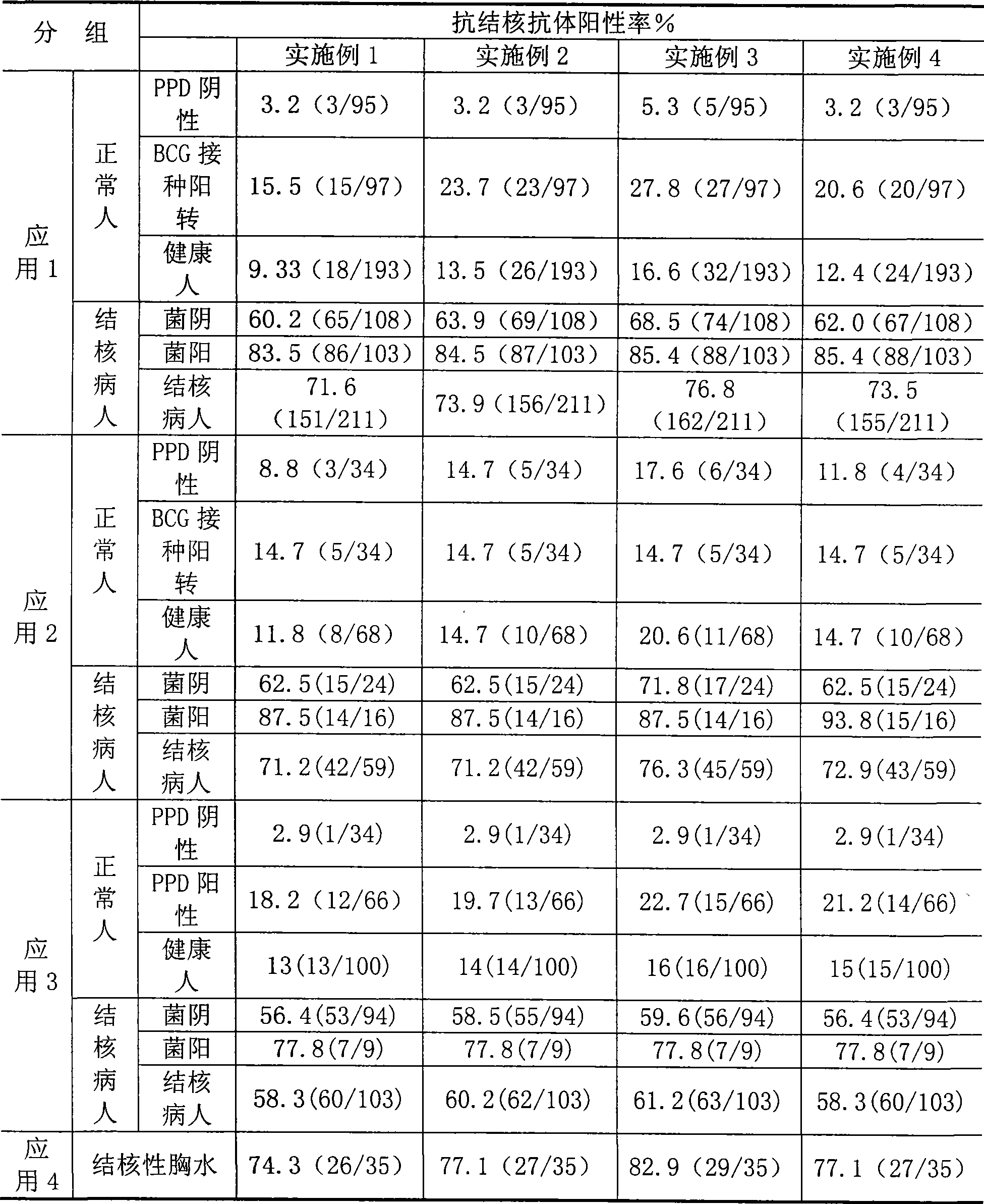
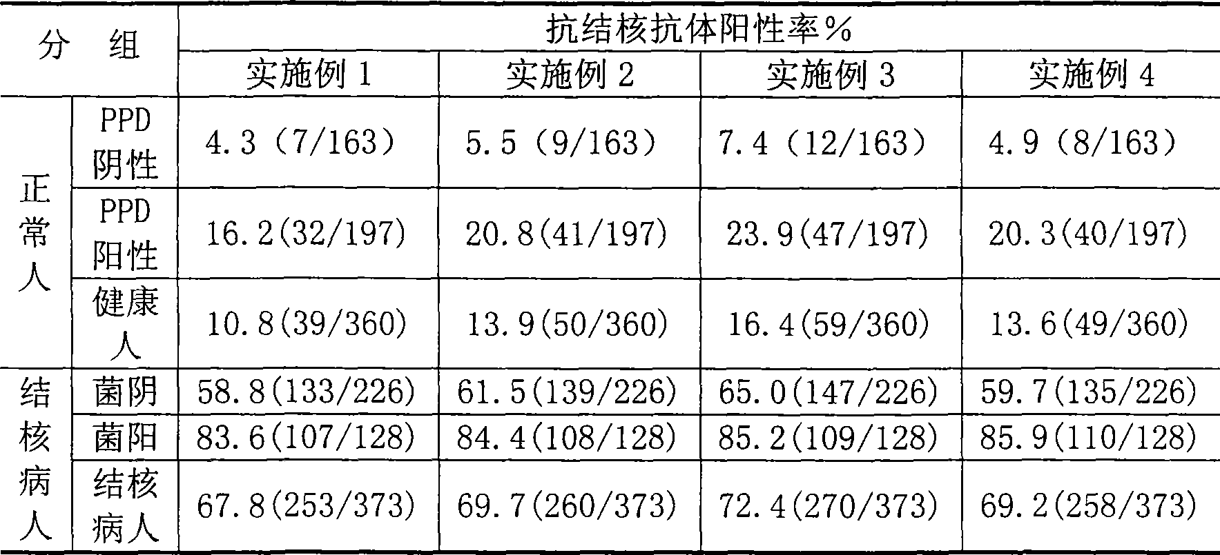
[0300] (1) Preparation of detection antigen: Dilute Mycobacterium tuberculosis LAM, 38kD, and 16kD recombinant proteins with diluent (0.5M sodium chloride-20mM disodium hydrogen phosphate-2% mannitol, pH7.4) respectively, so that each tuberculosis The concentration of mycobacterial recombinant protein is 1mg / ml, and then the diluted solution of each Mycobacterium tuberculosis recombinant protein is mixed, sterilized and filtered, divided into a container, 1ml / cartridge, and freeze-dried for later use;

[0301] (2) Kit assembly: 1mg / ml Mycobacterium tuberculosis LAM, 38kD, 16kD recombinant protein freeze-dried tubes, horseradish peroxidase-labeled anti-human IgG antibody, tuberculosis patient positive control serum, normal control 1 vial of serum and calf serum, 1 vial of 30% hydrogen peroxide, 1 vial of o-phenylenediamine, and 1 polystyrene microwell reaction plate were put into the packaging box, sealed, and stored at 4°C in the dark.

Embodiment 2

[0303] (1) Preparation of detection antigen: Dilute Mycobacterium tuberculosis LAM, 38kD, 16kD, and MPT63 recombinant proteins with diluent (phosphate buffer) respectively, so that the concentration of each Mycobacterium tuberculosis recombinant protein is 1mg / ml, and then Mix each diluted solution of Mycobacterium tuberculosis recombinant protein, sterilize and filter, divide into a container, 1ml / bottle, and freeze-dry for later use;

[0304](2) Assembly of the kit: freeze-dried tubes of 1mg / ml Mycobacterium tuberculosis LAM, 38kD, 16kD, MPT63 recombinant protein, horseradish peroxidase-labeled anti-human IgG antibody, tuberculosis patient positive control serum, normal Human control serum, 1 calf serum, 1 vial of 30% hydrogen peroxide, 1 o-phenylenediamine, and 1 polystyrene microporous reaction plate were placed in a packaging box, sealed, and stored at 4°C in the dark.


PUM

PropertyMeasurementUnit
Sensitivityaaaaaaaaaa

Abstract

The invention relates to a tuberculosis antibody multiple antigen ELISA detection kit and a preparation method thereof, which pertains to the field of tuberculosis medical immunology diagnostic techniques and mainly uses detection antigen, enzyme-linked antihuman IgG antibodies, substrates, positive control serum of tuberculosis patients, control serum of normal person, calf serum and polystyrene microplates to form the kit, wherein, the detection antigen adopts the mycobacterium tuberculosis complex strains of lipid Arabian mannose (LAM), 38kD and 16kD to be combined with arbitrary one or more than one mycobacterium tuberculosis recombinant proteins in the recombinant proteins of MPT63, MTB48 and CFP10-ESAT6. The mycobacterium tuberculosis has high sensitivity, strong specificity and complementarity, can be used for detecting specific antitubercular antibodies in such body fluid samples as serum, hydrothorax and the like, and assisting the diagnosis and differential diagnosis of tuberculosis.

Description

technical field [0001] The invention belongs to the technical field of tuberculosis medical immunology detection, and in particular relates to a tuberculosis antibody multi-antigen ELISA detection kit composed of different combinations of LAM, 38kD, 16kD, MPT63, MTB48, and CFP10-ESAT6 recombinant proteins as detection antigens. Background technique [0002] Tuberculosis is still one of the major infectious diseases that endanger human health in the world. Since 1985, the prevalence of AIDS, tuberculosis-infected immigrants and some people living in poverty have caused the incidence of tuberculosis to rise in the United States and other developed countries in Europe and the United States, especially tuberculosis. The problem of bacterial drug resistance and the incidence of non-tuberculous mycobacterial disease are increasing year by year, which makes the treatment of tuberculosis even worse. At present, there are about 20 million tuberculosis patients in the world, 8 to 10 m...

Claims


Application Information

Patent Timeline
Patent Type & Authority Applications(China)
IPC IPC(8): G01N33/545G01N33/531
Inventor 吴雪琼阳幼荣张俊仙李邦印梁艳李洪敏张翠英李娟
Owner 中国人民解放军总医院第二附属医院
Learn More