Method for detecting gentamicin through clinical magnetic resonance imaging

A technology of magnetic resonance imaging and gentamicin, applied in the field of immunoassay, achieves the effect of easy operation

Inactive Publication Date: 2009-10-28
JIANGNAN UNIV
View PDF0 Cites 20 Cited by
  • Summary
  • Abstract
  • Description
  • Claims
  • Application Information

AI Technical Summary

Problems solved by technology

So far in China, there has been no report on the use of magnetic resonance signals of superparamagnetic nanoparticles to detect in vitro targets. The present invention makes up for this gap

Method used


Image

  • Method for detecting gentamicin through clinical magnetic resonance imaging
  • Method for detecting gentamicin through clinical magnetic resonance imaging
  • Method for detecting gentamicin through clinical magnetic resonance imaging

Examples


Embodiment 1

[0027] Embodiment 1 The coupling of gentamicin and magnetic nanoparticles:

[0028] Add 1 μL carbodiimide EDC and 2.4 mg N-hydroxysulfosuccinimide sulfo-NHS to 0.5 mL, 1.3 mg / mL Fe 3 o 4 In aqueous solution, the reaction takes about 15 minutes to activate. Activated Fe 3 o 4 Add 1mg of gentamycin (GM drug), react at room temperature for 4 hours, and dialyze in 0.01M, pH 7.4 PBS for 24 hours to remove unreacted small drug molecules.

Embodiment 2

[0029] Embodiment 2 magnetic resonance:

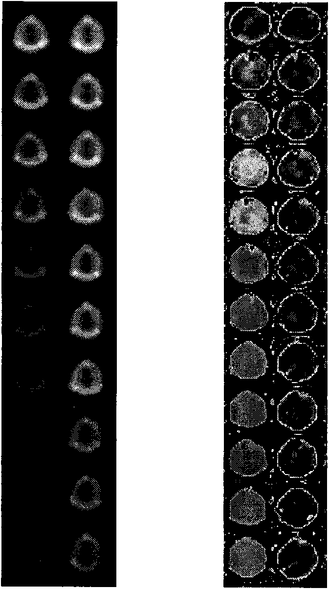
[0030] 20 μL, 150 μg / mL of gentamicin-modified Fe 3 o 4 Add 20 μL of different concentrations of gentamicin standard, add 1 μL of gentamicin antibody (5 mg / mL), then add 1 μL of goat anti-rabbit secondary antibody, incubate for 2 hours, dilute to a volume of 250 μL, and add to the microwell plate, measured by MRI.

[0031] Control group settings: gentamicin antibody was replaced by dexamethasone antibody, and other operations were the same as above.

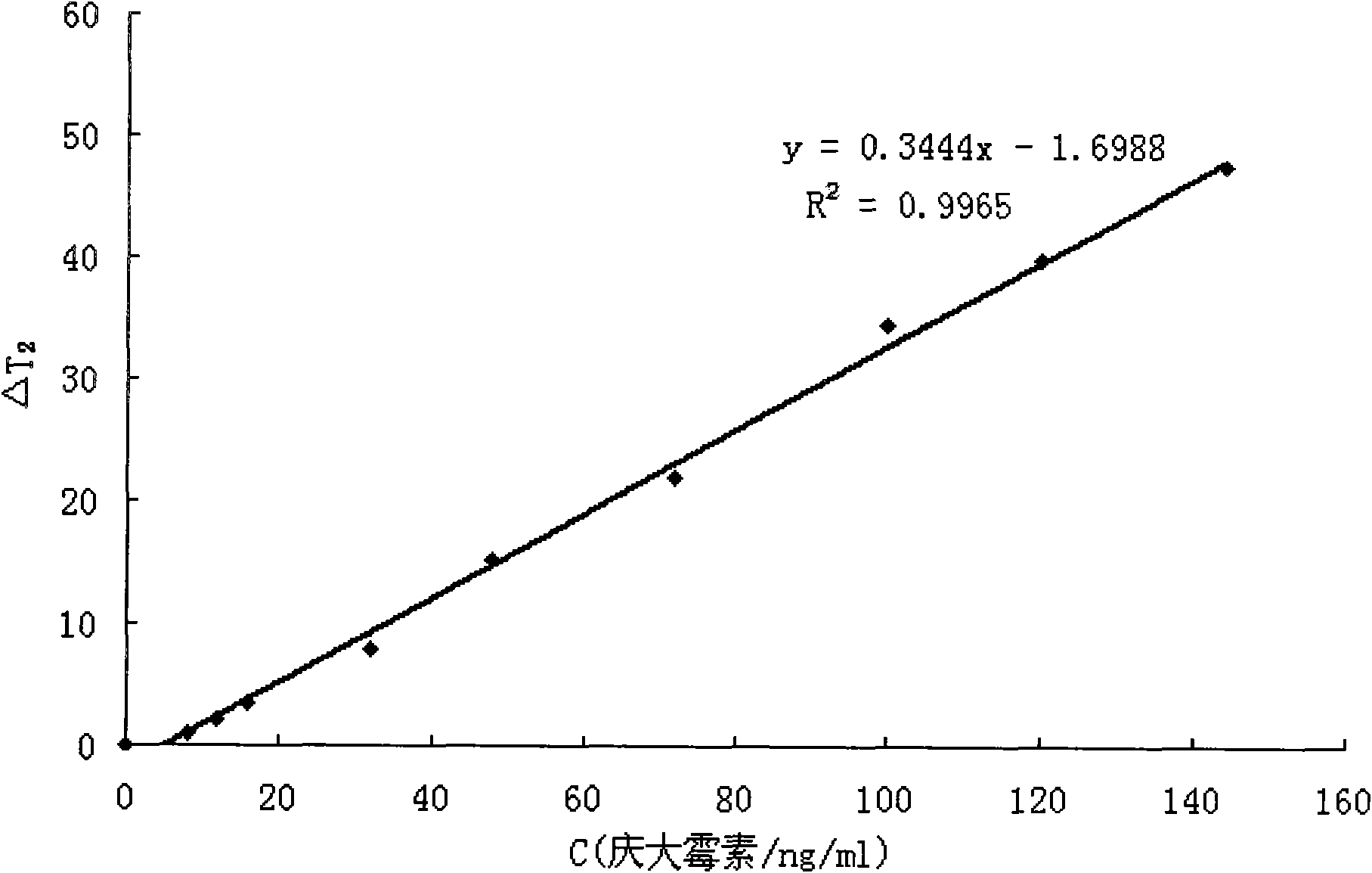
[0032] The solutions used were all 0.01M PBS buffer, pH 7.4. The concentrations of gentamicin standard substances were 0, 8, 12, 16, 32, 48, 72, 100, 120, 144ng / mL, respectively.

Embodiment 3

[0033] Embodiment 3 Magnetic resonance quantitative detection

[0034] The magnetic resonance equipment is MAGNETOM Avanto 1.5T MRI, Siemens clinical magnetic resonance imaging equipment.

[0035] The scanning time parameters used in this experiment are: echo time (TE) is 20-120ms, pulse repetition time (repetition time) TR is 2000ms, and the coil used is the head coil.

[0036] The principle of magnetic resonance detection of target is to induce water proton T through the formation of antibody and drug-modified magnetic nanoparticle immune polymer 2 The size of the value can achieve the purpose of detecting the target object. The more gentamicin added to the water, the less polymer is formed, T 2 The value is larger. established by T 2 ~C / gentamicin concentration standard curve can be compared to obtain the concentration of gentamicin in the sample. See attached for standard curve image 3 .


PUM


Abstract

The invention relates to a method for detecting gentamicin through clinical magnetic resonance imaging, and belongs to the technical field of immunodetection. The method is to add standard gentamicin supernatant or gentamicin supernatant to be measured, gentamicin antibodies and goat-anti and rabbit-anti antibodies into gentamicin modified magnetic nano particles, and use the antibodies and the gentamicin modified magnetic nano particles to form immune polymers so as to generate magnetic resonance signals and perform magnetic resonance detection. The magnetic resonance signal detection method utilizes the antibodies and the gentamicin modified magnetic nano particles to form the immune polymers, and effectively shortens the transverse relaxation time T2 value of protons in water so as to generate signals. In general, when the concentration of the gentamicin in a sample is larger, the quantity of the gentamicin caught by the antibodies is more, the quantity of the antibodies which are combined with the gentamicin on the surface of the magnetic nano particles is fewer, and the measured T2 value is larger. The method does not need complex sample pretreatment, is simple and convenient to operate, only needs to add the sample to be measured for incubation and directly measure the T2 value, and can complete the process by one step. Moreover, the particle diameter of superparamagnetic nano particles for marking is approximately 10 nanometers, so that the water solubility is high and the dispersity and the stability are good.

Description

technical field [0001] The invention relates to a method for quantitatively detecting gentamicin with a magnetic nanoparticle-labeled antibody, which belongs to the technical field of immune detection. Background technique [0002] Gentamicin is a multicomponent antibiotic produced by Micromonospora, containing C 1 、C 2 、C 1a and other components, the structure is shown in the appendix figure 1 . Gentamicin sulfate is a white powder, hygroscopic, easily soluble in water, insoluble in common organic solvents, stable to temperature, acid and alkali, and its antibacterial spectrum is basically the same as that of kanamycin. It is characterized by its inhibitory effect on Pseudomonas aeruginosa, Proteus and penicillin-resistant grapes. There is cross-resistance with neomycin and kanamycin. It is mainly used for the treatment of severe infections of the above-mentioned sensitive bacteria, such as sepsis, respiratory tract infection, pneumonia, intestinal infection, biliary ...

Claims


Application Information

Patent Timeline
IPC IPC(8): G01N24/08G01N33/551G01N33/536
CPCY02A90/30
Inventor 胥传来马伟陈伟徐丽广李灼坤彭池方
Owner JIANGNAN UNIV
Why Eureka
Try Eureka