Supercharge Your Innovation With Domain-Expert AI Agents!

Method for preparing and siliconizing DNA nano optical fibers and characterization

A nano-optical fiber and optical fiber technology, which is applied in biochemical equipment and methods, measurement/inspection of microorganisms, material analysis through optical means, etc., can solve the problem of inability to detect the directional distribution and bonding of nano-fiber bioactive molecules or silanized molecules Quantity, the inability to detect the surface composition of nano-fibers, etc., to achieve the effect of improving detection sensitivity and simple operation

Inactive Publication Date: 2009-12-30
UNIV OF ELECTRONIC SCI & TECH OF CHINA
View PDF0 Cites 0 Cited by
  • Summary
  • Abstract
  • Description
  • Claims
  • Application Information

AI Technical Summary

Problems solved by technology

[0006] 1. The directional distribution and bonding amount of nanofiber bioactive molecules or silanized molecules cannot be detected;
[0007] 2. The surface composition of different micro-regions of nano-fibers cannot be detected, indicating the uniformity of bonding of nano-fiber surface active molecules or silanized molecules

Method used


Image

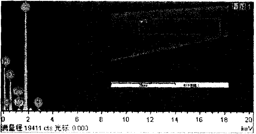
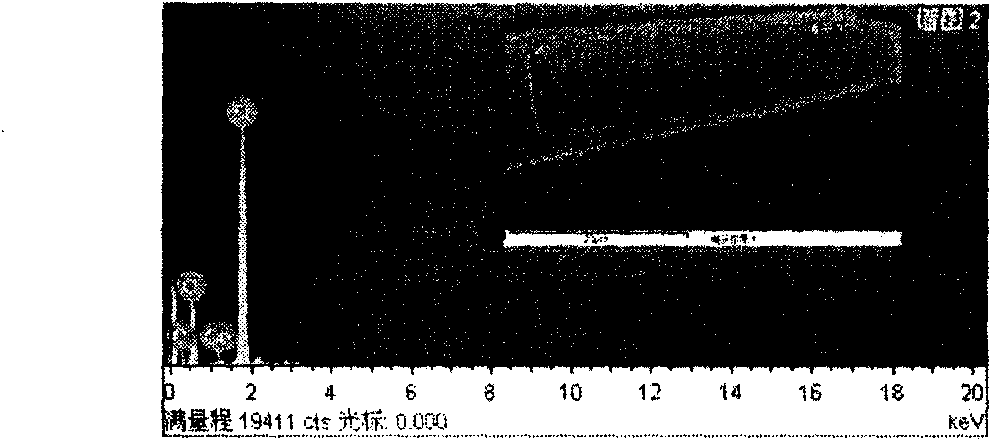
  • Method for preparing and siliconizing DNA nano optical fibers and characterization
  • Method for preparing and siliconizing DNA nano optical fibers and characterization
  • Method for preparing and siliconizing DNA nano optical fibers and characterization

Examples


Embodiment 1

[0035] (1) Preparation of silylating reagent

[0036] Take 3 parts by weight of the silylating agent, add 36 parts by weight of double distilled water, and adjust the pH to 3.5;

[0037] (2) Preparation of etching solution

[0038] Take 45 parts by weight of hydrofluoric acid, 10 parts by weight of ammonium fluoride, and 45 parts by weight of water, stir, mix evenly, and set aside.

[0039] (3) Preparation of surface pretreatment solution

[0040] Take 30 parts by weight of hydrogen peroxide, add 50 parts by weight of concentrated sulfuric acid, stir and dissolve to form a uniform solution, and cool it for later use.

[0041] (4) Preparation of nanofibers

[0042] The optical fiber with a length of about 10cm is immersed in the etching solution for etching, and the corrosion of the optical fiber is observed under a microscope until a good tapered tip appears. Take it out. Wash with distilled water until there is no hydrofluoric acid, remove the protective layer, and dry fo...

Embodiment 2

[0050] (1) Preparation of silylating reagent

[0051] Take 10 parts by weight of the silylating agent, add 90 parts by weight of double distilled water, and adjust the pH to 3.5;

[0052] (2) Preparation of etching solution

[0053] Take 45 parts by weight of hydrofluoric acid, 10 parts by weight of ammonium fluoride, and 45 parts by weight of water, stir, mix evenly, and set aside.

[0054] (3) Preparation of surface pretreatment solution

[0055] Take 30 parts by weight of hydrogen peroxide, add 50 parts by weight of concentrated sulfuric acid, stir and dissolve to form a uniform solution, and cool it for later use.

[0056] (4) Preparation of nanofibers

[0057] The optical fiber with a length of about 10cm is immersed in the etching solution for etching, and the corrosion of the optical fiber is observed under a microscope until a good tapered tip appears. Take it out. Wash with distilled water until there is no hydrofluoric acid, remove the protective layer, and dry f...


PUM


Abstract

The invention discloses a method for preparing and siliconizing DNA nano optical fibers and characterizing the surface microstructural components of the siliconized nano optical fibers, which can visually represent the covalent grafting on the head parts of the optical fibers, and is characterized in that: micron-sized optical fibers are etched in an etchant for 5 hours to form the nano optical fibers; the optical fibers are placed in 10 weight portions of silane solution to react for 1 to 5 hours under the conditions of a temperature of between 60 and 90 DEG C and a pH value of between 2 and 4.5; and the optical fibers are taken out, washed by distilled water and dried for 10 to 12 hours under vacuum and at 90 to 100 DEG C. The result of nano optical fiber surface microstructural component detection by the method shows that the method can be used for detecting the surface microstructural components of the siliconized optical fibers and can visually characterize the coupling between bioactive molecules and the siliconized optical fibers.

Description

technical field [0001] The invention relates to a preparation and characterization technology of a core sensitive element of a micro-nano optical fiber biosensor and a technical route for its realization. The technology is applied in the fields of environmental monitoring, biomedicine and clinical testing equipment manufacturing and the like. Background technique [0002] As the medium or sensitive element of the sensor, nano-fibers together constitute nano-fiber sensors, such as nano-fiber biosensors, which can be applied to non-invasive or micro-trauma in vivo or intracellular, real-time and remote online medical diagnosis, online Quality monitoring and environmental testing. For example, nano-fiber DNA biosensors can quickly and effectively detect special gene sequences and point mutations. Due to the strong anti-electromagnetic interference and optical signals of optical fibers, no additional reference devices are required for measurement [Mara Dolores Marazuela, Mara C...

Claims


Application Information

Patent Timeline
IPC IPC(8): G01N21/00C12Q1/68
Inventor 庞小峰王一明
Owner UNIV OF ELECTRONIC SCI & TECH OF CHINA
Features
  • R&D
  • Intellectual Property
  • Life Sciences
  • Materials
  • Tech Scout
  • Unparalleled Data Quality
  • Higher Quality Content
  • 60% Fewer Hallucinations
Learn More