Method for rapidly detecting neutralizing antibody of virus and kit therefor
A virus and antibody technology, applied in biological testing, measuring devices, material inspection products, etc., can solve the problems of difficult screening requirements, heavy workload, and inability to accurately detect neutralizing antibodies
- Summary
- Abstract
- Description
- Claims
- Application Information
AI Technical Summary
Problems solved by technology
Method used
Image
Examples
Embodiment 1
[0138] Example 1. Cultivation of enterovirus
[0139] The enterovirus strain used as an example in this embodiment is JS06-52-3 (preserved by the National Research Center for Infectious Disease Diagnostic Reagents and Vaccine Engineering Technology), and a nearly full-length 7312bp sequence (Genbank No: FJ 600325) was obtained by RT-PCR. ), belongs to the EV71C4 subtype, has a nucleotide sequence homology of 97.7% and an amino acid sequence homology of 97.6% with the EU703814 virus strain. Other enteroviruses can also be used.
[0140] The present invention infects cells in RD cells (ATCC, CCL-136 TM ) (ATCC is the supplier) produces EV71 virus.
[0141] experimental method:
[0142] The RD cells were cultured in a 10cm cell culture dish with a MEM medium (10% FBS, 2mM L-glutamine, 0.1mM MEM Non-Essential Amino Acids and 1% penicillin-streptomycin), and the confluence rate was about 80%.
[0143] After 10 hours, the cells in each dish were replaced with serum-free MEM medium (additi...
Embodiment 2
[0145] Example 2. Enterovirus infection of cells
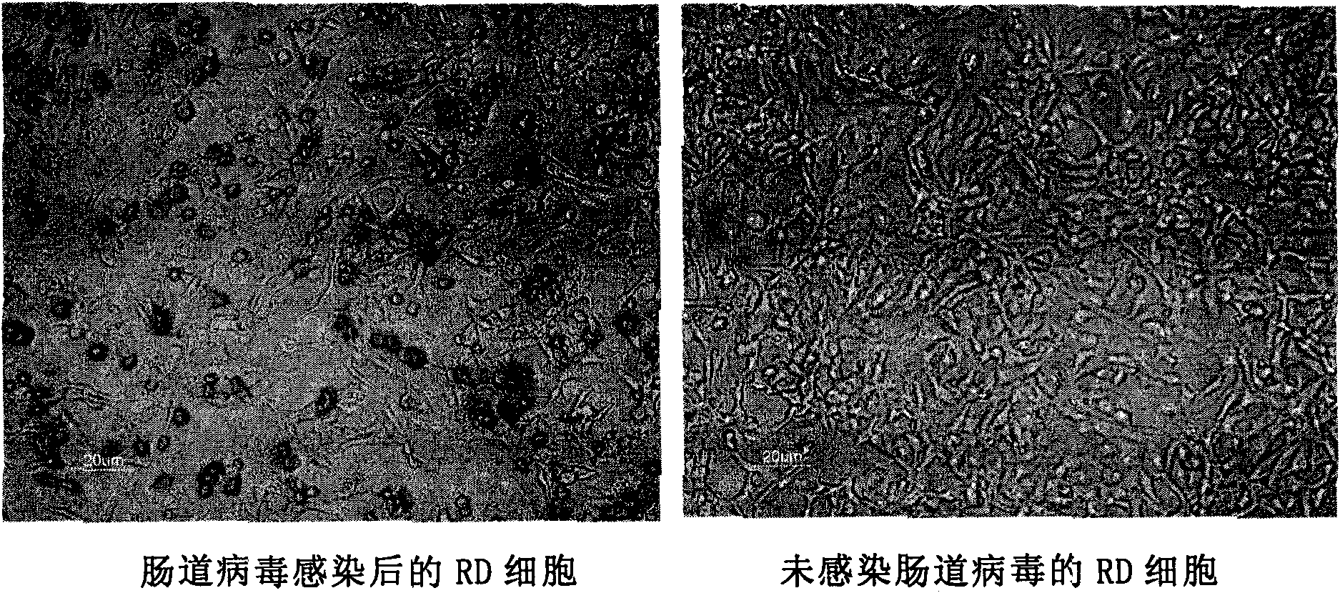
[0146] The harvested enterovirus was infected with RD cells, and the infection of RD cells was detected. In this embodiment, the EV71 virus infection is taken as an example, and the detection of other enterovirus infections may also be included.
[0147] experimental method:
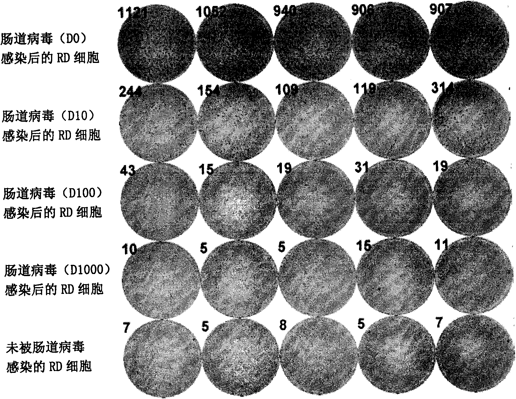
[0148] RD cells are cultured in 96-well cell culture plates, about 2×10 4 / Well, the medium is MEM medium (add 10% FBS, 2mM L-glutamine, 0.1mM MEM Non-EssentialAmino Acids and 1% penicillin-streptomycin), the confluence rate is about 80%. After culturing for 10 hours, 50 μL of EV71 virus (diluted by 0, 10, 100, 1000, 10000 times) was added to each well. After culturing for 14 hours, aspirate the medium in each well, add 100μL of fixative (PBS solution containing 0.2% glutaraldehyde) to each well, and let stand for 1h at room temperature in the dark; after 1h, aspirate the fixative. Add 100μL of 1% TritonX-100 solution to permeabilize the cells and let stand ...
Embodiment 3
[0151] Example 3. Using Elispot, a spot detection instrument, to detect cell infection by enterovirus
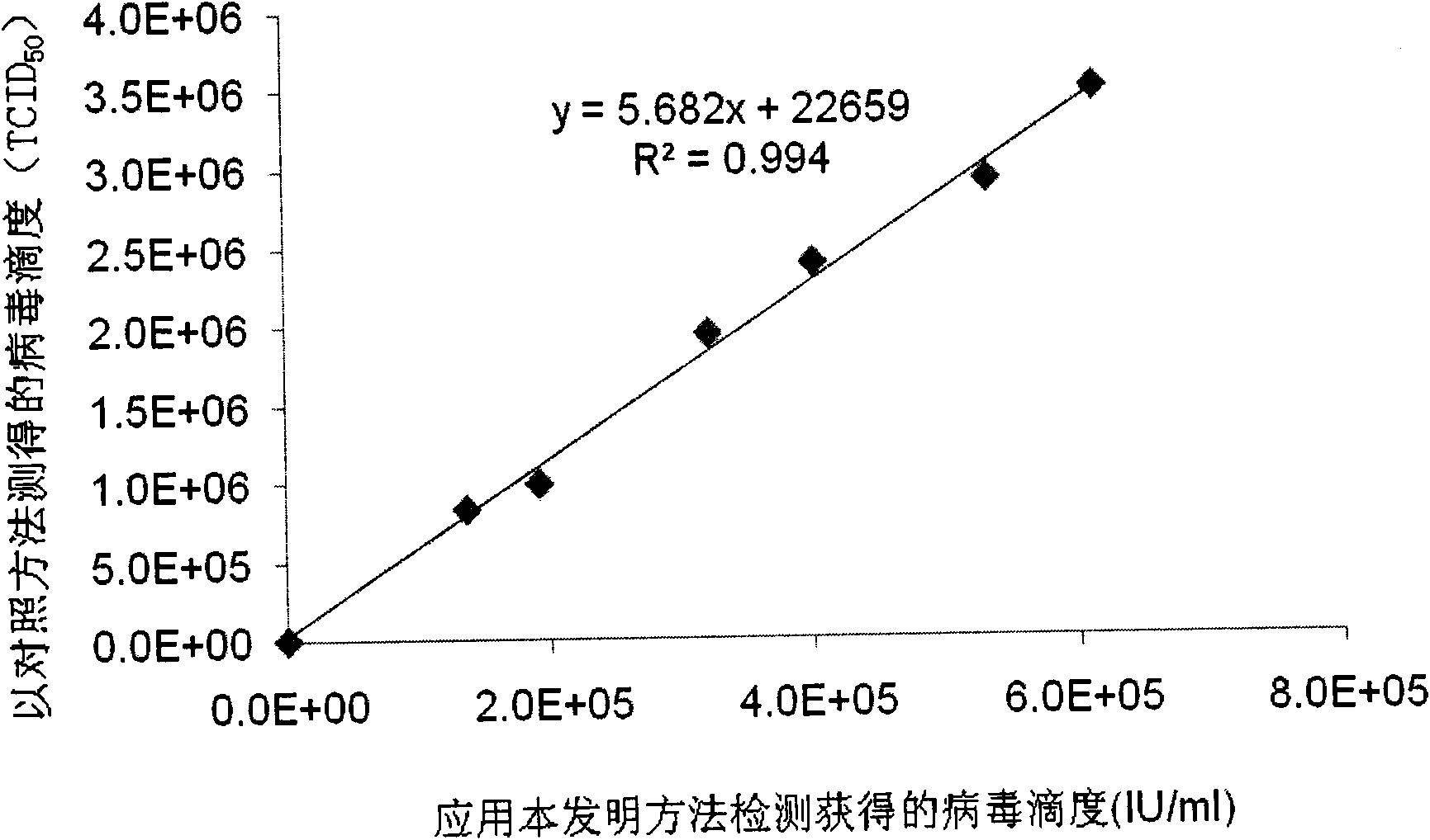
[0152] In the present invention, the EV71 virus is taken as an example. The EV71 virus is infected with RD cells. After infection, the detection by the enzyme-linked immunospot method can make the virus-infected cells produce a signal that is different from the uninfected cells. This signal can be observed under visible light, and it does not require excitation by a special light source (such as a laser, a mercury lamp with a limited wavelength range, etc.), and is suitable for detection with a spot detection instrument Elispot. Currently, the traditional TCID 50 The method is to observe the diseased cells with naked eyes, which is laborious and time-consuming, and is not suitable for high-throughput detection experiments; and lack of objectivity, there are certain subjective errors, which easily affect the reproducibility of experimental results. Elispot, a spot detection inst...
PUM
 Abstract
Description
Claims
Application Information
 - R&D
- Intellectual Property
- Life Sciences
- Materials
- Tech Scout
- Unparalleled Data Quality
- Higher Quality Content
- 60% Fewer Hallucinations
Browse by: Latest US Patents, China's latest patents, Technical Efficacy Thesaurus, Application Domain, Technology Topic, Popular Technical Reports.
© 2025 PatSnap. All rights reserved.Legal|Privacy policy|Modern Slavery Act Transparency Statement|Sitemap|About US| Contact US: help@patsnap.com


