Biotin labeling method of envelope viruses

A technology of biotinylation and biotin derivatives, applied in the field of biotin labeling, can solve the problems of loss of virus activity, complicated operation process, unfavorable application, etc., and achieve the effect of short time consumption and less damage to virus activity.

Inactive Publication Date: 2010-11-24
WUHAN UNIV
View PDF0 Cites 3 Cited by
  • Summary
  • Abstract
  • Description
  • Claims
  • Application Information

AI Technical Summary

Problems solved by technology

Although these methods have been widely used in various fields, their operation process is often very complicated and technically difficult, especially in the virus coupling and subsequent purification, which easily lead to the loss of virus activity, which is not conducive to its further application.
Especially for enveloped viruses, the envelope structure is very easy to be destroyed, which urgently requires us to find a fast and effective method for biotin-labeling enveloped viruses

Method used


Image

  • Biotin labeling method of envelope viruses
  • Biotin labeling method of envelope viruses
  • Biotin labeling method of envelope viruses

Examples


Embodiment 2

[0041] Biotinylation labeling of embodiment 2 poxvirus (Beijing Tiantan strain)

[0042] (1) Preparation of Vero cell growth medium: Dissolve DMEM medium dry powder in 1000ml of ultrapure water, add 10% fetal bovine serum and 100000 units of penicillin and streptomycin, and store at 4°C.

[0043] (2) Preparation of poxvirus adsorption solution: dissolve DMEM medium dry powder in 1000 ml of ultrapure water, 100000 units of penicillin and streptomycin, and store at 4°C.

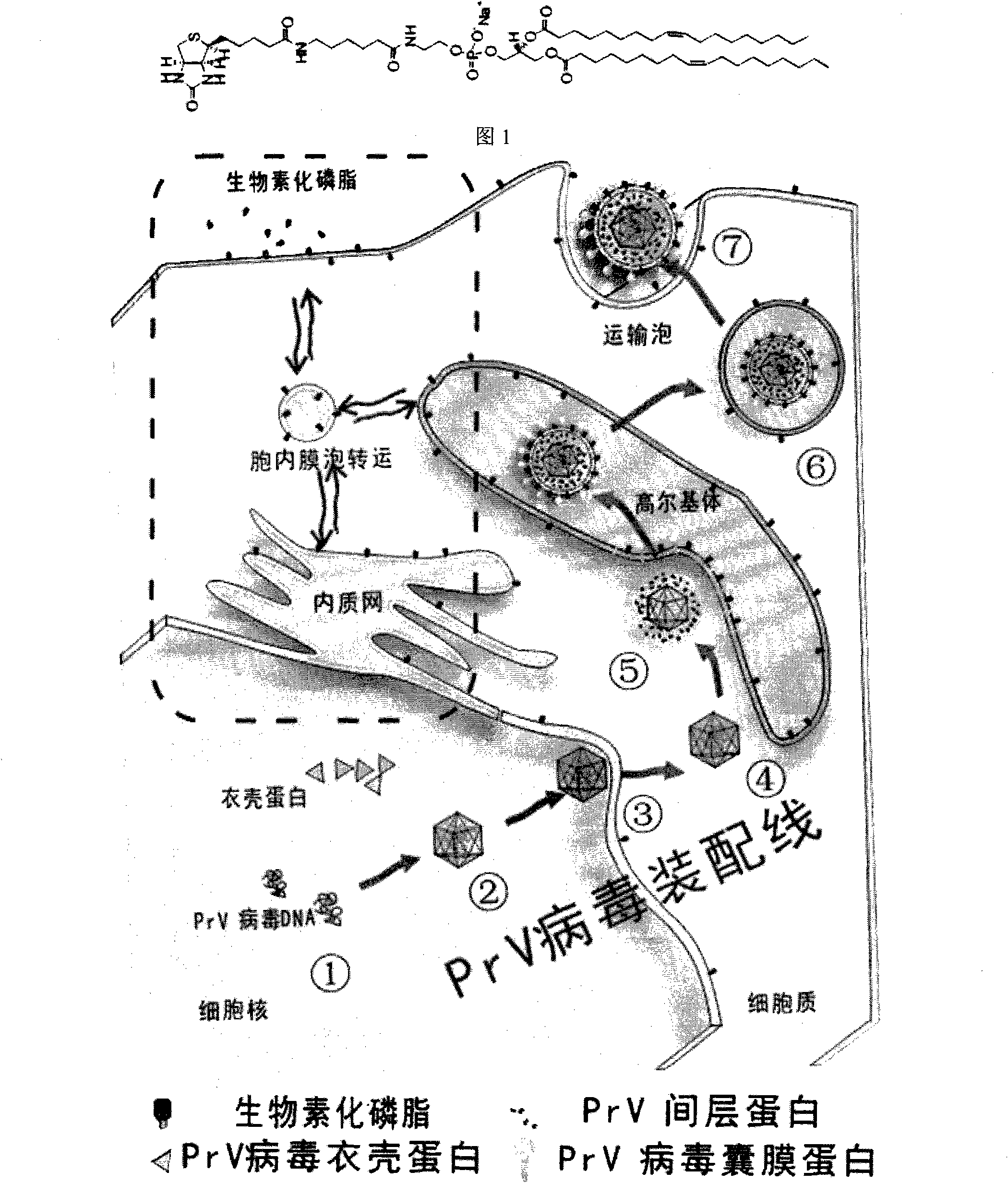
[0044] (3) Prepare biotinylated cell culture medium: add 0.02g / l Biotinyl Cap PE ((1,2-dioleoyl-sn-glycero-3-phosphoethanolamine-N-(cap biotinyl) (sodium salt), Avantilipids), stored at 4°C.

[0045] (4) Preparation of biotinylated cell maintenance solution (for virus growth): Dissolve DMEM medium dry powder in 1000ml of ultrapure water, add 2% fetal bovine serum and 100,000 units of penicillin and streptomycin, 0.02g / 1 Biotinyl Cap PE ((1,2-dioleoyl-sn-glycero-3-phosphoethanolamine-N-(cap biotinyl)(sodium...

Embodiment 3

[0048] Example 3 Biotinylation labeling of baculovirus

[0049] (1) Preparation of Sf9 cell growth medium: dissolve Grace insect medium dry powder in 1000ml of ultrapure water, and use NaHCO 3 Adjust the pH to 6, add 10% fetal bovine serum, and store at 4°C.

[0050] (2) Prepare biotinylated Sf9 cell culture medium: add 0.02g / l Biotinyl Cap PE ((1,2-dioleoyl-sn-glycero-3-phosphoethanolamine-N-( cap biotinyl) (sodium salt), Avantilipids), stored at 4°C. The adsorption and maintenance solution of baculovirus are the same as the cell culture medium

[0051] (3) To cultivate biotinylated Sf9 cells, just add 0.02g / l Biotinyl Cap PE ((1,2-dioleoyl-sn-glycero-3-phosphoethanolamine-N-(cap biotinyl)( sodium salt), Avantilipids). Usually, in order to make Biotinyl Cap PE balance in each inner membrane system of cells, cells need to be maintained in cell culture medium supplemented with Biotinyl Cap PE for at least 7 days. The required balance time Compared with Vero cells, it is lo...

Embodiment 4

[0053] Biotinylation Characterization of Example 4 Cells

[0054] In order to confirm that Biotin Cap PE can be incorporated into the membrane system of cells through this method, we used Cy3-SA (Molecular Probes) to carry out labeling experiments on cells. The specific method is as follows: First, cells and biotinylated cells were mixed with 2% The PBS solution of BSA (bovine serum albumin) was blocked for half an hour, and then incubated with 10 μg / mL Cy3-SA for half an hour, and then the excess Cy3-SA was washed and observed with a fluorescence microscope and a laser confocal microscope, respectively.

[0055] image 3 It is the result of labeling Biotin Cap PE on the cell membrane with Cy3-SA. Obviously, the cell membrane of the biotinylated cells had obvious fluorescence, but it was not observed in the cells without Biotin Cap PE. It can be further found by confocal microscopy that Cy3-SA on the surface of these cells can be swallowed by the cells and transferred to v...


PUM


Abstract

The invention relates to a biotin labeling method of envelope viruses. In the method, the biotinylation of a host cell membrane system is realized first by adding biotin phospholipid into a culture medium of host cells of the viruses, and then the biotin labeling of the envelope viruses is realized by using the characteristic that the envelope viruses use the host cell membrane components to construct envelopes when budding. In the invention, the labeling and organic culture of the envelope viruses are combined organically, complex genetic modification and chemical processing are avoided, and the method can be widely used in fields related to genetic treatment, virus purification, tumor treatment, biosensors, virus tracking and the like.

Description

technical field [0001] The invention relates to the fields of chemistry, virology, biological science and the like, in particular to a biotin labeling method of enveloped viruses. Background technique [0002] In recent years, the role of viruses has been quietly changing, from being a nonsense to an important tool for human beings to understand and transform nature. As the most primitive biological species in nature, the application fields of viruses continue to expand. Viruses are widely used in fields from materials science to biomedicine due to their small size and nanoscale, symmetrical structure, genetic manipulation and good dispersion in aqueous phase. In the field of materials, scientists often use the unique symmetrical structure of viruses to construct multifunctional nanomaterials with unique structures based on various functionalized viruses or viral proteins. In the fields of medicine and life sciences, viruses (adenovirus, etc.) have been widely used as gene...

Claims


Application Information

Patent Timeline
Patent Type & Authority Applications(China)
IPC IPC(8): C12N7/00C12N7/02
Inventor 庞代文黄碧海谢敏刘安安
Owner WUHAN UNIV
Why Eureka
Try Eureka