System and method for realizing total internal reflection fluorescence microscopy by using concentric double conical surface lens

A total internal reflection, biconical surface technology, used in microscopes, fluorescence/phosphorescence, optics, etc., can solve problems such as low transmittance, achieve weak light damage effect, reduce the possibility of light damage, and facilitate switching. Effect

Inactive Publication Date: 2011-04-06
XI'AN INST OF OPTICS & FINE MECHANICS - CHINESE ACAD OF SCI
View PDF5 Cites 24 Cited by
  • Summary
  • Abstract
  • Description
  • Claims
  • Application Information

AI Technical Summary

Problems solved by technology

[0014] In order to solve the problem of low transmittance of the existing objective-type total internal reflection fluorescence microscopy method, the present invention provides a system and method for realizing total internal reflection fluorescence microscopy using concentric biconical mirrors

Method used


Image

  • System and method for realizing total internal reflection fluorescence microscopy by using concentric double conical surface lens
  • System and method for realizing total internal reflection fluorescence microscopy by using concentric double conical surface lens
  • System and method for realizing total internal reflection fluorescence microscopy by using concentric double conical surface lens

Examples


Embodiment 1

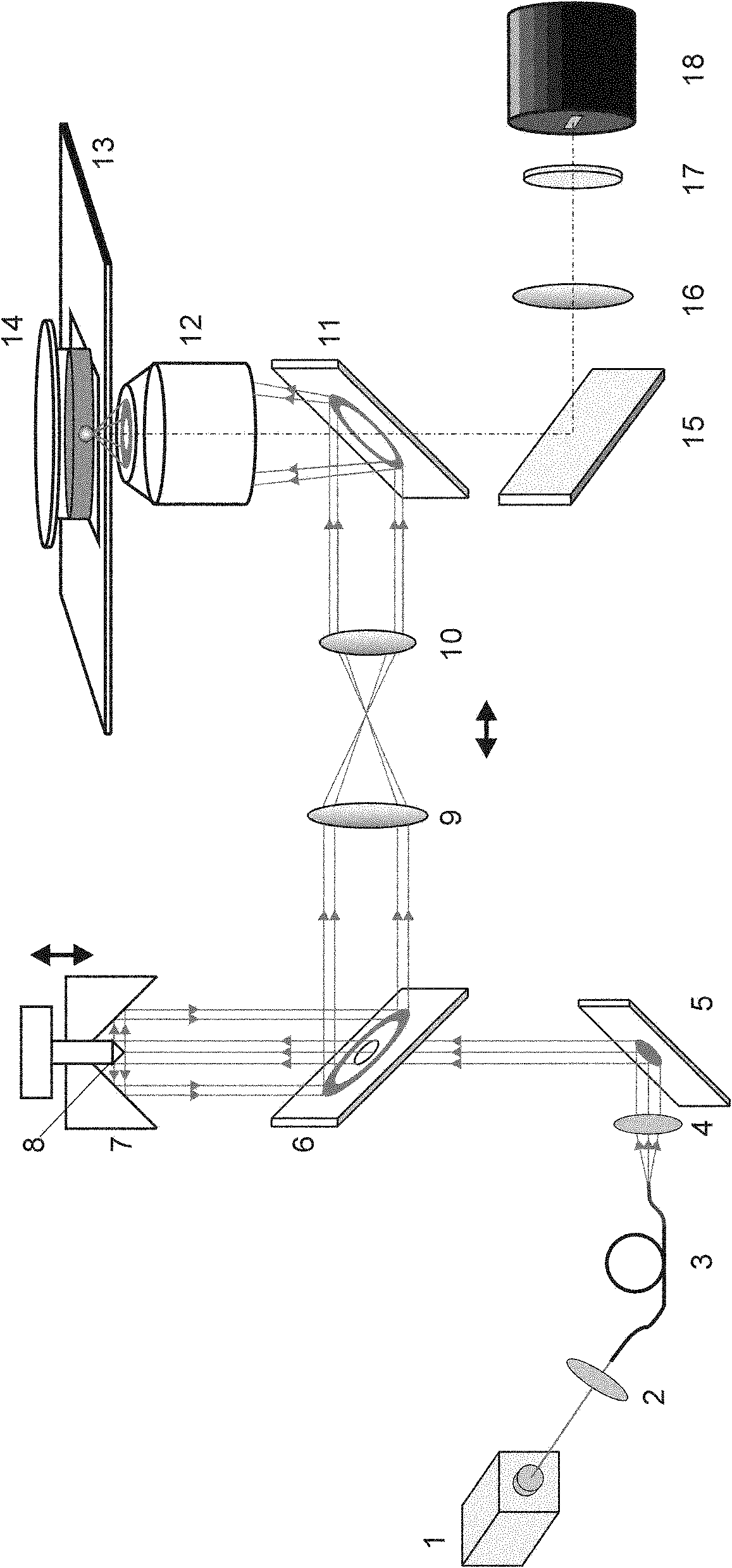
[0051] Example 1: Figure 4 The apparatus of the present invention compares the imaging experiments of wide-field fluorescence microscopy and total internal reflection fluorescence microscopy on fluorescent beads with a diameter of 1 μm. The scale is 10 μm, the microscopic objective lens in the experiment is 63X, NA=1.4, and the laser is frequency-doubled YAG laser with a wavelength of 532nm. During the experiment, adjusting the relative position of the convex axicon 8 in the concave axicon 7 can realize the switching between the total internal reflection fluorescence microscope and the wide-field fluorescence microscope. Figure 4 (a) is a wide-field fluorescence microscope image, where the background noise caused by the fluorescent beads at the out-of-focus position can be seen. Figure 4 (b) is a total internal reflection fluorescence microscope image, only the fluorescent spheres at the focal plane can be seen, and the contrast is very high.

Embodiment 2

[0052] Example 2: Figure 5 The apparatus of the present invention compares the imaging experiments of wide-field fluorescence microscopy and total internal reflection fluorescence microscopy on lily of the valley slices. The scale is 10 μm, the microscopic objective lens in the experiment is 100X, NA=1.45, and the laser is frequency-doubled YAG laser with a wavelength of 532nm. During the experiment, adjusting the relative position of the convex axicon 8 in the concave axicon 7 can realize the switching between the total internal reflection fluorescence microscope and the wide-field fluorescence microscope. Figure 4 (a) is a wide-field fluorescence microscopy image, where the autofluorescence of the sample at the out-of-focus position can be seen. Figure 4 (b) is a total internal reflection fluorescence microscope image, only the fluorescence signal at the interface can be seen, and the image contrast is very high.


PUM

PropertyMeasurementUnit
refractive indexaaaaaaaaaa

Abstract

The invention relates to a system and a method for realizing total internal reflection fluorescence microscopy by using a concentric double conical surface lens. The system comprises a parallel light generating device, and an annular light beam generating device, a fluorescence excitation device and an imaging device which are arranged on a light path in turn, wherein the parallel light generating device generates parallel light, and the annular light beam generating device is arranged on the path of the parallel light; and the annular light beam generating device comprises a hollow reflector which is arranged at a 45-degree angle to the incident direction of the parallel light, a concave surface conical lens arranged in the same axial with the parallel light, and a convex surface conical lens which is arranged in the center of the concave surface conical lens. The system and the method solve the low-transmittance of the conventional objective lens type total internal reflection fluorescence microscopy, have the advantage of an optical energy utilization rate as high as nearly 100 percent, and can realize the switching between total internal reflection fluorescence microscopy and common wide field fluorescence microscopy conveniently. The system and the method can image a single cell or a whole organelle and meet the requirements of most in-vivo biological experiments.

Description

technical field [0001] The invention relates to a system and method for realizing total internal reflection fluorescence microscopy by using a concentric double-cone mirror, which can be widely used in research in the fields of biology, medicine, biophysics, material chemistry and the like. Background technique [0002] Fluorescent probes are a general term for a class of substances that can convert the absorbed light into light of a different wavelength and emit it after absorbing light of a specific wavelength. Different fluorescent probes can be used to mark different positions inside the sample, so that they can be used to detect the microstructure of the sample, and can also observe the activities and responses of fluorescently labeled genes and cells in living animals in real time. Fluorescence microscopy has become a powerful tool for imaging chemical and biological samples. [0003] A major problem plaguing fluorescence microscopy is background interference from flu...

Claims


Application Information

Patent Timeline
Patent Type & Authority Applications(China)
IPC IPC(8): G02B21/18G02B21/36G02B17/06G02B23/02G01N21/64
Inventor 雷铭姚保利严绍辉叶彤
Owner XI'AN INST OF OPTICS & FINE MECHANICS - CHINESE ACAD OF SCI
Why Eureka
Try Eureka