Method for analyzing epitope of protein

An antigenic epitope and protein technology, applied in the direction of analyzing materials, instruments, and using carriers to introduce foreign genetic material, etc., can solve the problems that bacteriophages are easy to be airborne, cannot be well simulated, difficult to express and display, etc., to improve accuracy Performance and screening throughput, good simulation space conformation, and not easy to cross-infection

Inactive Publication Date: 2011-04-20
TSINGHUA UNIV
View PDF2 Cites 3 Cited by
  • Summary
  • Abstract
  • Description
  • Claims
  • Application Information

AI Technical Summary

Problems solved by technology

However, the phage display peptide library technology also has great limitations: (1) Most of the short peptide libraries that can be generated are linear epitopes, which cannot well mimic the conformational epitopes that account for more than 80% of antigenic epitopes; (2) The foreign molecules that can be accommodated by the capsid protein of phage are small and simple in structure, and lack of modification of foreign proteins. Many cytotoxic molecules and eukaryotic proteins in this system are difficult to express and display; (3) The individual phage is too small , mostly using affinity chromatography columns or affinity polystyrene plates for screening, the screening efficiency is low; (4) After each round of screening, the phage must re-infect the host cells in order to obtain enough virus particles for the next round of screening, recombinant virus The difference in infection ability between particles will result in certain amplification advantages; (5) phages are easily spread by air, thus easily causing cross-contamination

Method used


Image

  • Method for analyzing epitope of protein
  • Method for analyzing epitope of protein
  • Method for analyzing epitope of protein

Examples


Embodiment 1

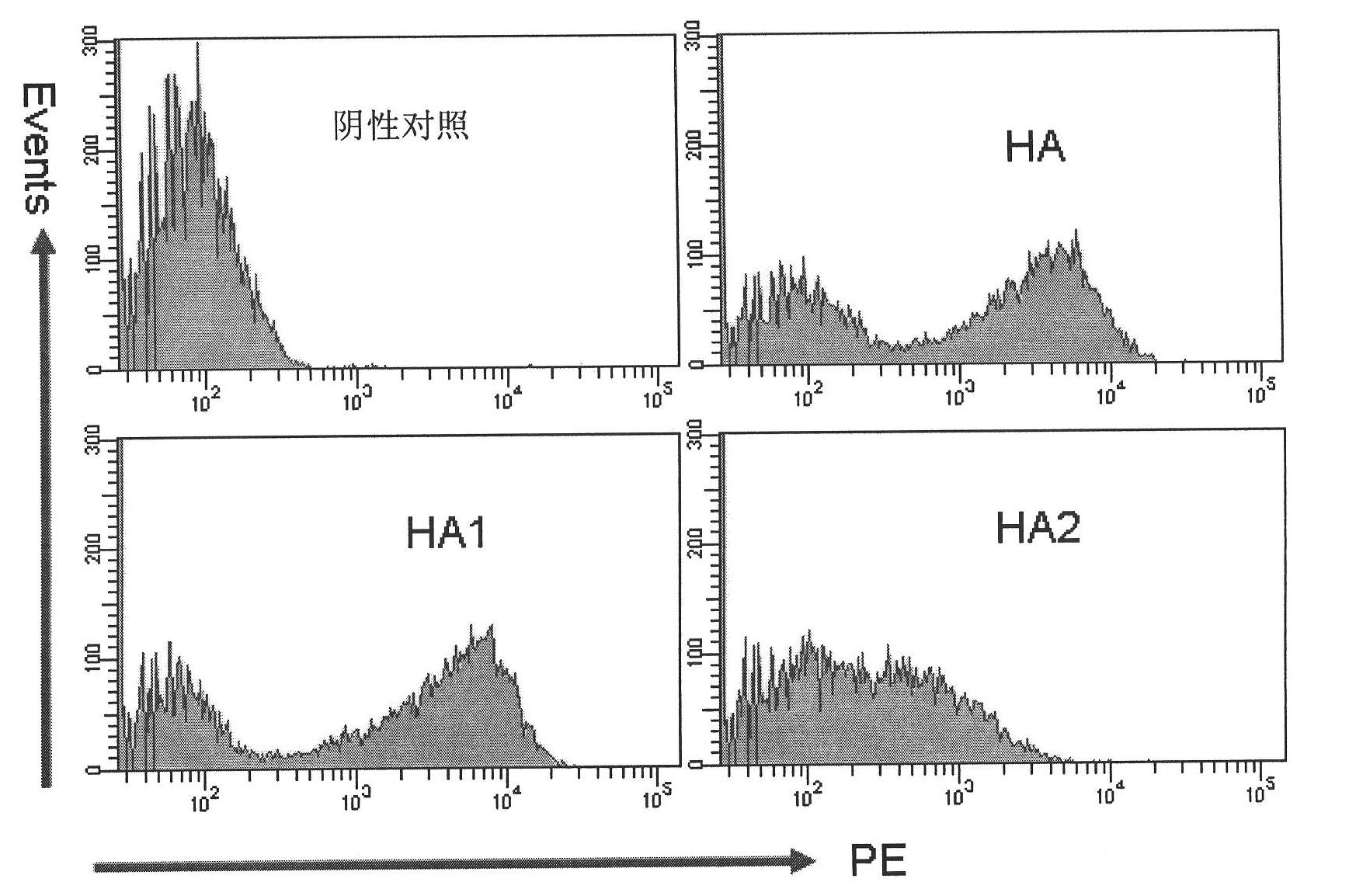
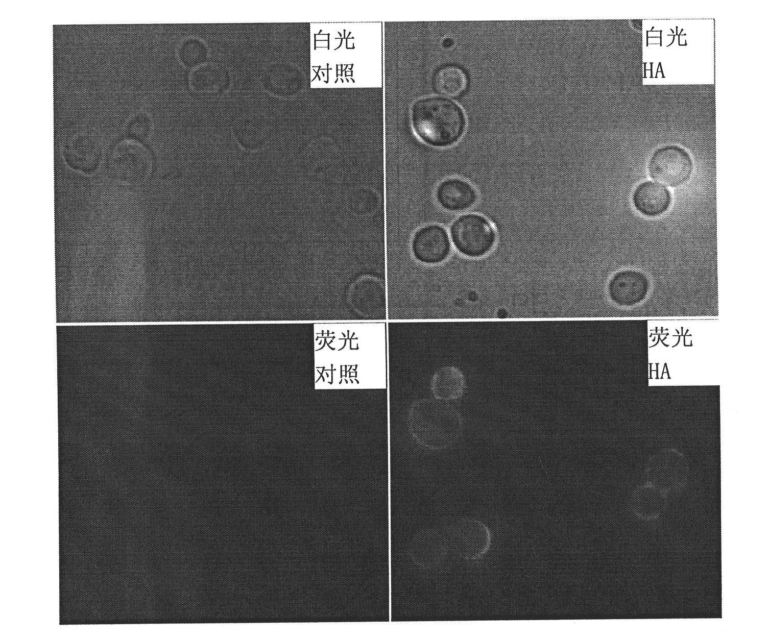
[0048] Example 1. Evaluation of the ability of pCTCON2 plasmid and yeast to display foreign protein

[0049] 1. Construction of recombinant plasmids

[0050] The H5N1-HA protein coding gene (the HA protein of H5N1 avian influenza virus, as shown in Sequence 1 of the Sequence Listing, and its coding gene as shown in Sequence 2 of the Sequence Listing) was inserted between the Nhe I and BamH I sites of the pCTCON2 plasmid. During this time, the recombinant plasmid A was obtained.

[0051] The H5N1-HA1 protein coding gene (as shown in the sequence 2 of the sequence listing from the 1-987th nucleotide at the 5' end, and the sequence 1 of the coding sequence listing from the amino acid residue shown at the 1st to the 329th position at the N-terminal. ) was inserted between the Nhe I and BamH I sites of the pCTCON2 plasmid to obtain a recombinant plasmid B.

[0052] The H5N1-HA2 protein encoding gene (as shown in the sequence 2 of the sequence listing from the 988th to 1545th nucl...

Embodiment 2

[0060] Example 2. Construction of pCTCON2-T plasmid (recombinant yeast display vector)

[0061] In order to facilitate library construction, the pCTCON2 plasmid needs to be transformed into a T vector, and the steps are as follows:

[0062] 1. Synthesize the DNA (double-stranded) shown in Sequence 3 of the Sequence Listing.

[0063] 5’-AGTCGCTAGC GGATCCGGGT-3' (sequence 3).

[0064] The Nhe I recognition sequence is underlined on the left, the BamH I recognition sequence is underlined on the right, and the two Xcm I recognition sequences are marked in bold.

[0065] 2. The DNA synthesized in step 1 was double digested with restriction enzymes Nhe I and BamH I, and the digested product was recovered.

[0066] 3. The pCTCON2 plasmid was digested with restriction endonucleases Nhe I and BamH I, and the vector backbone was recovered.

[0067] 4. Connect the enzyme digestion product of step 2 and the vector backbone of step 3 to obtain pCTCON2-T plasmid (the backbone vector is...

Embodiment 3

[0068] Example 3. Application of the method of the present invention to analyze the antigenic epitope of H5N1-HA protein

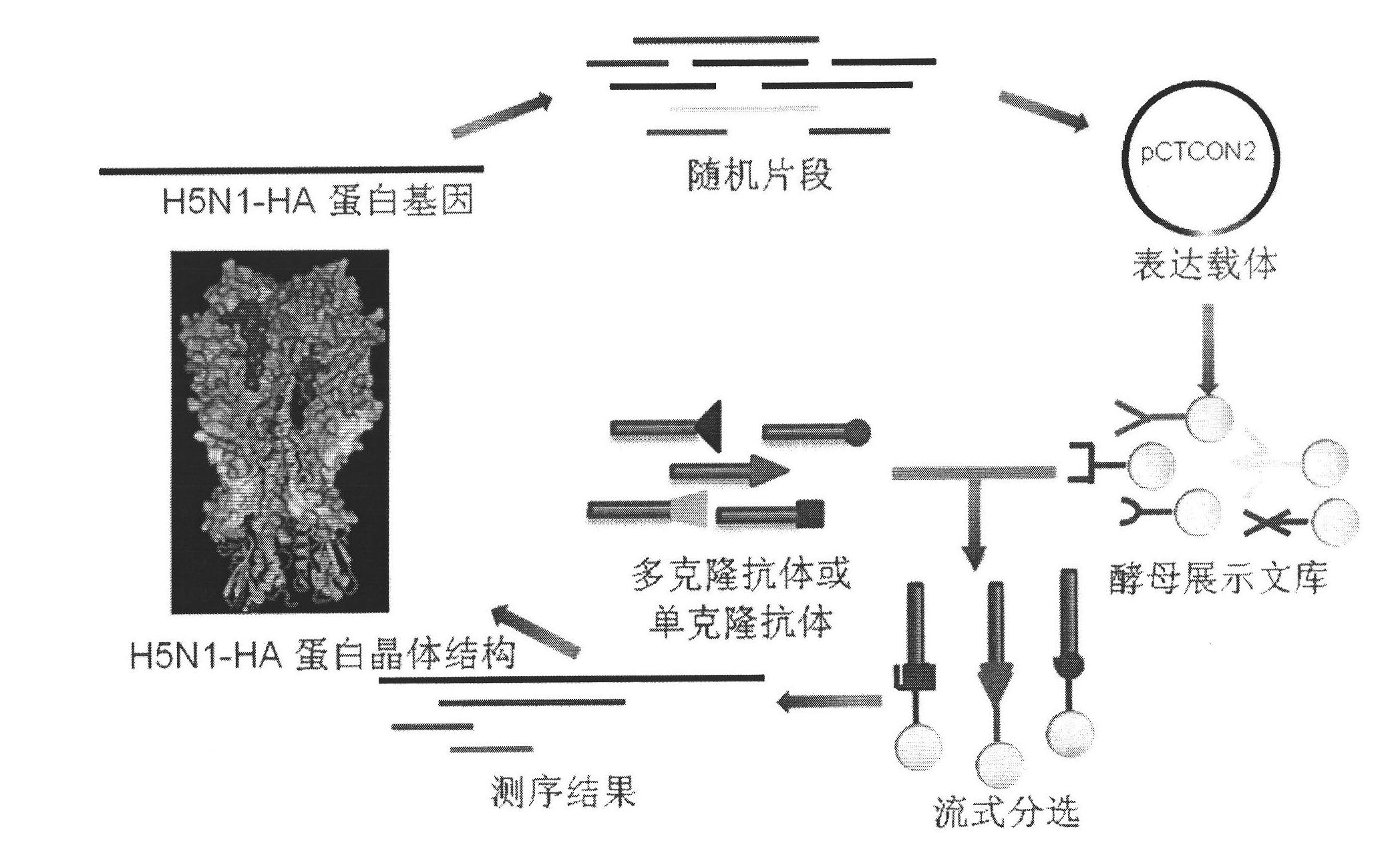
[0069] Apply the method of the present invention to screen the antigenic epitope of the H5N1-HA protein, the process is as follows image 3 shown. Firstly, the gene encoding H5N1-HA protein was randomly cut and recombined to obtain random fragments; the random fragments were inserted into the pCTCON2-T plasmid through T-A ligation; then Saccharomyces cerevisiae was transformed to induce expression of various fusion polypeptides (polypeptides encoded by random fragments and Aga2 The C-terminal fusion of the protein) is displayed on the surface of Saccharomyces cerevisiae (yeast display library); FACS sorting is performed by the antibody (polyclonal antibody or monoclonal antibody) of the H5N1-HA protein to obtain recombinant yeast bound to the antibody; The recombinant yeast plasmid was sequenced, and the antigen-antibody interaction was studied according ...


PUM


Abstract

The invention discloses a method for analyzing epitope of a protein, comprising the following steps of: (1) constructing a deoxyribonucleic acid (DNA) library of a target protein; inserting the DNA segment of the coding gene of the protein into a T carrier, wherein the T carrier contains the coding gene of yeast a lectin Aga2p; fusing a peptide coded by the DNA segment with the C end of the yeast a lectin Aga2p; (2) displaying the DNA library on yeast surfaces to obtain a yeast display library; (3) separating out yeasts combined with the antibody of the target protein; and (4) extracting plasmid sequencing and analyzing the epitope of the protein. The invention has the advantages that (1) yeast cells have multiple modification paths on the protein, can conveniently display the complicated high molecular weight protein and better simulates a space conformation; (2) the yeasts can be screened one by one by adopting flow cytometry (FACS) so as to improve the accuracy and the screening flux; (3) the yeasts can independently complete the self growth reproduction process, the operation is simple and convenient and the interference factors are few; and (4) the yeasts are not easy to propagate in the air or generate cross infection.

Description

technical field [0001] The present invention relates to a method for analyzing antigenic epitopes of proteins. Background technique [0002] The host's antibody response plays an important role in resisting pathogen infection, while the in vivo polyclonal antibody response consists of monoclonal antibodies directed against different antigens or different epitopes of the same antigen. An in-depth understanding of this complex process can provide key information for studying the pathogenesis of diseases, and can also provide an important theoretical basis for the development of efficient vaccines and disease treatment. [0003] Among the many research methods of antigenic epitopes, the phage display peptide library technology that emerged in 1990 (Scott, J.K.and G.P.Smith, Searching for peptide ligands with an epitope library. Science, 1990.249(4967): p.386-90. ), that is, use a randomly synthesized short peptide library (usually 6-12 amino acids, the library capacity can rea...

Claims


Application Information

Patent Timeline
IPC IPC(8): C12Q1/68C12N15/79C12N15/70G01N33/569
Inventor 张林琦林章凛史宣玲刘中华左腾徐望晖
Owner TSINGHUA UNIV
Why Eureka
Try Eureka