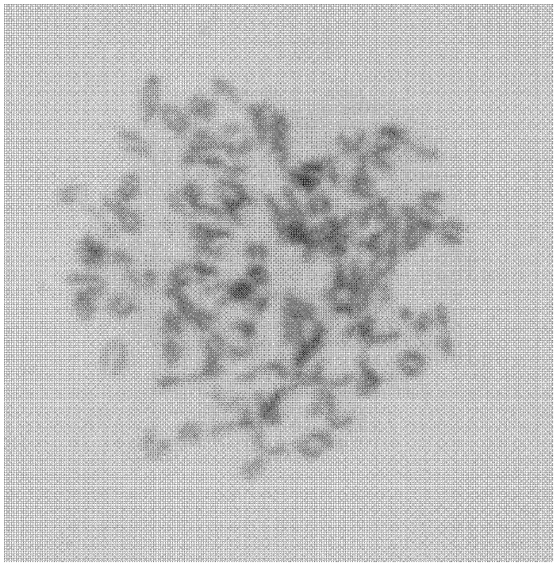
Monoclonal antibody of outer membrane protein of chlamydia abortus and application thereof
A monoclonal antibody, outer membrane protein technology, applied in anti-bacterial immunoglobulin, biochemical equipment and methods, instruments, etc., can solve the problems of high cost, narrow scope of application, unfavorable clinical popularization, etc., to achieve strong specificity, The effect of good fluorescence properties
- Summary
- Abstract
- Description
- Claims
- Application Information
AI Technical Summary
Problems solved by technology
Method used
Image
Examples
preparation example Construction
[0044] Preparation of monoclonal antibody ascites:
[0045] The screened hybridoma cells were inoculated into the peritoneal cavity of 10-week-old healthy Balb / c mice treated with inducer silica gel H (or sterilized liquid paraffin, 0.5mL / mouse) for 10 days in advance, 0.5mL / mouse (containing 5 ×10 5 ~1×10 6 hybridoma cells), after 14 days, it can be seen that the abdomen of the mouse increased significantly. At this time, the ascites was collected and the collected ascites was centrifuged at 3000r / min for 10min. Save for later.
[0046] Identification of monoclonal antibodies:
[0047] Using the Sigma mouse monoclonal antibody typing kit, the agar diffusion test was used to determine that 5 of the monoclonal antibodies secreted by the obtained 6 hybridoma cells were IgG1 type, and one was IgG3 type.
[0048] The potency of hybridoma cell culture supernatant was detected by indirect ELISA method. The results show that the C.abortus POMP18D-1 protein monoclonal antibody 1F...
Embodiment 2
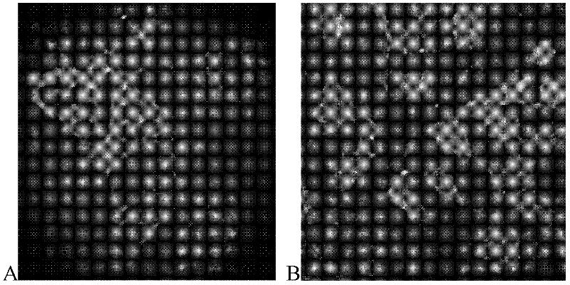
[0054] Example 2C. Establishment of abortus POMP18D-1 protein monoclonal antibody direct immunofluorescence detection method
[0055] Tissue sample processing: Take suspected abortion chlamydia disease materials, take appropriate amount of fresh lung, spleen, uterine mucosa, and vaginal mucosa, make 0.1-0.3mm frozen sections, fix with cold acetone for 10 minutes, and keep away from light for inspection.
[0056] Cell sample processing: Grind the diseased tissue at 4°C, treat with 200UI / mL gentamicin at room temperature for 30min, centrifuge at 1,000r / min for 10min, discard the precipitate, and use the supernatant to infect McCoy cells that have grown to a monolayer (24 Coverslips were pre-placed in the well cell plate), infected for 48 hours, the coverslips were taken out, fixed with pre-cooled methanol or absolute ethanol for 10 minutes, taken out to dry, and stored at -20°C for later use;
[0057]Take out the above prepared glass slides, add PBS dropwise to infiltrate for 10...
Embodiment 3
[0061] Example 3C. Establishment of abortus POMP18D-1 protein monoclonal antibody indirect immunofluorescence detection method
[0062] Take suspected C.abortus disease materials, cut out appropriate amount of lung, spleen, uterine mucosa, vaginal mucosa, grind at 4°C, treat with 200UI / mL gentamicin at room temperature for 30min, centrifuge at 1.000r / min for 10min, discard the precipitate, and use The supernatant was used to infect McCoy cells that had grown to a monolayer (the coverslips were placed in the 24-well cell plate in advance), for 48 hours after infection, the coverslips were taken out, fixed with pre-cooled methanol or absolute ethanol for 10 minutes, taken out and dried in the air at -20 Store at ℃ for later use.
[0063] Take out the prepared glass slide, add 25 μL 10 μg / ml C.abortus POMP18D-1 protein monoclonal antibody 1F10D4 dropwise, put it into a wet box, incubate at 37°C for 1 hour, take it out and wash it with PBST for 3 times, each time for 5 minutes, an...
PUM
 Abstract
Description
Claims
Application Information
 - R&D
- Intellectual Property
- Life Sciences
- Materials
- Tech Scout
- Unparalleled Data Quality
- Higher Quality Content
- 60% Fewer Hallucinations
Browse by: Latest US Patents, China's latest patents, Technical Efficacy Thesaurus, Application Domain, Technology Topic, Popular Technical Reports.
© 2025 PatSnap. All rights reserved.Legal|Privacy policy|Modern Slavery Act Transparency Statement|Sitemap|About US| Contact US: help@patsnap.com


