
Fusion protein capable of inducing and activating cancer-targeted T cells, preparation method and use thereof

A fusion protein and cancer cell technology, applied in the field of biomedicine, to achieve the effect of inhibiting tumor growth

Active Publication Date: 2012-03-21
孙嘉琳 +1
View PDF3 Cites 1 Cited by
  • Summary
  • Abstract
  • Description
  • Claims
  • Application Information

AI Technical Summary

Problems solved by technology

Using these T cells without cancer cell specificity to target cancer cells is a new and powerful method in the field of anti-cancer. Such drugs are different from antibodies and have not been reported yet

Method used


Image

  • Fusion protein capable of inducing and activating cancer-targeted T cells, preparation method and use thereof
  • Fusion protein capable of inducing and activating cancer-targeted T cells, preparation method and use thereof
  • Fusion protein capable of inducing and activating cancer-targeted T cells, preparation method and use thereof

Examples


Embodiment 1

[0031] Example 1 Construction of B7.2 (CD86) expression vector

[0032] According to the information (NM_175862 and L25259) of the B7.2 gene (also known as CD86) of human origin in the GenBank database, TAKARA was commissioned to synthesize a nucleic acid sequence fragment including a connecting peptide and a B7.2 gene (222 extracellular parts of the code Amino acid) and a few bases of HindIII in front of the whole fragment and XhoI restriction enzyme cutting point in the back, insert this synthesized nucleic acid fragment into T vector and carry out DNA sequencing identification, and then use HindIII and XhoI by double restriction method After treatment, this fragment was inserted into the pET22b plasmid, thus producing the expression vector pET22b-B7.2, which can express the B7.2 protein. The sequence listing (SEQ ID NO.2) is only the B7.2 protein, and the previous connecting peptide can be found in another sequence listing (SEQ ID NO.14).

Embodiment 2

[0033] Example 2 Construction of TGF-α-B7.2 expression vector

[0034] According to the TGF-α gene information (NM_003236) of human origin in the GenBank database, TAKARA was commissioned to synthesize a nucleic acid sequence fragment including the TGF-α gene and several restriction endonuclease sites BamHI and EcoRI in front of TGF-α. bases, the end of the fragment contains the restriction endonuclease sites of SalI and HindIII. The synthesized nucleic acid fragment was inserted into the T vector and identified by DNA sequencing, and then treated with BamHI and HindIII by double enzyme digestion, and then inserted into the pET22b-B7.2 plasmid, thus producing the expression vector pET22b - TGF-α-B7.2, capable of expressing TGF-α-B7.2 fusion protein (see SEQ ID NO.4 in the sequence listing).

Embodiment 3

[0035] Example 3 Construction of EGF-B7.2 expression vector

[0036] According to the information of the EGF gene (NM_001963 and NM_001178130) of the human origin of GenBank database, entrust TAKARA company to synthesize a nucleic acid sequence fragment and comprise EGF gene and several bases of restriction endonuclease site BamHI and EcoRI in front of EGF again, in The rear of the fragment contains restriction endonuclease sites for SalI and HindIII. The synthesized nucleic acid fragment was inserted into the T vector and identified by DNA sequencing, and then treated with BamHI and HindIII by double enzyme digestion, and then inserted into the pET22b-B7.2 plasmid, thus producing the expression vector pET22b - EGF-B7.2, capable of expressing EGF-B7.2 fusion protein (see SEQ ID NO.6 in the sequence listing).


PUM


Abstract

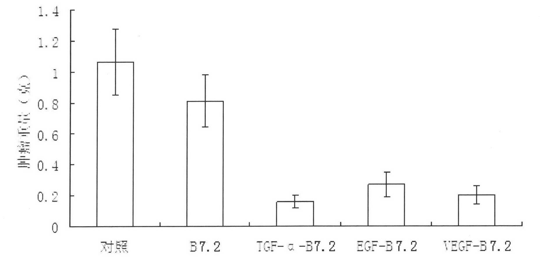
The present invention discloses a fusion protein capable of inducing and activating cancer-targeted T cells, a preparation method and use thereof. The protein comprises peptides which function on the cancer cells and costimulatory molecules B7.2. The peptides which function on the cancer cells are selected from transforming growth factor-alpha, epidernal growth factor, vascular endothelial growthfactor, gonadotropin-releasing hormone or gastrin-releasing peptide. The fusion protein of the invention has a cancer targeting function. On one hand, the fusion protein can respectively function with the VEGFR, EGFR, GnRH-R or GRP-R; and on the other hand, the fusion protein interacts with corresponding receptors CD28 and CTLA-4 which are expressed on the T cell. Thus, the T cell is targetedly positioned to the periphery of cancer cells which express VEGFR, EGFR, GnRH-R or GRP-R in great numbers. Experiments prove that the fusion protein of the invention can restrain tumor growth and causes cancer cell apoptosis.

Description

technical field [0001] The invention relates to a fusion protein, its preparation method and application, and belongs to the field of biomedicine. Background technique [0002] Epidermal growth factor (EGF), vascular endothelial cell growth factor (VEGF), gonadotropin-releasing hormone (GnRH) and gastrin-releasing peptide (GRP) ) and other receptors are abundantly expressed in various tumor tissues, for example, EGF receptors in intestinal mucosal tumors are 300 times higher than those in normal tissues (Gastroenterology, 98, 961-967, 1990). Therefore, abnormally high-expressed EGF receptors, VEGF receptors, GnRH receptors, GRP receptors, etc. have become attractive targets for cancer therapy. The receptor of transforming growth factor-α (Transforming growth factor-α, TGF-α) is the same as that of EGF. [0003] The above cytokines, hormones and peptides can interact with the corresponding receptors (Receptor) on cancer cells. Therefore, an effector such as a toxin protein...

Claims


Application Information

Patent Timeline
Patent Type & Authority Applications(China)
IPC IPC(8): C07K19/00C12N15/62C12N15/63C12N1/15C12N1/19C12N1/21C12N5/10C12N5/0783C12P21/00A61K38/16A61K47/48A61P35/00
Inventor 孙嘉琳
Owner 孙嘉琳
Learn More