Immunofluorescent reagent used for detecting Muscovy duck gosling blast diseases
An immunofluorescence and goose plague technology, applied in fluorescence/phosphorescence, measuring devices, instruments, etc., can solve the problems of lack of rapid diagnostic reagents for gosling plague in Muscovy ducks, difficulties in clinical diagnosis, etc., and achieves innovative, practical, and accurate Highly specific and highly specific effects
- Summary
- Abstract
- Description
- Claims
- Application Information
AI Technical Summary
Problems solved by technology
Method used
Image
Examples
preparation example Construction
[0020] 3.1 Preparation of GPV-infected cells: Muscovy duck gosling plague virus (GPV) was inoculated on a 96-well MDEF cell culture plate that had grown into a single layer, and normal MDEF cells were set as negative controls; cultured at 37°C and 5% CO2 When the cell lesion reaches about 30%, discard the culture supernatant, wash once with sterile PBS or normal saline, add 100 μL of cold methanol to each well, fix at 4°C for 30 min, discard the fixative and wash once at -40 ℃ frozen for later use.
[0021] 3.2 Preparation of tissue frozen sections: take the liver, spleen and pancreas of Muscovy duck artificially infected with Muscovy duck gosling plague virus (GPV) or the embryo heart of dead Muscovy duck inoculated with GPV, and cut into 0.5cm 3 About the size, placed on the tissue fixer of the cryostat, cut into 6-8um thin slices after the tissue was frozen, adhered to the glass slide, fixed in cold acetone for 10-15 minutes, and frozen at -20°C for later use.
[0022] 3.3...
experiment example
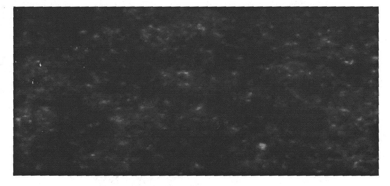
[0036] According to the operation method of direct immunofluorescence assay (DFA), after appropriately diluting the fluorescent antibody, take GPV, MPV, MDRV, ARV, PMV and other infected cells and normal control cells respectively, add 1:150 diluted fluorescent reagent 30ul to each well, and carry out DFA test. The results show that weak positive fluorescence can be seen in the nucleus of MDEF cells infected with GPV at the initial stage, and strong bright green fluorescence appears in the nucleus 36 hours after infection, and then the fluorescence gradually appears in the whole cell and goes out with the disintegration of the cell. Diffusion; while other virus-infected cells and normal MDEF cells did not show fluorescence. It shows that GPV immunofluorescence reagent can be used for the identification and antigen localization of GPV-infected cells.
[0037] Table 2. Identification of GPV antigens by immunofluorescence assay (DFA)
[0038]
[0039] Note: Judgment criteria...
PUM
 Abstract
Description
Claims
Application Information
 - R&D
- Intellectual Property
- Life Sciences
- Materials
- Tech Scout
- Unparalleled Data Quality
- Higher Quality Content
- 60% Fewer Hallucinations
Browse by: Latest US Patents, China's latest patents, Technical Efficacy Thesaurus, Application Domain, Technology Topic, Popular Technical Reports.
© 2025 PatSnap. All rights reserved.Legal|Privacy policy|Modern Slavery Act Transparency Statement|Sitemap|About US| Contact US: help@patsnap.com


