Preparation method of p75NTR-ED-Fc fusion protein and application of p75NTR-ED-Fc fusion protein in regeneration and functional recovery of injured central nerve
A p75ntr-ed-fc, 1.p75ntr-ed-fc technology, applied in the preparation method of peptides, nervous system diseases, peptide/protein components, etc. Purification and regeneration effect
- Summary
- Abstract
- Description
- Claims
- Application Information
AI Technical Summary
Problems solved by technology
Method used
Image
Examples
Embodiment 1
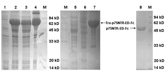
[0041] Implementation 1: The preparation method of p75NTR-ED-Fc fusion protein is realized by the following steps:
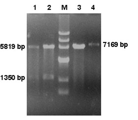
[0042] (1) Construction and identification of p75NTR-ED-Fc expression vector
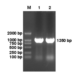
[0043] In order to obtain a large number of naturally expressed p75NTR-ED-Fc fusion proteins, the present invention uses the eukaryotic expression vector pcDNA-p75NTR-ED-Fc plasmid (preserved in our laboratory) as a template, and is based on the p75NTR nucleoside numbered NM_002507.1 in Genebank The acid sequence and the nucleotide sequence of the Fc segment of IgG numbered XM_002348257.1 are designed to design specific p75NTR forward primers and semi-specific p75NTR / Fc reverse primers, and introduce Kpn I and Xho I digestion at both ends of the primers. The site and corresponding protective bases were amplified by PCR to obtain the p75NTR-ED-Fc fragment. The primer sequence is as follows:
[0044] Forward primer: 5'CGG GGTACC ATGGGGGCAGGTGCCACC 3'
[0045] KpnI restriction site
[0046] Rev...
Embodiment 2
[0069] Example 2: In vitro observation of p75NTR-ED-Fc against NgR signal to promote the growth of neuronal processes
[0070] Isolate and culture the Dorsal root ganglia (DRG) neurons of newborn SD rats by conventional methods. 6 Cell density per ml was seeded in a 24-well culture plate coated with polylysine. Adjust the concentration of MAG, p75NTR-ED-Fc fusion protein and bovine serum albumin (BSA) to 100ng / ml with PBS in advance. After the above-mentioned cultured DRG neurons adhered to the wall, the fluid was changed, and they were randomly divided into 3 groups, and each group was repeated twice. The first group: add only 1ml MAG; the second group: add 1ml each of MAG and p75NTR-ED-Fc fusion protein; the third group: normal control, only add 1ml BSA. The cells in the above groups were fixed with 4% paraformaldehyde after 24 hours of culture, and the projection length after 24 hours of culture was measured by Image-Pro Plus 5.0 software, and neurite outgrowth assay was perf...
Embodiment 3
[0072] Example 3: In vivo detection of p75NTR-ED-Fc on the repair of injured spinal cord
[0073] The dorsal column of the spinal cord of adult SD rats was transected (1mm deep) with ophthalmic scissors to establish a rat spinal cord injury model. Immediately after injury, p75NTR-ED-Fc fusion protein (2mm each) was injected on both sides of the transversal point ( A total of 25μl, 0.4mg / ml), with spinal cord injury (SCI) as a negative control, while sham operation and normal rats as a positive control, retrograde tracing after 1, 2, 4, and 6w The technique detects the regeneration of axons. The detailed operation process is as follows: After spinal cord injury, each group of animals was injected with nuclear yellow (NY) into the sciatic nerve 16 to 18 hours before perfusion. The number of neurons indicates the regeneration of axons. At the same time, behavioral (BBB score, footprint test) and neuro-electrophysiological methods were used to detect the functional recovery of the i...
PUM
| Property | Measurement | Unit |
|---|---|---|
| Pre-denatured | aaaaa | aaaaa |
| Molecular weight | aaaaa | aaaaa |
Abstract
Description
Claims
Application Information
 - R&D
- Intellectual Property
- Life Sciences
- Materials
- Tech Scout
- Unparalleled Data Quality
- Higher Quality Content
- 60% Fewer Hallucinations
Browse by: Latest US Patents, China's latest patents, Technical Efficacy Thesaurus, Application Domain, Technology Topic, Popular Technical Reports.
© 2025 PatSnap. All rights reserved.Legal|Privacy policy|Modern Slavery Act Transparency Statement|Sitemap|About US| Contact US: help@patsnap.com


