Optical clearing agent for bone tissue

A technology of optical clearing agent and bone tissue, applied in the field of biomedical optical imaging, can solve problems such as restricting the development and application of cortical optical imaging in transcranial in vivo imaging, transparency of hard bone tissue, etc., so as to reduce the internal scattering of the tissue and improve the imaging depth. Effect

Active Publication Date: 2012-10-24
HUAZHONG UNIV OF SCI & TECH
View PDF1 Cites 20 Cited by
  • Summary
  • Abstract
  • Description
  • Claims
  • Application Information

AI Technical Summary

Problems solved by technology

However, due to the differences in the composition and structure of soft tissue and bone tissue, these light clearing agents that have been proven to be equally effective f

Method used


Image

  • Optical clearing agent for bone tissue
  • Optical clearing agent for bone tissue
  • Optical clearing agent for bone tissue

Examples


Embodiment 1

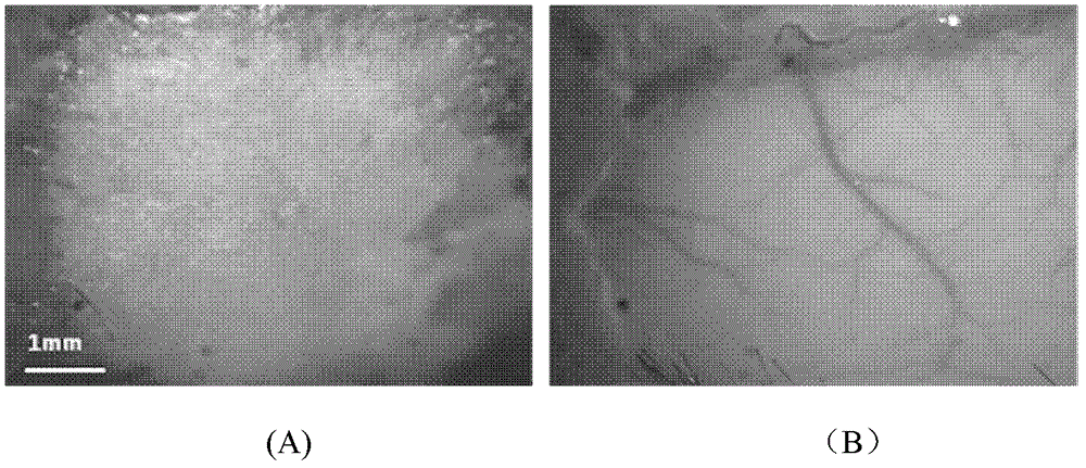
[0015] The scalp of a C57 mouse (20 weeks old) was cut open to expose the skull of about 1cm×1cm square. The optical transparency agent is prepared into a mixed solution according to the proportions of the components of the solution in Example 1 in Table 1, and is directly dripped onto the skull of a living mouse, smeared evenly (0.5-0.8ml / cm 2 ). figure 1 The CCD images taken before and after treatment with the transparent agent of the live mouse skull are given. among them figure 1 (A) is the case of a normal C57 mouse with bare skull. Due to the turbidity of the skull, it is impossible to see the intracranial cortical blood vessels; figure 1 (B) is the situation 5 minutes after the application of the transparent agent. At this time, the skull becomes transparent to light, the cortical blood vessels below the skull become clearly visible, and some small branches can also be distinguished in the visual field.

Embodiment 2

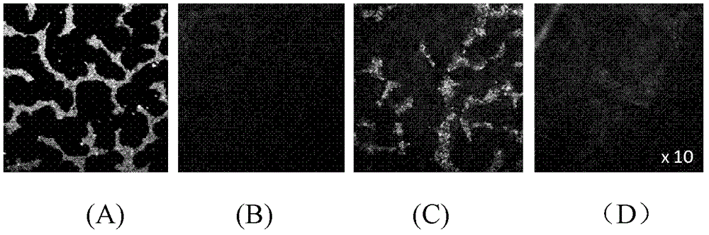
[0017] The isolated skull was taken from a 4-week-old SD rat, covered it on the encapsulated fluorescent bead solution, and observed with a fluorescence microscope. The transparent agent is prepared according to the ratio of the components of the solution in Example 2 in Table 1. The rat skull is immersed in vitro (1.5-2ml / cm 2 ) In a light transparent agent, 5 minutes later, cover the encapsulated fluorescent bead solution for imaging. Rinse the skull on which the optical transparency has been acted with saline to reverse the optical transparency effect, and then image under a fluorescent microscope. figure 2 The fluorescence signals observed without skull, with skull, phototransparency on the skull and after the phototransparency effect of normal saline is reversed are respectively given. figure 2 (A) gives the fluorescence signal observed without skull, the fluorescence information at this time is very strong; figure 2 It can be seen in (B) that the skull almost completely ...

Embodiment 3

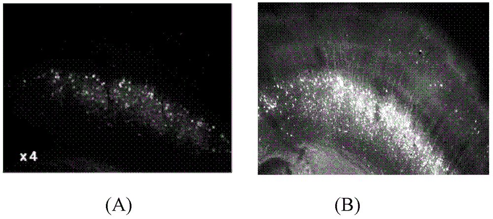
[0019] Cut the GFP-labeled transgenic mouse brain slices to a thickness of 100 μm. One group was not treated with a transparent agent and observed directly under a fluorescent microscope; the other group, the transparent agent was mixed according to the proportions of the components of the solution in Example 3 in Table 1. Solution, directly apply to mouse brain slices (0.2-0.4ml / cm 2 ), observe with a fluorescence microscope after 1 minute. image 3 Respectively given is the 4x objective lens before the optical transparency agent ( image 3 (A)) after ( image 3 (B)) Fluorescence signal of mouse brain slice. Only a small amount of neurocytes can be observed in the mouse brain slices that have not been treated with a transparent agent; but after a short period of treatment with a transparent agent, the cell bodies of the nerve cells are significantly brighter and the dendritic structure is now clearly distinguishable.

[0020] The different transparent agent formulations shown in ...


PUM


Abstract

The present invention discloses an optical clearing agent for bone tissue. The optical clearing agent comprises dimethyl sulfoxide, sodium bicarbonate, and at least two materials selected from polyethylene glycol-400, methyl salicylate, ethylenediaminetetraacetic acid, sodium diatrizoate, glycerol, glucose, sodium dodecylbenzene sulfonate, sorbitol and laurinol, wherein a volume percentage of the dimethyl sulfoxide in the optical clearing agent for bone tissue is 40-80%, a mass percentage of the sodium bicarbonate in the optical clearing agent for bone tissue is 1-2%, the balance is other materials, and the pH value of the optical clearing agent for bone tissue is 6-11. After the optical clearing agent for bone tissue in the present invention acts on bone tissue, a refractive index inside the tissue can be quickly homogenized, and scattering inside the tissue can be reduced, such that the tissue provides improved transparency for light, the imaging depth is increased, and a new method for obtaining cortex neuron subcellular structures and microvascular information is provided. After the optical clearing agent for bone tissue is locally coated on the bone tissues or is adopted to soak the bone tissues, the bone tissue can provide improved transparency for light.

Description

Technical field [0001] The invention belongs to the technical field of biomedical optical imaging, and specifically relates to a bone tissue optical transparency agent, which is a mixed solution that can change the optical transparency of bone tissue. Background technique [0002] With the development of the discipline of biomedical photonics, modern optical imaging technology provides a new method for obtaining high-resolution three-dimensional microstructures at the molecular level of biological tissues. However, the high scattering of visible and near-infrared light by biological tissues limits the penetration depth of light, making these technologies only capable of imaging superficial tissues, and the image contrast decreases significantly with the imaging depth. In recent years, the optical transparency technology for biological tissues has been proposed, which can effectively enhance the penetration depth of light in biological tissues by introducing hypertonic and high-re...

Claims


Application Information

Patent Timeline
IPC IPC(8): G01N1/28G01N1/30
Inventor 张洋
Owner HUAZHONG UNIV OF SCI & TECH
Why Eureka
Try Eureka