Preparation and application of tumor marker immunosensor built by putamen nanometer materials

A tumor marker and immunosensor technology, applied in the field of new nano functional materials and biosensing, can solve the problems of unreproducible sample luminescence and poor stability, and achieve the effect of batch sample determination, improved performance and high catalytic performance.

Active Publication Date: 2013-11-13
UNIV OF JINAN
View PDF3 Cites 0 Cited by
  • Summary
  • Abstract
  • Description
  • Claims
  • Application Information

AI Technical Summary

Problems solved by technology

However, there are many factors that affect the detection results of chemiluminescence analysis, so its stability is poor, and after the chemical reaction occurs, the luminescence of the sample cannot be reproduced

Method used


Image

  • Preparation and application of tumor marker immunosensor built by putamen nanometer materials
  • Preparation and application of tumor marker immunosensor built by putamen nanometer materials
  • Preparation and application of tumor marker immunosensor built by putamen nanometer materials

Examples


Embodiment 1

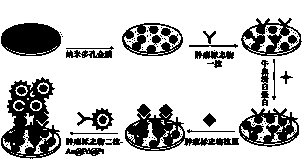
[0057] The preparation of a tumor marker immunosensor constructed of shell-core nanomaterials comprises the following steps.

[0058] (1) Float the silver-gold alloy film in 15 mol L -1 On the surface of the nitric acid solution for 0.5 min, the silver was etched away with concentrated nitric acid to form a nanoporous gold film. The nanoporous gold membrane was washed with ultrapure water until its pH = 7.0.

[0059] (2) Add 20 mmol·L to the flask -1 Chlorauric acid 2.5 mL; 20 mmol L -1 Sodium tetrachloropalladate 4.0 mL; 20 mmol L -1 Potassium tetrachloroplatinate 4.0 mL; block polyether F-127 0.1 g, mix well and add 0.4 mol·L -1 Ascorbic acid 1.0 mL. Stir at room temperature for 1 h. Centrifuged and washed 3 times with water, AuPdPt core-shell nanomaterials were made after drying. The morphology of the nanomaterials is shown in figure 1 ,Depend on figure 1 It can be seen from the transmission electron microscope that AuPdPt has a shell-core structure with a particl...

Embodiment 2

[0064] The preparation of a tumor marker immunosensor constructed of shell-core nanomaterials comprises the following steps.

[0065] (1) Float the silver-gold alloy film in 15 mol L -1 On the surface of the nitric acid solution for 3 min, the silver was etched away with concentrated nitric acid to form a nanoporous gold film. The nanoporous gold membrane was washed with ultrapure water until its pH = 7.0.

[0066] (2) Add 20 mmol·L to the flask -1 Chlorauric acid 2.5 mL; 20 mmol L -1 Sodium tetrachloropalladate 4.0 mL; 20 mmol L -1 Potassium tetrachloroplatinate 4.0 mL; block polyether F-127 0.1 g, mix well and add 0.4 mol·L -1 Ascorbic acid 1.0 mL. Stir at room temperature for 1 h. Centrifugal washing 3 times, after drying, the AuPdPt core-shell nanomaterials were made, and the TEM morphology was shown in figure 1 .

[0067] (3) Mix AuPdPt core-shell nanomaterials and tumor marker secondary antibodies into pH = 7.4 phosphate buffer solution to make a mixed solution,...

Embodiment 3

[0071] The preparation of a tumor marker immunosensor constructed of shell-core nanomaterials comprises the following steps.

[0072] (1) Float the silver-gold alloy film in 15 mol L -1 On the surface of the nitric acid solution for 5 min, the silver was etched away with concentrated nitric acid to form a nanoporous gold film. The nanoporous gold membrane was washed with ultrapure water until its pH = 7.0.

[0073] (2) Add 20 mmol·L to the flask -1 Chlorauric acid 2.5 mL; 20 mmol L -1 Sodium tetrachloropalladate 4.0 mL; 20 mmol L -1 Potassium tetrachloroplatinate 4.0 mL; block polyether F-127 0.1 g, mix well and add 0.4 mol·L -1 Ascorbic acid 1.0 mL. Stir at room temperature for 1 h. Centrifugal washing 3 times, after drying, the AuPdPt core-shell nanomaterials were made, and the TEM morphology was shown in figure 1 .

[0074] (3) Mix AuPdPt core-shell nanomaterials and tumor marker secondary antibodies into pH = 7.4 phosphate buffer solution to make a mixed solution,...


PUM

PropertyMeasurementUnit
recovery rateaaaaaaaaaa

Abstract

The invention relates to preparation and application of a tumor marker immunosensor built by putamen nanometer materials and belongs to the technical fields of novel nanometer functional materials and biosensors. The preparation and the application are characterized by comprising (1) preparation of nanometer multihole gold film; (2) preparation of a secondary antibody composite of an Au@Pd@Pt putamen nanometer material-tumor marker; and (3) building of an electrochemistry immunosensor. The preparation and application have the advantages of being high in sensitivity, good in specificity, easy to operate, low in limit of detection and capable of achieving high sensitivity, specificity and rapid and accurate detection of a plurality of tumor markers.

Description

technical field [0001] The invention relates to the preparation and application of a tumor marker immunosensor constructed by shell core nanomaterials. Specifically, it is based on nanoporous gold film and AuPdPt shell core nanomaterials, the preparation method and application of a sandwich-type electrochemical immunosensor for detecting multiple tumor markers, which is used to detect tumor markers in serum. It belongs to the field of new nano functional material and biosensing technology. Background technique [0002] Tumor markers have important practical value in the early diagnosis of cancer. Tumor markers are a class of substances produced by tumor cells themselves or by the body's response to tumor cells during the occurrence and proliferation of tumor cells, reflecting the existence and growth of tumors. Including proteins, hormones, enzymes, polyamines and oncogene products. [0003] Tumor markers are widely used in clinical practice, and play an extremely importa...

Claims


Application Information

Patent Timeline
Patent Type & Authority Patents(China)
IPC IPC(8): G01N33/574G01N33/76G01N27/327
Inventor 魏琴张勇张潇月吴丹于海琴李贺杜斌罗川南曹伟马洪敏李玉阳朱宝存闫良国李燕孙蒙
Owner UNIV OF JINAN
Why Eureka
Try Eureka