Ribonuclease detection method and applications thereof, and kit

A technology of ribonuclease and detection method, which is applied in the field of tumor detection kits, can solve the problems that are not suitable for high-throughput detection of sample ribonuclease activity, achieve high use value, improve specificity and accuracy, and be simple to operate Effect

Inactive Publication Date: 2013-04-03
SUZHOU RIBO LIFE SCIENCE CO LTD
View PDF0 Cites 7 Cited by
  • Summary
  • Abstract
  • Description
  • Claims
  • Application Information

AI Technical Summary

Problems solved by technology

Although these different technical schemes and their improved schemes improve the sensitivity and accuracy of ribonuclease activity detection to a certain extent, they all have certain defects and are not suitable as a general research method for the detection of ribonuclease activity in samples. High-throughput detection, there is still a lot of room for improvement in technology

Method used


Image

  • Ribonuclease detection method and applications thereof, and kit
  • Ribonuclease detection method and applications thereof, and kit
  • Ribonuclease detection method and applications thereof, and kit

Examples


Embodiment 1

[0062] This example is used to illustrate the sequence structure of the oligoribonucleotide used in the present invention and the optical parameters of the fluorescent group.

[0063] Entrust Guangzhou Ruibo Biotechnology Co., Ltd. to synthesize oligoribonucleotides with the sequence shown in Table 1, and the fluorescent group is labeled at the 5' end of the oligoribonucleotides. Complementary oligoribonucleotides can be annealed to form double-stranded ribonucleic acid according to the method in "Molecular Cloning: A Laboratory Manual". Table 1 shows the sequences of the synthesized oligoribonucleotides. Wherein, in the oligoribonucleotide represented by SEQ ID NO: 11, the pentose sugar of the guanylic acid residue at position 3 is modified with 2'-oxymethyl (2'-OME). In the oligoribonucleotide represented by SEQ ID NO: 12, the pentose sugar of the uridine acid residue at position 14 was modified by 2'-fluoro substitution (2'-Fluro substitution). In the oligoribonucleotide ...

Embodiment 2

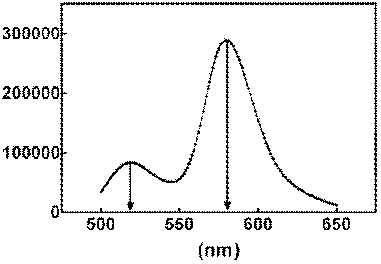
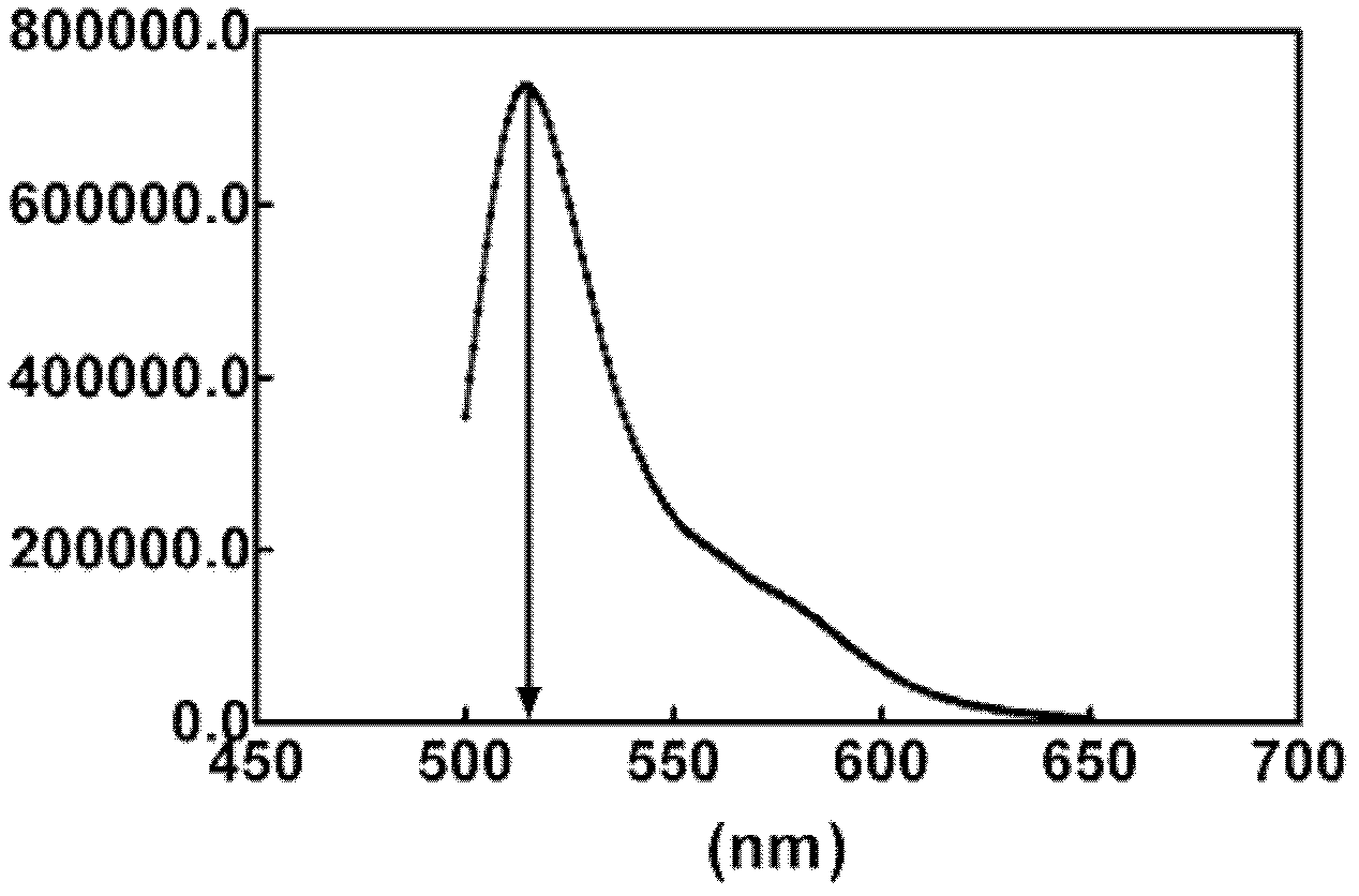
[0069] This example is used to illustrate the identification of fluorescence resonance energy transfer phenomenon. In the technical scheme of the application of fluorescence resonance energy transfer method detection ribonuclease level provided by the present invention, a key condition is to be able to Fluorescence resonance energy transfer occurs.

[0070] In order to verify this, at first the oligoribonucleotide (5'-FAM-AUGAGCCUGAUUU) shown in the synthetic SEQ ID NO:1 and the oligoribonucleotide (UACUCGGACUAAA-TAMRA) shown in SEQ ID NO:2 were synthesized -5') anneal to form complementary double-stranded ribonucleic acid substrate DS1. Take 4 μl of DS1 and add it to 2ml fluorescence resonance energy transfer buffer (0.01M Tris-HCl, pH 7.4, 0.002M MgCl 2 ), the final concentration of DS1 was 10 nM, and then the reaction system was added into the quartz detection cup of the fluorescence spectrometer, and a micro-magnetic rotor was placed in the quartz cup. Put the quartz cu...

Embodiment 3

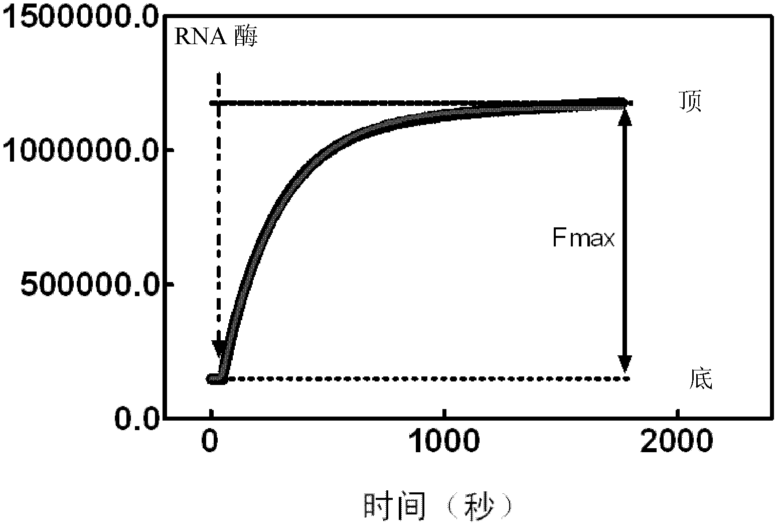
[0073] This example is used to illustrate the method of the present invention for detecting the level of ribonuclease in a sample by using fluorescence resonance energy transfer.

[0074] In order to utilize the FRET method to detect the activity and content of ribonuclease in the sample, take 4 μ l of double-stranded ribonucleic acid substrate DS1 and join in 2 ml fluorescence resonance energy transfer buffer (0.01M Tris-HCl, pH7.4, 0.002M MgCl 2 ), the final concentration of DS1 was 10 nM, and then the reaction system was added into the quartz detection cup of the fluorescence spectrometer, and a micro-magnetic rotor was placed in the quartz cup. After adding 20 μl of a certain concentration of RNase A to the reaction system, the reaction system is continuously excited at 480nm and detected at 515nm at the same time to obtain the fluorescence intensity values ​​at intervals of seconds, and use these values ​​to draw the detection time-fluorescence intensity curve .

[0075]...


PUM


Abstract

The present invention provides a ribonuclease detection method, which comprises the following steps that: 1) a body fluid sample is acquired from an individual subject; 2) the body fluid sample contacts double-stranded nucleic acid, such that the double-stranded nucleic acid can be cut by ribonuclease possibly existing in the body fluid sample; and 3) products after contacting in the step 2) are detected so as to detect content of the ribonuclease in the body fluid sample. The present invention further provides applications of the detection method and a tumor detection kit. With the ribonuclease detection substrate and the detection method, activities and contents of ribonucleases in sera derived from different tumor patients can be sensitively and accurately detected, such that operation is convenient, and detection cost is substantially reduced. In addition, high application values are provided for early detection, efficacy evaluation, population mass screening and other large-scale applications of a plurality of diseases.

Description

technical field [0001] The invention relates to the fields of medicine and molecular diagnosis, in particular to a method for detecting ribonuclease in body fluid of a subject, an application of the method in tumor detection and a tumor detection kit. Background technique [0002] Early diagnosis is a key link in the treatment of diseases, especially tumors. Generally, people detect various tumors by measuring the substances corresponding to each tumor in the blood (often referred to as tumor markers). The tumor markers can be defined as "in the substances produced by cancer cells or in the substances produced by the interaction between normal cells and cancer cells in the body, which can be detected by blood, tissue and excreta (urine and feces) Substances that are examined for use as markers for the diagnosis or treatment of cancer". [0003] Ribonuclease, or RNase, is a large class of enzymes including dozens of species that can hydrolyze RNA substrates into small molec...

Claims


Application Information

Patent Timeline
Patent Type & Authority Applications(China)
IPC IPC(8): C12Q1/68C12Q1/44
Inventor 不公告发明人
Owner SUZHOU RIBO LIFE SCIENCE CO LTD
Why Eureka
Try Eureka