Separate detection system and method of rare cells based on centrifugal micro-fluidic technology

A microfluidic technology and detection system technology, applied in biological testing, material inspection products, material excitation analysis, etc., can solve the problems of restricting large-scale applications, time-consuming, expensive instruments, etc., and achieve faster separation and detection speed and efficiency , Improve the separation purity, fast and efficient separation effect

Active Publication Date: 2013-04-03
SHANGHAI INST OF MICROSYSTEM & INFORMATION TECH CHINESE ACAD OF SCI
View PDF4 Cites 37 Cited by
  • Summary
  • Abstract
  • Description
  • Claims
  • Application Information

AI Technical Summary

Problems solved by technology

Nevertheless, this method still has major deficiencies: on the one hand, in order to ensure the capture efficiency of cells, the blood sample must maintain a relatively slow speed (~1mL /h), so the detection process of this method takes a long time (5~7 hours), and the efficiency is low
On the other hand, the chip used is made of opaque silicon base, which is not conducive to optical detection, and the plasma etching process of the micropillars in the chip requires expensive instruments and high production costs, which limits its large-scale clinical application
Although, some people ha...

Method used


Image

  • Separate detection system and method of rare cells based on centrifugal micro-fluidic technology
  • Separate detection system and method of rare cells based on centrifugal micro-fluidic technology
  • Separate detection system and method of rare cells based on centrifugal micro-fluidic technology

Examples


Embodiment 1

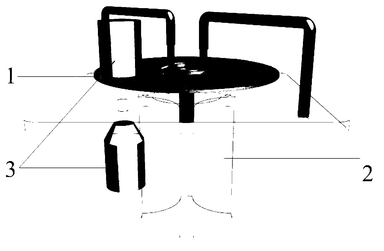
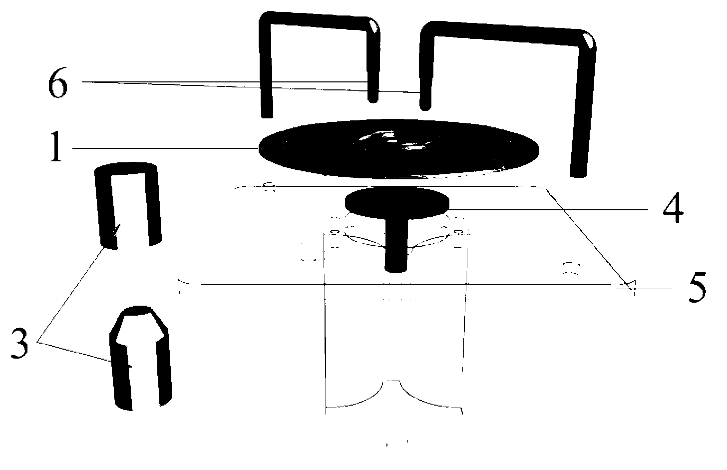
[0023] Collect about 5mL of peripheral blood from tumor patients, add EpCAM-labeled immune microspheres that can bind to tumor cells into the blood sample, and then inject it into the liquid storage cavity of the microfluidic chip of the rare cell separation detection system; place the chip after adding the sample Put it on the centrifugal platform, and assemble the elastic micro-column, rotate the centrifugal platform at 60~100 rpm to realize automatic and rapid mixing reaction; after 5 minutes, increase the rotational speed of the rotating centrifugal platform to 500 rpm to achieve the separation of target cells; 10 After 10 minutes, turn off the centrifugal platform, tear off the sealing tape of the sample port above each cell collection area, and add CD45-FITC that can identify blood cells and CK-PE antibody that specifically marks tumor cells into the cell collection area in turn, and the liquid addition is complete. Then seal each sample port with tape again; incubate for...

Embodiment 2

[0025] Collect about 100mL of human bone marrow, add CD71-labeled immune microspheres that can bind to fetal red blood cells into the blood sample, and then inject it into the liquid storage chamber of the microfluidic chip of the rare cell separation detection system; place the chip after adding the sample in a centrifuge On the platform, assemble the elastic micro-column, rotate the centrifuge platform at 60~100 rpm to realize automatic and rapid mixing reaction; after 5 minutes, increase the speed of the rotating centrifugal platform to 500 rpm to achieve the separation of target cells; after 10 minutes , turn off the operation of the centrifugal platform, and tear off the sealing tape of the sample port above each cell collection area, add the fluorescently labeled antibody solution that can recognize fetal red blood cell hemoglobin into the cell collection area, and seal the sample port again with tape after the liquid addition is completed; Incubate for 20 minutes, inject...

Embodiment 3

[0027] Collect about 1mL of human umbilical cord blood, add immune microspheres that can selectively bind to hematopoietic stem cells into the blood sample, and then inject it into the liquid storage chamber of the microfluidic chip of the rare cell separation detection system; place the added chip in a centrifuge On the platform, assemble the elastic micro-column, rotate the centrifuge platform at 60~100 rpm to realize automatic and rapid mixing reaction; after 5 minutes, increase the speed of the rotating centrifugal platform to 500 rpm to achieve the separation of target cells; after 10 minutes , turn off the operation of the centrifugal platform, and tear off the sealing tape of the sample port above each cell collection area, add the CD45-FITC solution that can identify hematopoietic stem cells into the cell collection area, and seal the sample port again with tape after the liquid addition is completed; For 20 minutes, inject phosphate buffer solution (PBS) into the chip ...


PUM

PropertyMeasurementUnit
Apertureaaaaaaaaaa

Abstract

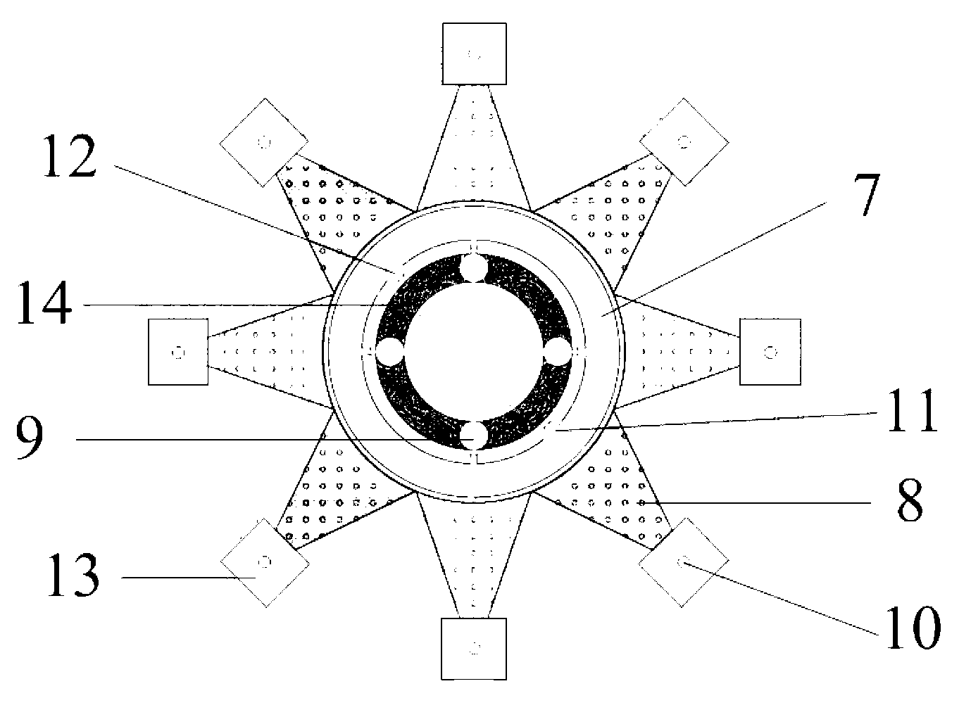
The invention discloses a separate detection system and method of rare cells based on a centrifugal micro-fluidic technology. The system comprises a micro-fluidic chip similar to an optical disk, a centrifugal drive module and an optical detection module, wherein the micro-fluidic chip comprises a plurality of groups of micro-channels and micro-cavities arranged in a radiation manner; and the entire chip structure is composed of an elastic micro-column guide rail layer, a deformable film layer, a pipeline/cavity layer, a filtering membrane layer and a liquid waste collecting layer. The method comprises the steps of: firstly, leading a sample solution and immune modified microspheres into a liquid storage cavity through an injection port of the micro-fluidic chip in use, putting the sample solution and the immune modified microspheres on a centrifugal platform of the centrifugal drive module, assembling an elastic micro-column, and rotating at a low speed, so as to achieve fully mixing and reacting of the sample solution and immune modified microsphere liquid in the liquid storage cavity, separating by a high-speed rotary chip, and then dropwise adding a specifically recognized fluorescently-labeled antibody solution in each separate cell collection area, carrying out incubate reaction, adding a buffer solution and centrifuging, and finally identifying and analyzing through the optical detection module.

Description

technical field [0001] The invention relates to a rare cell separation and detection system based on centrifugal microfluidic technology and its application method, which can be applied to the fields of non-invasive prenatal diagnosis, early tumor diagnosis and prognosis monitoring, stem cell research and the like. Background technique [0002] Selective isolation of rare cells from complex mixtures is of great significance in both cell biology and clinical aspects, such as isolation of fetal red blood cells from maternal peripheral blood, isolation of circulating tumor cells (Circulating Tumor Cell, CTC) from peripheral blood of patients, and bone marrow or Separation of stem cells in tissues, etc. The analysis of these cells can often provide very valuable information for clinical diagnosis and treatment of diseases, but the content of such cells in samples is very rare, usually only 1-10 cells / mL, separation and extraction very difficult, thus greatly limiting its clinica...

Claims


Application Information

Patent Timeline
IPC IPC(8): G01N21/64G01N1/28G01N33/53
Inventor 李刚贾春平赵建龙
Owner SHANGHAI INST OF MICROSYSTEM & INFORMATION TECH CHINESE ACAD OF SCI
Why Eureka
Try Eureka