Confocal laser scanning microscope

A confocal microscope and laser scanning technology, applied in fluorescence/phosphorescence, material excitation analysis, etc., can solve problems such as impossible, inability to judge the type of phase, inability to visually observe the distribution of tiny impurities or doped phases, etc., to promote development Effect

Inactive Publication Date: 2013-04-17
JIANGSU UNIV
View PDF7 Cites 7 Cited by
  • Summary
  • Abstract
  • Description
  • Claims
  • Application Information

AI Technical Summary

Problems solved by technology

[0002] The current main methods for characterizing the microscopic morphology of the surface or interface of material samples include scanning electron microscopy (SEM), atomic force microscopy (AFM), transmission electron microscopy (TEM), etc.: SEM cannot test water-containing samples, and for some tiny (particle size <1nm) Impurities or doped phases must be increased in magnification, and the increase in magnification reduces the field of view, so it is impossible to visually observe the distribution of tiny impurities or doped phases in a larger area (about 1mm×1mm); AFM is difficult and easy Judgment of impurity phases; TEM has high requirements on samples, sample preparation is difficult, and like SEM, it is impossible to visually observe the distribution of tiny impurities or doped phases in a larger area
The current non-destructive testing methods for the composition of material samples include X-ray diffraction (XRD), energy dispersive spectrometer (EDS) or spectrometer (WDS) added to the scanning electron microscope, etc.: XRD is powerless for specific microphases or phases with small content; EDS or WDS can only obtain the relative content of elements, but cannot judge the phase type (for example, the ratio of Ti to O can only be known as 1:2, but rutile or anatase cannot be judged), and there is nothing to be done about the composition inside the cavity
Therefore, there is currently no material sample characterization method that can make up for the shortcomings of the above-mentioned characterization techniques at the same time, and it is even more impossible to dynamically study the heat and mass transfer process of various materials when the temperature changes using the existing testing methods in the field of materials.

Method used


Image

  • Confocal laser scanning microscope
  • Confocal laser scanning microscope
  • Confocal laser scanning microscope

Examples


Embodiment 1

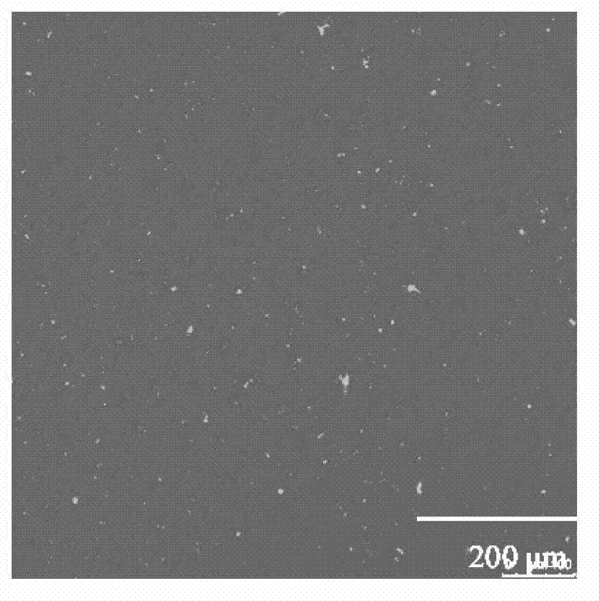
[0021] Bright-field microscopic imaging of pure copper calcium titanate thin films prepared on silicon substrates was carried out by laser scanning confocal microscopy. From figure 2 It can be seen that the inorganic thin film prepared on the opaque substrate can be well imaged, and the thin film is dense and uniform in a wide range, containing a small amount of impurities.

Embodiment 2

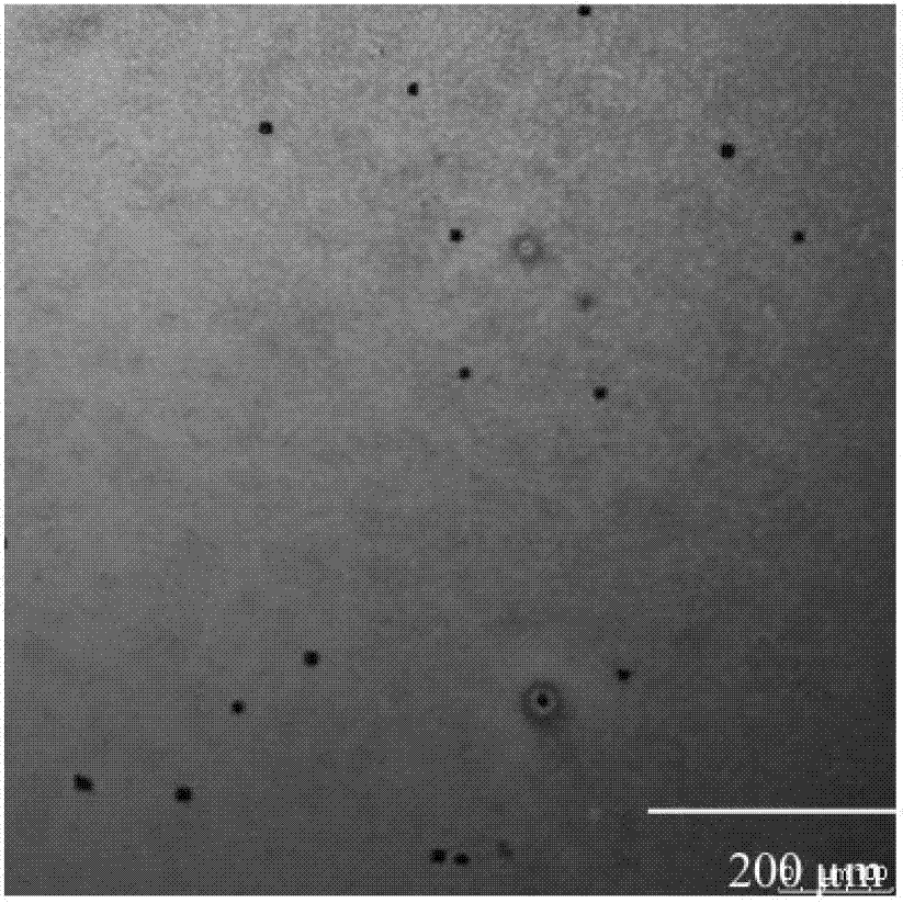
[0023] Bright-field microscopic imaging of ZnO-doped copper calcium titanate thin films prepared on silicon substrates was carried out by laser scanning confocal microscopy. From image 3 It can be seen that the laser scanning confocal microscope has a high resolution for material characterization, the tiny cracks in the film are clearly discernible, and the impurity phase in the film can be easily identified through the difference between light and dark.

Embodiment 3

[0025] Using a 380 nm laser as the light source, the pure copper calcium titanate film and the ZnO-doped copper calcium titanate film were imaged by laser scanning confocal microscope. From Figure 4 and Figure 5 It can be seen that there are no fluorescent bright spots in the pure copper calcium titanate film, but blue fluorescent bright spots of ZnO are found in the ZnO-doped copper calcium titanate thin film. In a large field of view, the doped ZnO and the parent phase copper calcium titanate can be clearly distinguished, and the distribution, migration direction and growth process of ZnO in the parent phase are clear at a glance.


PUM


Abstract

The invention relates to a confocal laser scanning microscope. An objective table of the confocal laser scanning microscope is additionally provided with a heating device; a temperature measuring device and an infrared detector are additionally arranged between an objective lens and a sample platform of the confocal laser scanning microscope; and heat image processing software is additionally arranged in a computer system of the confocal laser scanning microscope. The confocal laser scanning microscope relates to the following novel detection characterization techniques: (1) the microstructures of materials can be researched; (2) the distribution condition in a large range is directly observed by applying impurity or doped phase fluorescence excitation; (3) fluorescence is captured to dynamically track; (4) a system is focused for many times to obtain images with different depths; and three-dimensional imaging can be implemented for samples through computer processing; and (5) the heat and mass transfer processes of various materials can be synchronously dynamically researched when the temperature is changed, so that material phase changed thermodynamics and dynamics mechanisms can be built.

Description

technical field [0001] The invention relates to a novel analysis and characterization device for material samples, in particular to a laser scanning confocal microscope, which belongs to the technical field of material characterization. Background technique [0002] The current main methods for characterizing the microscopic morphology of the surface or interface of material samples include scanning electron microscopy (SEM), atomic force microscopy (AFM), transmission electron microscopy (TEM), etc.: SEM cannot test water-containing samples, and for some tiny (particle size <1nm) Impurities or doped phases must be increased in magnification, and the increase in magnification reduces the field of view, so it is impossible to visually observe the distribution of tiny impurities or doped phases in a larger area (about 1mm×1mm); AFM is difficult and easy Judgment of impurity phases; TEM has high requirements for samples, sample preparation is difficult, and like SEM, it is i...

Claims


Application Information

Patent Timeline
Patent Type & Authority Applications(China)
IPC IPC(8): G01N21/64
Inventor 徐东程晓农
Owner JIANGSU UNIV
Why Eureka
Try Eureka